

zalambdalestes - American Museum of Natural History
zalambdalestes - American Museum of Natural History
zalambdalestes - American Museum of Natural History

You also want an ePaper? Increase the reach of your titles
YUMPU automatically turns print PDFs into web optimized ePapers that Google loves.
46 BULLETIN AMERICAN MUSEUM OF NATURAL HISTORY NO. 281<br />
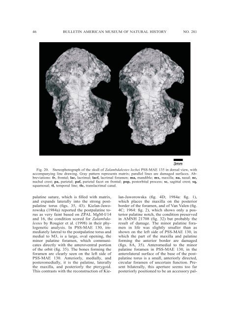
Fig. 20. Stereophotograph <strong>of</strong> the skull <strong>of</strong> Zalambdalestes lechei PSS-MAE 135 in dorsal view, with<br />
accompanying line drawing. Gray pattern represents matrix; parallel lines are damaged surfaces. Abbreviations:<br />
fr, frontal; lac, lacrimal; lacf, lacrimal foramen; ma, mandible; mx, maxilla; na, nasal; nc,<br />
nuchal crest; pa, parietal; paf, parietal facet on frontal; pop, postorbital process; sc, sagittal crest; sq,<br />
squamosal; tl, temporal line; tlc, translacrimal canal.<br />
palatine suture, which is filled with matrix,<br />
and expands laterally into the strong postpalatine<br />
torus (figs. 35, 43). Kielan-Jaworowska<br />
(1984a) reported the postpalatine torus<br />
as very faint based on ZPAL MgM-I/14<br />
and 16, the condition scored for Zalambdalestes<br />
by Rougier et al. (1998) in their phylogenetic<br />
analysis. In PSS-MAE 130, immediately<br />
lateral to the postpalatine torus and<br />
medial to M3, is a large, oval opening, the<br />
minor palatine foramen, which communicates<br />
directly with the anteroventral portion<br />
<strong>of</strong> the orbit (fig. 35). The bones forming the<br />
foramen are clearly seen on the left side <strong>of</strong><br />
PSS-MAE 130. Anteriorly, medially, and<br />
posteromedially, it is the palatine, laterally<br />
the maxilla, and posteriorly the pterygoid.<br />
This contrasts with the reconstruction <strong>of</strong> Kielan-Jaworowska<br />
(fig. 4D; 1984a: fig. 1),<br />
which places the maxilla on the posterior<br />
border <strong>of</strong> the foramen, and <strong>of</strong> Van Valen (fig.<br />
4C; 1964: fig. 2), which shows only a posterior<br />
palatine notch, the condition preserved<br />
in AMNH 21708 (fig. 32) but probably the<br />
result <strong>of</strong> damage. The minor palatine foramen<br />
in life was slightly smaller than as<br />
shown on the left side <strong>of</strong> PSS-MAE 130, in<br />
which the part <strong>of</strong> the maxilla and palatine<br />
forming the anterior border are damaged<br />
(figs. 8A, 35). Anteromedial to the minor<br />
palatine foramen in PSS-MAE 130, in the<br />
anterolateral surface <strong>of</strong> the base <strong>of</strong> the postpalatine<br />
torus is a small, anteriorly directed,<br />
circular foramen <strong>of</strong> uncertain function. Present<br />
bilaterally, this aperture seems too far<br />
posteriorly positioned to be an accessory pal-


