

zalambdalestes - American Museum of Natural History
zalambdalestes - American Museum of Natural History
zalambdalestes - American Museum of Natural History

Create successful ePaper yourself
Turn your PDF publications into a flip-book with our unique Google optimized e-Paper software.
20 BULLETIN AMERICAN MUSEUM OF NATURAL HISTORY NO. 281<br />
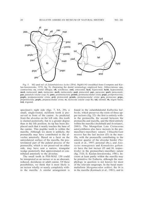
Fig. 5. M2 amd m3 <strong>of</strong> Zalambdalestes lechei ZPAL MgM-I/43 (modified from Crompton and Kielan-Jaworowska,<br />
1978: fig. 9), illustrating the dental terminology employed here. Abbreviations: cec,<br />
centrocrista; co, cristid obliqua; efl, ect<strong>of</strong>lexus; end, entoconid; hyd, hypoconid; hyld, hypoconulid;<br />
med, metaconid; met, metacone; metl, metaconule; pad, paraconid; par, paracone; parl, paraconule;<br />
pas, parastyle (stylar cusp A); pmc, postmetacrista; pomlc, postmetaconular crista; popc, postprotocrista;<br />
poplc, postparaconular crista; prd, protoconid; prmlc, premetaconular crista; pro, protocone; prpc,<br />
preprotocrista; prplc, preparaconular crista; st, stylocone (stylar cusp B); tal, talonid; tb, trigon basin;<br />
trd, trigonid.<br />
specimen’s right side (figs. 7, 8A, 29), a<br />
small, single-rooted, styliform tooth is preserved<br />
in front <strong>of</strong> the canine. As predicted<br />
from the alveolus on the left side, this tooth<br />
is slanted posteriorly, but to a greater degree<br />
than in the life position; its tip has been displaced<br />
such that it nearly touches the base <strong>of</strong><br />
the canine. This peglike tooth is within the<br />
maxilla. Although we deem it unlikely, the<br />
premaxilla may have contributed to the alveolus<br />
anteriorly. Based on a facet on the<br />
anterolateral surface <strong>of</strong> the maxilla, the posterolateral<br />
part <strong>of</strong> the palatal process <strong>of</strong> the<br />
premaxilla, which is not preserved on either<br />
side, may have sent a narrow, triangular<br />
wedge posteriorly that approximated or contributed<br />
anteriorly to the alveolus.<br />
The small tooth in PSS-MAE 130 could<br />
be interpreted as an incisor or as an aberrant,<br />
reduced, deciduous or adult canine. Of these<br />
possibilities, we think that it most likely is<br />
an incisor wholly or nearly completely within<br />
the maxilla. A similar arrangement is<br />
found in the zalambdalestid Kulbeckia kulbecke,<br />
which preserves the roots <strong>of</strong> three upper<br />
incisors (fig. 52): the first is entirely within<br />
the premaxilla, the second between the<br />
premaxilla and maxilla, and the third entirely<br />
within the maxilla (Archibald and Averianov,<br />
2003). The Mongolian Late Cretaceous<br />
asioryctitheres also have incisors in the premaxillary-maxillary<br />
suture; Ukhaatherium<br />
nessovi has the last incisor (I5) in the maxilla,<br />
with the premaxilla contributing to the<br />
anterior quarter <strong>of</strong> the alveolar border (Novacek<br />
et al., 1997; personal obs.), and Asioryctes<br />
nemegtensis and Kennalestes gobiensis<br />
have the last incisor (I5 and I4, respectively)<br />
in the premaxillary-maxillary suture<br />
(fig. 51D, E; Kielan-Jaworowska, 1981). Incisors<br />
at least partly within the maxilla may<br />
be primitive for Eutheria, although the morphology<br />
in question is not known for most<br />
<strong>of</strong> the relevant outgroups. In the basal mammaliaform<br />
Morganucodon oehleri, the I4 is<br />
in the maxilla (Kermack et al., 1981), and in


