

zalambdalestes - American Museum of Natural History
zalambdalestes - American Museum of Natural History
zalambdalestes - American Museum of Natural History

Create successful ePaper yourself
Turn your PDF publications into a flip-book with our unique Google optimized e-Paper software.
48 BULLETIN AMERICAN MUSEUM OF NATURAL HISTORY NO. 281<br />
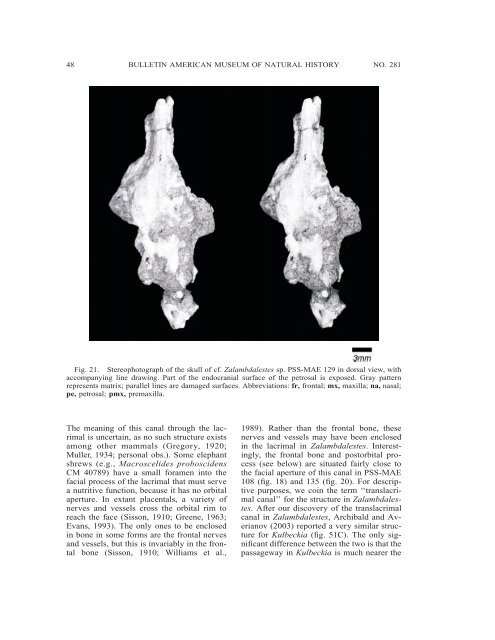
Fig. 21. Stereophotograph <strong>of</strong> the skull <strong>of</strong> cf. Zalambdalestes sp. PSS-MAE 129 in dorsal view, with<br />
accompanying line drawing. Part <strong>of</strong> the endocranial surface <strong>of</strong> the petrosal is exposed. Gray pattern<br />
represents matrix; parallel lines are damaged surfaces. Abbreviations: fr, frontal; mx, maxilla; na, nasal;<br />
pe, petrosal; pmx, premaxilla.<br />
The meaning <strong>of</strong> this canal through the lacrimal<br />
is uncertain, as no such structure exists<br />
among other mammals (Gregory, 1920;<br />
Muller, 1934; personal obs.). Some elephant<br />
shrews (e.g., Macroscelides proboscidens<br />
CM 40789) have a small foramen into the<br />
facial process <strong>of</strong> the lacrimal that must serve<br />
a nutritive function, because it has no orbital<br />
aperture. In extant placentals, a variety <strong>of</strong><br />
nerves and vessels cross the orbital rim to<br />
reach the face (Sisson, 1910; Greene, 1963;<br />
Evans, 1993). The only ones to be enclosed<br />
in bone in some forms are the frontal nerves<br />
and vessels, but this is invariably in the frontal<br />
bone (Sisson, 1910; Williams et al.,<br />
1989). Rather than the frontal bone, these<br />
nerves and vessels may have been enclosed<br />
in the lacrimal in Zalambdalestes. Interestingly,<br />
the frontal bone and postorbital process<br />
(see below) are situated fairly close to<br />
the facial aperture <strong>of</strong> this canal in PSS-MAE<br />
108 (fig. 18) and 135 (fig. 20). For descriptive<br />
purposes, we coin the term ‘‘translacrimal<br />
canal’’ for the structure in Zalambdalestes.<br />
After our discovery <strong>of</strong> the translacrimal<br />
canal in Zalambdalestes, Archibald and Averianov<br />
(2003) reported a very similar structure<br />
for Kulbeckia (fig. 51C). The only significant<br />
difference between the two is that the<br />
passageway in Kulbeckia is much nearer the


