Novel FGF8 Mutations Associated with Recessive ...
Novel FGF8 Mutations Associated with Recessive ...
Novel FGF8 Mutations Associated with Recessive ...

Create successful ePaper yourself
Turn your PDF publications into a flip-book with our unique Google optimized e-Paper software.
J Clin Endocrinol Metab, October 2011, 96(10):0000–0000 jcem.endojournals.org 3<br />
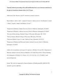
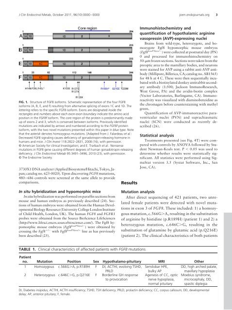
FIG. 1. Structure of <strong>FGF8</strong> isoforms. Schematic representation of the four <strong>FGF8</strong><br />
isoforms (A, B, E, and F) resulting from alternative splicing of exons 1C and 1D. The<br />
lettering refers to the specific <strong>FGF8</strong> isoform. Exons are designated inside the<br />
rectangles and numbers above each exon-exon boundary indicate the amino acid<br />
position in the <strong>FGF8</strong>f isoform. The core region of the protein is predominantly made<br />
up of exons 2 and 3, which is conserved between isoforms. Previously identified<br />
mutations are indicated by arrows and numbered according to the <strong>FGF8</strong>f protein<br />
isoform, <strong>with</strong> the two novel mutations presented <strong>with</strong>in this paper in blue type. Note<br />
that the asterisk denotes homozygous mutations. [Adapted from J. Falardeau et al.:<br />
Decreased <strong>FGF8</strong> signaling causes deficiency of gonadotropin-releasing hormone in<br />
humans and mice. J Clin Invest 118:2822–2831, 2008 (16), <strong>with</strong> permission.<br />
© American Society for clinical Investigation; and E. Trarbach et al.: Nonsense<br />
mutations in <strong>FGF8</strong> gene causing different degrees of human gonadotropin-releasing<br />
deficiency. J Clin Endocrinol Metab 95:3491–3496, 2010 (23), <strong>with</strong> permission.<br />
© The Endocrine Society.<br />
3730X1 DNA analyzer (Applied Biosystems/Hitachi, Tokyo, Japan;<br />
catalog no. 625-0020). Upon discovering <strong>FGF8</strong> mutations,<br />
480–686 controls were screened at the same allele to provide<br />
comparisons.<br />
Results<br />
Immunohistochemistry and<br />
quantification of hypothalamic arginine<br />
vasopressin (AVP)-expressing nuclei<br />
Brains from wild-type, heterozygote, and homozygote<br />
Fgf8 hypomorphic mouse embryos<br />
(Fgf8 loxPNeo/ ) were collected at postnatal day (PN)<br />
0 and processed for immunohistochemistry on<br />
50-m frozen sections. Sections were taken from the<br />
preoptic area to the mamillary bodies, and neurons<br />
were stained for AVP using a rabbit anti-AVP antibody<br />
(Millipore, Billerica, CA; catalog no. AB1565)<br />
for 48 h at 4 C. These were then sequentially incubated<br />
<strong>with</strong> a biotinylated donkey antirabbit secondary<br />
antibody (1:500; Jackson ImmunoResearch,<br />
West Grove, PA) and the avidin-biotin complex<br />
(Vector Laboratories, Burlingame, CA). Immunoreactivity<br />
was visualized <strong>with</strong> diaminobenzidine as<br />
the chromagen before counterstaining <strong>with</strong> methyl<br />
green.<br />
Quantification of AVP-immunoreactive paraventricular<br />
nuclei (PVN) and suprachiasmatic<br />
nuclei (SCN) were conducted as recently described<br />
(26).<br />
Statistical analysis<br />
Treatments presented (see Fig. 4T) were compared<br />
<strong>with</strong> controls by ANOVA followed by Student<br />
Newman-Keuls test. P 0.05 was used to<br />
determine whether results were statistically significant.<br />
All statistics were performed using SigmaStat<br />
version 3.5 (Systat Software, Inc., San<br />
Jose, CA).<br />
In situ hybridization and hypomorphic mice<br />
In situ hybridization was performed on paraffin sections from<br />
mouse and human embryos as previously described (24). Sections<br />
of human embryos were obtained from the Human Developmental<br />
Biology Resource (University College London Institute<br />
of Child Health, London, UK). The human <strong>FGF8</strong> and FGFR1<br />
probes were obtained from the Source BioScience LifeSciences<br />
(http://www.lifesci.ences.sourcebioscience.com/). The Fgf8 hypomorphic<br />
mouse embryos (Fgf8 loxPNeo/ ) were obtained by<br />
crossing the Fgf8 / <strong>with</strong> Fgf8 loxPNeo/ line as has previously<br />
been described (25).<br />
Mutation analysis<br />
After direct sequencing of 421 patients, two unrelated<br />
female patients were detected <strong>with</strong> novel mutations<br />
in exon 3 of <strong>FGF8</strong>. These included: 1) a homozygous<br />
mutation, c.566GA, resulting in the substitution<br />
of arginine by histidine (p.R189H) (patient 1) and 2) a<br />
heterozygous mutation, c.646CG, resulting in the<br />
substitution of glutamine by glutamic acid (p.Q216E)<br />
(patient 2). The clinical characteristics of both patients<br />
TABLE 1. Clinical characteristics of affected patients <strong>with</strong> <strong>FGF8</strong> mutations<br />
Patient<br />
no. Mutation Position Sex Hypothalamo-pituitary MRI Other<br />
1 Homozygous c.566GA, p.R189H F DI, ACTHI, evolving TSHD, Semilobar HPE, DD, high arched palate,<br />
PRLD<br />
2 Heterozygous c.646CG, p.Q216E F Borderline GH response<br />
to provocation<br />
bulky AP<br />
Agenesis of CC, optic<br />
nerve hypoplasia,<br />
normal pituitary<br />
maxillary hypoplasia<br />
Moebius syndrome,<br />
microcephaly, DD,<br />
spastic diplegia<br />
DI, Diabetes insipidus; ACTHI, ACTH insufficiency; TSHD, TSH deficiency; PRLD, prolactin deficiency; CC, corpus callosum; DD, developmental<br />
delay; AP, anterior pituitary; F, female.