A Study of Endometrial Pathology by Transvaginal Color ... - medIND
A Study of Endometrial Pathology by Transvaginal Color ... - medIND
A Study of Endometrial Pathology by Transvaginal Color ... - medIND
Create successful ePaper yourself
Turn your PDF publications into a flip-book with our unique Google optimized e-Paper software.
134 Indian Medical Gazette — APRIL 2013<br />
Original <strong>Study</strong><br />
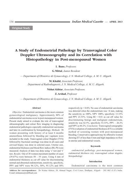
A <strong>Study</strong> <strong>of</strong> <strong>Endometrial</strong> <strong>Pathology</strong> <strong>by</strong> <strong>Transvaginal</strong> <strong>Color</strong><br />
Doppler Ultrasonography and its Correlation with<br />
Histopathology in Post-menopausal Women<br />
I. Bano, Pr<strong>of</strong>essor,<br />
G. Mittal, Junior Resident<br />
— Department <strong>of</strong> Obstetrics & Gynaecology, J. N. Medical College, A. M. U. Aligarh.<br />
M. Khalid, Associate Pr<strong>of</strong>essor,<br />
Department <strong>of</strong> Radiodiagnosis, J. N. Medical College, A. M. U. Aligarh.<br />
Nishat Akhtar, Associate Pr<strong>of</strong>essor,<br />
Z. Arshad, Pr<strong>of</strong>essor<br />
— Department <strong>of</strong> Obstetrics & Gynaecology, J. N. Medical College, A. M. U. Aligarh.<br />
Abstract<br />
Objective: <strong>Endometrial</strong> carcinoma is the most common<br />
gynaecological malignancy. Approximately 80% <strong>of</strong><br />
endomentrial carcinoma occur in post-menopausal women.<br />
Present study aimed to evaluate the role <strong>of</strong> transvaginal<br />
ultrasonography and colour flow imaging in diagnosing<br />
endometrial pathologies especially endometrial carcinoma<br />
and later its confirmation <strong>by</strong> histopathology. Methods: 38<br />
women presenting with history <strong>of</strong> at least 6 months<br />
amenorrhoea followed <strong>by</strong> bleeding per vaginum were<br />
included in the study. <strong>Transvaginal</strong> colour Doppler (TVS)<br />
followed <strong>by</strong> fractional curettage was done in all cases and<br />
cervical biopsy was done in selected cases. Uterine size,<br />
endometrial thickness and blood flow indices (RI, PI) were<br />
measured. Analysis <strong>of</strong> data was done using ‘z’ test and ‘t’<br />
test. Results: Out <strong>of</strong> 38 women maximum number <strong>of</strong> cases<br />
(39.47%) were between 50 – 55 years. Using 4 mm <strong>of</strong><br />
endometrial thickness as cut <strong>of</strong>f value for discriminating<br />
normal and abnormal endometrium, sensitivity, specificity,<br />
PPV and NPV were 94.12%, 50%, 95.12% and 50%<br />
respectively (p < 0.05). No case <strong>of</strong> endometrial carcinoma<br />
was detected when the endometrium was
Indian Medical Gazette — APRIL 2013 135<br />
gynaecological malignancy. It is the seventh leading cause<br />
<strong>of</strong> death form malignancy in women, yet also the one with<br />
best survival statistics. There has been a steady increase in<br />
incidence over recent years which may be related to<br />
increased longevity, increased cholesterol in the diet,<br />
exogenous estrogen supplementation and probably better<br />
diagnostic methods.<br />
Approximately, 80% <strong>of</strong> endometrial carcinoma occur<br />
in postmenopausal women, with bleeding as the initial<br />
symptom in 90% <strong>of</strong> cases. To diagnose endometrial<br />
pathology these women are subjected to diagnostic<br />
curettage. However, diagnostic curettage is an invasive<br />
procedure and not without danger in the elderly. The false<br />
negative rate for curettage is 2-6% because curettage may<br />
not empty the uretine cavity completely. Therefore, non<br />
invasive technique like ultrasonography is being utilized<br />
nowadays to detect endometrial carcinoma. Recently<br />
transvaginal colour and pulsed Doppler ultrasound has<br />
increased the reliability <strong>of</strong> ultrasonographic diagnosis <strong>of</strong><br />
women with certain endometrial pathologies. It is able to<br />
detect subtle changes in the endometrium and it has been<br />
observed that endometrial thickness 6 cm in size)<br />
whereas only 3 cases (7.89%) had normal sized uterus. In<br />
our study, duration <strong>of</strong> menopause ranged from 6 months<br />
to 25 years with median as 2 years.<br />
Correlation <strong>of</strong> Ultrasound with Histopathology<br />
Report<br />
The category normal endometrium comprised the<br />
histologic diagnosis <strong>of</strong> atrophic endometrium and those<br />
reported as insufficient sample. The category abnormal<br />
endometrium included hyperplasia, proliferative<br />
endometrium, endometritis and endometrial carcinoma.<br />
Table 1 shows that no case <strong>of</strong> abnormal endometrium<br />
was found when the size <strong>of</strong> uterus small i.e. < 6 cm whereas<br />
34 cases <strong>of</strong> abnormal endometrium and one case <strong>of</strong> normal<br />
endometrium was found when the size <strong>of</strong> uterus was bulky<br />
i.e. >6 cm. Correlation between the size <strong>of</strong> uterus and<br />
endometrial pathology was highly significant.<br />
Mean <strong>of</strong> Malignant endometrium = 16.36±7.1 mm, t =<br />
3.4 (p
136 Indian Medical Gazette — APRIL 2013<br />
were <strong>of</strong> atrophic, proliferative, hyperplastic endometrium<br />
and insufficient sample as reported on histopathological<br />
examination. Thus, when endometrial thickness was more<br />
than 4 mm, incidence <strong>of</strong> endometrial carcinoma was high<br />
which was even more when endometrial thickness was<br />
>10 mm. Using Z test, significant difference was found<br />
between the mean <strong>of</strong> benign and malignant endometrium.<br />
Table 3 shows that using 4 mm <strong>of</strong> endometrial thickness<br />
as cut <strong>of</strong>f value for discriminating normal and abnormal<br />
endometrium, sensitivity was 94.12%, specificity 50%,<br />
positive predictive value 95.12%, and negative predictive<br />
value 50%. Using ‘Z’ test, we found significant correlation<br />
between endometrial pathology and endometrial thickness.<br />
Above table depicts that no case <strong>of</strong> endometrial<br />
carcinoma was detected when the endometrium was 4 mm <strong>of</strong><br />
endometrial thickness and all the 8 cases <strong>of</strong> endometrial<br />
carcinoma had more than 4 mm thick endometrium. Thus<br />
using endometrial thickness we can exclude malignant<br />
endometrium with confidence but cannot differentiate<br />
between benign and malignant endometrium when<br />
endometrial thickness (ET) is > 4 mm (p < 0.05).<br />
Using PI = 1.83 as cut <strong>of</strong>f value for discriminating<br />
between benign and malignant endometrium, sensitivity was<br />
75%, specificity 56.67%, positive predictive value 31.56%<br />
and negative predictive value 89.47% using ‘t’ test, no<br />
significant difference was observed between mean PI values<br />
<strong>of</strong> benign and malignant endometrium. Benign endometrium<br />
was associated with high mean RI value (0.95±0.21) ranging<br />
from 0.57 to 1.8 indicating high resistance to blood flow in<br />
uterine artery whereas malignant endometrium was<br />
associated with low value <strong>of</strong> mean RI (0.77±0.14) ranging<br />
from 0.55 to 0.96 suggesting a low resistance to blood<br />
flow in uterine artery in presence <strong>of</strong> malignancy. Using ‘t’
Indian Medical Gazette — APRIL 2013 137<br />
effort has been made to predict the efficacy <strong>of</strong> transvaginal<br />
color Doppler sonography in discriminating between benign<br />
and malignant endometrium and whether ultrasonographic<br />
evaluation could help avoid an unnecessary curettage in<br />
these patients.<br />
test, we observed significant difference between mean RI<br />
values <strong>of</strong> benign and malignant endometrium.<br />
Table 5 shows that using RI = 0.81 as cut <strong>of</strong>f value for<br />
discrimination between benign and malignant endometrium,<br />
sensitivity was 62.5% specificity = 53.33%, positive<br />
predictive value = 26.3% and negative predictive value was<br />
84.2%.<br />
Discussion<br />
It is well known fact that cancer does not develop<br />
suddenly from normal but is preceded <strong>by</strong> various histological<br />
changes such as hyperplasia. If these changes are<br />
recognized and treated early then it will be possible to<br />
decrease significantly the frequency <strong>of</strong> invasive cancer. An<br />
Results <strong>of</strong> previous studies have shown that endometrial<br />
thickness is a reliable method <strong>of</strong> detecting pathological<br />
changes in the endometrium. The maximum thickness <strong>of</strong><br />
the atrophic endometrium as measured histologically in the<br />
previous studies is 3 mm. Based on these findings, we have<br />
chosen cut <strong>of</strong>f limit <strong>of</strong> 4 mm <strong>of</strong> endometrial thickness for<br />
discriminating between normal and abnormal endometrium<br />
or between benign and malignant endometrium.<br />
The maximum number <strong>of</strong> cases (50%) with benign<br />
endometrium were immediately postmenopausal whereas<br />
those with malignant endometrium were usually more than<br />
10 years postmenopausal. Thus endometrial carcinoma in<br />
the present study was found to be more common after 10<br />
years <strong>of</strong> attaining menopause. This is in accordance with<br />
Novak’s Gynaecology (12th Ed. 1996) 3 , that peak incidence<br />
<strong>of</strong> endometrial cancer occurs 10-15 years after average<br />
age <strong>of</strong> menopause.<br />
In the present study, we observed significant difference
138 Indian Medical Gazette — APRIL 2013<br />
between the mean endometrial thickness in cases with<br />
benign endometrium i.e. 9.2 mm (S.D. = 4.67 mm) and in<br />
cases with malignant endometrium i.e. 16.36 (S.D> = 7.1<br />
mm) whereas another study showed mean endometrial<br />
thickness as 8 mm ± 9 mm in absence <strong>of</strong> cancer and 20<br />
mm ± 9 mm in presence <strong>of</strong> cancer 4 .<br />
With 20 mm thick.<br />
Using 4 mm <strong>of</strong> endometrial thickness as a cut <strong>of</strong>f value<br />
for discriminating between normal and abnormal<br />
endometrium; sensitivity and positive predictive value were<br />
91% and 94% respectively in our study. Using ‘Z’ test<br />
significant correlation was found between endometrial<br />
pathology and endometrial thickness (p
Indian Medical Gazette — APRIL 2013 139<br />
neovascularization and decreased pulsatility index and<br />
resistive index in uterine and endometrial vessels in malignant<br />
conditions.<br />
Thus, a conservative approach may be <strong>of</strong>fered to<br />
women with post menopausal bleeding showing endometrial<br />
thickness <strong>of</strong>


