Get PDF (1214K) - Wiley Online Library
Get PDF (1214K) - Wiley Online Library
Get PDF (1214K) - Wiley Online Library

Create successful ePaper yourself
Turn your PDF publications into a flip-book with our unique Google optimized e-Paper software.
204 S.M. Küchler et al.<br />
Screening for the presence of Rickettsia sp.<br />
Six Deronectes species and one subspecies were examined<br />
using Rickettsia-specific primers in a diagnostic PCR assay<br />
(Table 1). A total of 45 D. platynotus were screened. Rickettsia<br />
sp. could be detected in 100% of all tested D. platynotus. In<br />
contrast, only 39.4% of 71 examined individuals of D. aubei<br />
were infected. Both species were collected in different seasons<br />
and populations. Just two individuals of D. delarouzei and<br />
one individual of D. semirufus werefoundtobepositivefor<br />
the Rickettsia symbiont. For elaboration of infection rates in<br />
these two species, more specimens from different populations<br />
have to be examined for rickettsiae. No rickettsiae were<br />
detected in seven individuals of D. latus. In the same way,<br />
three surveys of D. aubei sanfilippoi indicated a negative signal<br />
for Rickettsia. Two examined individuals of D. moestus<br />
inconspectus also did not show any infection of Rickettsia.<br />
However, only a few individuals of D. latus, D. aubei<br />
sanfilippoi and D. moestus inconspectus could be examined<br />
for the presence of rickettsiae. All examined species of<br />
D. platynotus and D. aubei exhibited a well-balanced sex ratio.<br />
Furthermore, there was no evidence for male killing, parthenogenesis<br />
or other sex ratio distortion in numerically examined<br />
species of D. platynotus and D. aubei. Currentstudies<br />
indicated that further water beetles are associated with<br />
Rickettsia sp. too (data not shown). Examined species of<br />
Agabus melanarius, Agabus wasastjernae, Agabus guttatus,<br />
Hydroporus gyllenhalii, Hydroporus tristis, Hydroporus umbrosus<br />
and Hydroporus obscurus also exhibited Rickettsia infections.<br />
In situ hybridization of Rickettsia symbiont<br />
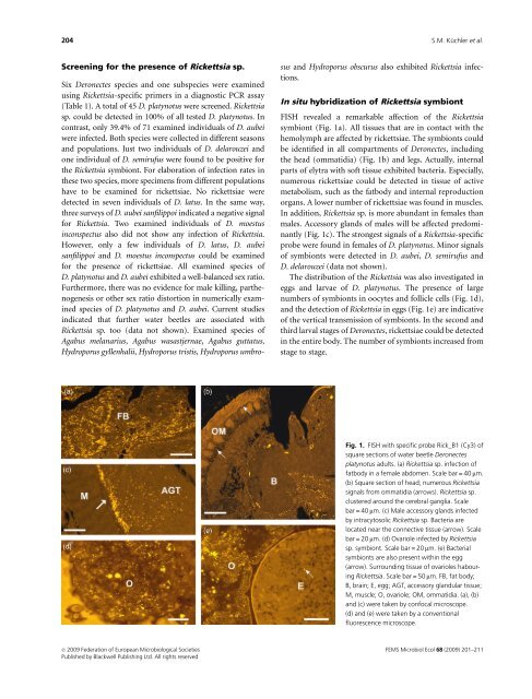
FISH revealed a remarkable affection of the Rickettsia<br />
symbiont (Fig. 1a). All tissues that are in contact with the<br />
hemolymph are affected by rickettsiae. The symbionts could<br />
be identified in all compartments of Deronectes, including<br />
the head (ommatidia) (Fig. 1b) and legs. Actually, internal<br />
parts of elytra with soft tissue exhibited bacteria. Especially,<br />
numerous rickettsiae could be detected in tissue of active<br />
metabolism, such as the fatbody and internal reproduction<br />
organs. A lower number of rickettsiae was found in muscles.<br />
In addition, Rickettsia sp. is more abundant in females than<br />
males. Accessory glands of males will be affected predominantly<br />
(Fig. 1c). The strongest signals of a Rickettsia-specific<br />
probe were found in females of D. platynotus. Minor signals<br />
of symbionts were detected in D. aubei, D. semirufus and<br />
D. delarouzei (data not shown).<br />
The distribution of the Rickettsia was also investigated in<br />
eggs and larvae of D. platynotus. The presence of large<br />
numbers of symbionts in oocytes and follicle cells (Fig. 1d),<br />
and the detection of Rickettsia in eggs (Fig. 1e) are indicative<br />
of the vertical transmission of symbionts. In the second and<br />
third larval stages of Deronectes, rickettsiae could be detected<br />
in the entire body. The number of symbionts increased from<br />
stage to stage.<br />
Fig. 1. FISH with specific probe Rick_B1 (Cy3) of<br />
square sections of water beetle Deronectes<br />
platynotus adults. (a) Rickettsia sp. infection of<br />
fatbody in a female abdomen. Scale bar = 40 mm.<br />
(b) Square section of head; numerous Rickettsia<br />
signals from ommatidia (arrows). Rickettsia sp.<br />
clustered around the cerebral ganglia. Scale<br />
bar = 40 mm. (c) Male accessory glands infected<br />
by intracytosolic Rickettsia sp. Bacteria are<br />
located near the connective tissue (arrow). Scale<br />
bar = 20 mm. (d) Ovariole infected by Rickettsia<br />
sp. symbiont. Scale bar = 20 mm. (e) Bacterial<br />
symbionts are also present within the egg<br />
(arrow). Surrounding tissue of ovarioles habouring<br />
Rickettsia. Scale bar = 50 mm. FB, fat body;<br />
B, brain; E, egg; AGT, accessory glandular tissue;<br />
M, muscle; O, ovariole; OM, ommatidia. (a), (b)<br />
and (c) were taken by confocal microscope.<br />
(d) and (e) were taken by a conventional<br />
fluorescence microscope.<br />
c 2009 Federation of European Microbiological Societies<br />
Published by Blackwell Publishing Ltd. All rights reserved<br />
FEMS Microbiol Ecol 68 (2009) 201–211