Get PDF (1214K) - Wiley Online Library
Get PDF (1214K) - Wiley Online Library
Get PDF (1214K) - Wiley Online Library

Create successful ePaper yourself
Turn your PDF publications into a flip-book with our unique Google optimized e-Paper software.
206 S.M. Küchler et al.<br />
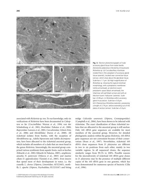
Fig. 3. Electron photomicrographs of male<br />
accessory gland tissue from water beetle<br />
Deronectes platynotus infected by intracytosolic<br />
Rickettsia sp. (a) Coccobacillary rickettsiae<br />
resided free in the cytoplasm of accessory gland<br />
tissue (asterisk), located near connective tissue<br />
(arrow), which close about the glandular tissue.<br />
Scale bar = 1.1 mm. (b) High magnification of<br />
Rickettsia sp. showing the typical rickettsial<br />
morphology, with a periplasmic membrane<br />
(white arrowhead), an electron-lucent<br />
periplasmic space (black arrowhead), the<br />
trilaminar cell wall (black arrow) and with an<br />
electron-lucent ‘halozone’ (asterisk). Scale<br />
bar = 0.09 mm. (c) Rickettsia within accessory<br />
gland musculature. Scale bar = 0.6 mm.<br />
(d) A filamentous Rickettsia (asterisk), possessing<br />
a length of 2.74 mm, before extending out of the<br />
plane of section (arrow). Scale bar = 0.4 mm.<br />
associated with Rickettsia sp. too. To our knowledge, only six<br />
verifications of Rickettsia have been documented in Coleoptera<br />
so far (Coccinellidae: Werren et al., 1994; von der<br />
Schulenburg et al., 2001; Bruchidae: Fukatsu et al., 2000;<br />
Buprestidae: Lawson et al., 2001; Curculionidae: Zchori-Fein<br />
et al., 2006; and Mordellidae: Duron et al., 2008). All<br />
rickettsial isolates from beetles, with the exception of<br />
Mordellistena sp., Adalia bipunctata and Adalia decempunctata,<br />
have been allocated to the rickettsial ancestral group,<br />
which includes all members of a clade that are most basal in<br />
the genus Rickettsia. Interestingly, the ancestral group comprised<br />
various symbionts from aquatic hosts, such as leeches<br />
(Kikuchi et al., 2002; Kikuchi & Fukatsu, 2005), amphizoic<br />
amoeba N. pattersoni (Dykova et al., 2003) and marine<br />
ciliate D. appendiculata (Vannini et al., 2005). Even insects<br />
that spend most of their development in water, i.e. the<br />
cranefly L. chorea (Diptera, Limoniidae; AF322443), mothfly<br />
L. apache (Diptera, Psychodidae; EU223247) and biting<br />
midge Culicoides sonorensis (Diptera, Ceratopogonidae)<br />
(Campbell et al., 2004), have been shown to be infected with<br />
rickettsiae. The exact classification of these rickettsial isolates<br />
that are allocated to the ancestral group is still unclear.<br />
Only 16S rRNA gene sequences are available for most<br />
members of the ancestral group. However, for detailed<br />
phylogenetic analysis within the genus Rickettsia, 16S rRNA<br />
gene sequences are not convenient because of their conservatism<br />
(Roux et al., 1997). Nevertheless, rickettsial 16S<br />
rRNA clone sequences from D. platynotus are different<br />
in two to six positions from each other, mainly in two<br />
variable regions. In all sequenced clones, the sequence<br />
similarity ranges from 99.1% to 100%. Possible reasons<br />
for the microdiversity of rickettsial 16S rRNA gene clones<br />
in D. platynotus may be the presence of multiple different<br />
copies of the 16S rRNA gene in one genome, which has<br />
been demonstrated for numerous species of bacteria (Cilia<br />
et al., 1996).<br />
c 2009 Federation of European Microbiological Societies<br />
Published by Blackwell Publishing Ltd. All rights reserved<br />
FEMS Microbiol Ecol 68 (2009) 201–211