
Create successful ePaper yourself
Turn your PDF publications into a flip-book with our unique Google optimized e-Paper software.
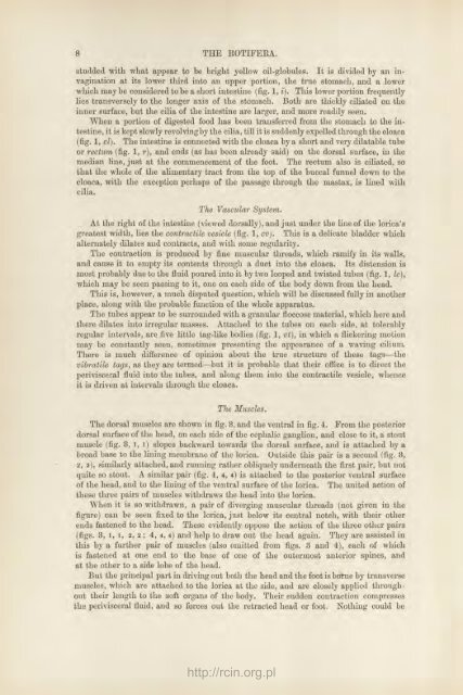
studded with what appear to be bright yellow oil-globules. It is divided by an invagination<br />
at its lower third into an upper portion, <strong>the</strong> true stomach, and a lower<br />
which may be considered to be a short intestine (fig. 1, i). This lower portion frequently<br />
lies transversely to <strong>the</strong> longer axis of <strong>the</strong> stomach. Both are thickly ciliated on <strong>the</strong><br />
inner surface, but <strong>the</strong> cilia of <strong>the</strong> intestine are larger, and more readily seen.<br />
When a portion of digested food has been transferred from <strong>the</strong> stomach to <strong>the</strong> intestine,<br />
it is kept slowly revolving by <strong>the</strong> cilia, till it is suddenly expelled through <strong>the</strong> cloaca<br />
(fig. 1, cl). The intestine is connected with <strong>the</strong> cloaca by a short and very dilatable tube<br />
or rectum (fig. 1, r), and ends (as has been already said) on <strong>the</strong> dorsal surface, in <strong>the</strong><br />
median line, just at <strong>the</strong> commencement of <strong>the</strong> foot. The rectum also is ciliated, so<br />
that <strong>the</strong> whole of <strong>the</strong> alimentary tract from <strong>the</strong> top of <strong>the</strong> buccal funnel down to <strong>the</strong><br />
cloaca, with <strong>the</strong> exception perhaps of <strong>the</strong> passage through <strong>the</strong> mastax, is lined with<br />
cilia.<br />
The Vascular<br />
System.<br />
At <strong>the</strong> right of <strong>the</strong> intestine (viewed dorsally), and just under <strong>the</strong> line of <strong>the</strong> lorica's<br />
greatest width, lies <strong>the</strong> contractile vesicle (fig. 1, cv). This is a delicate bladder which<br />
alternately dilates and contracts, and with some regularity.<br />
The contraction is produced by fine muscular threads, which ramify in its walls,<br />
and cause it to empty its contents through a duct into <strong>the</strong> cloaca. Its distension is<br />
most probably due to <strong>the</strong> fluid poured into it by two looped and twisted tubes (fig. 1, Ic),<br />
which may be seen passing to it, one on each side of <strong>the</strong> body down from <strong>the</strong> head.<br />
This is, however, a much disputed question, which will be discussed fully in ano<strong>the</strong>r<br />
place, along with <strong>the</strong> probable function of <strong>the</strong> whole apparatus.<br />
The tubes appear to be surrounded with a granular floccose material, which here and<br />
<strong>the</strong>re dilates into irregular masses. Attached to <strong>the</strong> tubes on each side, at tolerably<br />
regular inter\als, are five little tag-like bodies (fig. 1, vt), in which a flickering motion<br />
may be constantly seen, sometimes presenting <strong>the</strong> appearance of a waving cilium.<br />
There is much difference of opinion about <strong>the</strong> true structure of <strong>the</strong>se tags—<strong>the</strong><br />
vibratile tags, as <strong>the</strong>y are termed—but it is probable that <strong>the</strong>ir office is to direct <strong>the</strong><br />
perivisceral fluid into <strong>the</strong> tubes, and along <strong>the</strong>m into <strong>the</strong> contractile vesicle, whence<br />
it is driven at intervals through <strong>the</strong> cloaca.<br />
The Muscles.<br />
The dorsal muscles are shown in fig. 3, and <strong>the</strong> ventral in fig. 4. From <strong>the</strong> posterior<br />
dorsal surface of <strong>the</strong> head, on each side of <strong>the</strong> cephalic ganglion, and close to it, a stout<br />
muscle (fig. 3, l, l) slopes backward towards <strong>the</strong> dorsal surface, and is attached by a<br />
broad base to <strong>the</strong> lining membrane of <strong>the</strong> lorica. Outside this pair is a second (fig. 3,<br />
2, 2), similarly attached, and running ra<strong>the</strong>r obliquely underneath <strong>the</strong> first pair, but not<br />
quite so stout. A similar pair (fig. 4, 4, 4) is attached to <strong>the</strong> posterior ventral surface<br />
of <strong>the</strong> head, and to <strong>the</strong> lining of <strong>the</strong> ventral surface of <strong>the</strong> lorica. The united action of<br />
<strong>the</strong>se three pairs of muscles withdraws <strong>the</strong> head into <strong>the</strong> lorica.<br />
When it is so withdrawn, a pair of diverging muscular threads (not given in <strong>the</strong><br />
figure) can be seen fixed to <strong>the</strong> lorica, just below its central notch, with <strong>the</strong>ir o<strong>the</strong>r<br />
ends fastened to <strong>the</strong> head. These evidently oppose <strong>the</strong> action of <strong>the</strong> three o<strong>the</strong>r pairs<br />
(figs. 3, l, i, 2, 2 ; 4, 4, 4) and help to draw out <strong>the</strong> head again. They are assisted in<br />
this by a fur<strong>the</strong>r pair of muscles (also omitted from figs. 3 and 4), each of which<br />
is fastened at one end to <strong>the</strong> base of one of <strong>the</strong> outermost anterior spines, and<br />
at <strong>the</strong> o<strong>the</strong>r to a side lobe of <strong>the</strong> head.<br />
But <strong>the</strong> principal part in driving out both <strong>the</strong> head and <strong>the</strong> foot is borne by transverse<br />
muscles, which are attached to <strong>the</strong> lorica at <strong>the</strong> side, and are closely applied throughout<br />
<strong>the</strong>ir length to <strong>the</strong> soft organs of <strong>the</strong> body. Their sudden contraction compresses<br />
<strong>the</strong> perivisceral fluid, and so forces out <strong>the</strong> retracted head or foot. Nothing could be<br />
http://rcin.org.pl