Spinal Ataxia in Zebras
Spinal Ataxia in Zebras
Spinal Ataxia in Zebras

Create successful ePaper yourself
Turn your PDF publications into a flip-book with our unique Google optimized e-Paper software.
74 MONTALI et al.<br />
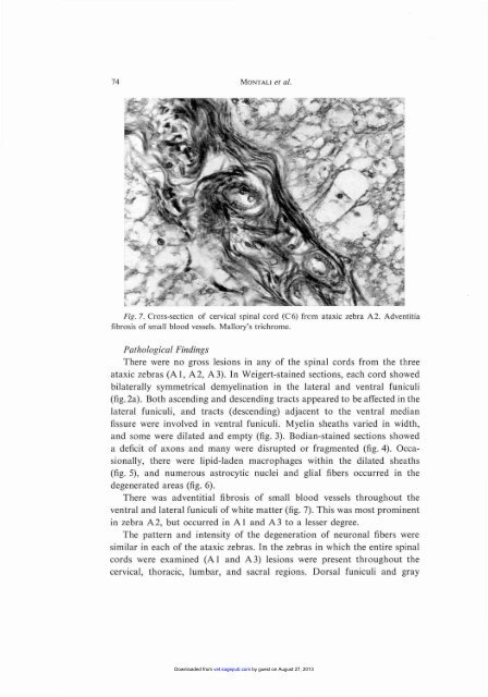
Fig. 7. Cross-section of cervica l sp<strong>in</strong>al co rd (C6) frcm ataxic zebra A2 . Advent itia<br />
fibrosis of small blood vessels. Ma llory's trichro me.<br />
Pathological F<strong>in</strong>d<strong>in</strong>gs<br />
Th ere were no gross lesion s <strong>in</strong> any of the sp<strong>in</strong>a l cord s from the three<br />
atax ic zebras (A I, A 2, A 3). In Weiger t-sta<strong>in</strong> ed sectio ns, eac h co rd showed<br />
bilaterally symmet rical demyel<strong>in</strong>ati on <strong>in</strong> the lat eral and ventral funi culi<br />
(fig.2a) . Both asce nd<strong>in</strong>g and descend<strong>in</strong> g tr acts ap pea red to be affected <strong>in</strong> th e<br />
lateral funiculi, a nd tracts (descend<strong>in</strong>g) adjacent to th e ventral median<br />
fissure were <strong>in</strong>volved <strong>in</strong> ventra l funi culi . Myel<strong>in</strong> sheaths varied <strong>in</strong> width,<br />
and some were dilated a nd empty (fig. 3). Bodi an- sta<strong>in</strong> ed sectio ns showed<br />
a deficit ofaxons and man y were disrupted or fragmented (fig. 4). Occasiona<br />
lly, there were lipid-lad en macrophages with<strong>in</strong> the dilated sheaths<br />
(fig. 5), a nd numerou s astrocytic nuclei and glial" fibers occurred <strong>in</strong> the<br />
degenerated a reas (fig. 6).<br />
Th ere was advent itia l fibrosis of sma ll blood vessels throu ghout the<br />
ventra l a nd lateral funiculi of white matter (fig. 7). This was most prom<strong>in</strong>ent<br />
<strong>in</strong> zeb ra A 2, but occ ur red <strong>in</strong> A I and A 3 to a lesser degree.<br />
The pattern a nd <strong>in</strong>ten sity of th e degeneratio n of neu ron al fiber s were<br />
similar <strong>in</strong> each of the ataxic zebras . In th e zebras <strong>in</strong> wh ich the entire sp<strong>in</strong>a l<br />
co rds were exa m<strong>in</strong>ed (A I and A 3) lesion s were prese nt thr ou ghout the<br />
cerv ical, th oracic, lumba r, and sac ra l regions . Dorsal funi cul i and gray<br />
Downloaded from vet.sagepub.com by guest on August 27, 2013