Spinal Ataxia in Zebras
Spinal Ataxia in Zebras
Spinal Ataxia in Zebras

You also want an ePaper? Increase the reach of your titles
YUMPU automatically turns print PDFs into web optimized ePapers that Google loves.
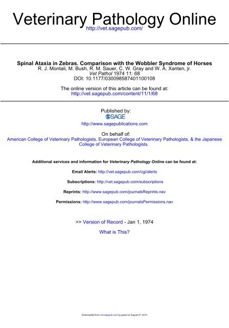
Veter<strong>in</strong>ary Pathology Onl<strong>in</strong>e<br />
http://vet.sagepub.com/<br />
<strong>Sp<strong>in</strong>al</strong> <strong>Ataxia</strong> <strong>in</strong> <strong>Zebras</strong>. Comparison with the Wobbler Syndrome of Horses<br />
R. J. Montali, M. Bush, R. M. Sauer, C. W. Gray and W. A. Xanten, jr.<br />
Vet Pathol 1974 11: 68<br />
DOI: 10.1177/030098587401100108<br />
The onl<strong>in</strong>e version of this article can be found at:<br />
http://vet.sagepub.com/content/11/1/68<br />
Published by:<br />
http://www.sagepublications.com<br />
On behalf of:<br />
American College of Veter<strong>in</strong>ary Pathologists, European College of Veter<strong>in</strong>ary Pathologists, & the Japanese<br />
College of Veter<strong>in</strong>ary Pathologists.<br />
Additional services and <strong>in</strong>formation for Veter<strong>in</strong>ary Pathology Onl<strong>in</strong>e can be found at:<br />
Email Alerts: http://vet.sagepub.com/cgi/alerts<br />
Subscriptions: http://vet.sagepub.com/subscriptions<br />
Repr<strong>in</strong>ts: http://www.sagepub.com/journalsRepr<strong>in</strong>ts.nav<br />
Permissions: http://www.sagepub.com/journalsPermissions.nav<br />
>> Version of Record - Jan 1, 1974<br />
What is This?<br />
Downloaded from vet.sagepub.com by guest on August 27, 2013
Vet. Path. Jl: 68-78 (1974)<br />
<strong>Sp<strong>in</strong>al</strong> <strong>Ataxia</strong> <strong>in</strong> <strong>Zebras</strong><br />
Comparison with the Wobbler Syndrome of Horses<br />
R. J. MONTALI, M. BUSH, R. M. SAUER, C. W. GRAY and W.A. XANTEN, jr.<br />
The Division of Laboratory Animal Medic<strong>in</strong>e and The Department of Pathology,<br />
The Johns Hopk<strong>in</strong>s University, School of Medic<strong>in</strong>e, Baltimore, Md.;<br />
and National Zoological Park, Smithsonian Institution, Wash<strong>in</strong>gton, D. C.<br />
Abstract. Eight of 17 zebra foals (Equus burchelli) born at the National Zoological<br />
Park, Wash<strong>in</strong>gton, D. c., developed an ataxic condition that cl<strong>in</strong>ically resembled the<br />
wobbler syndrome of horses. Four were males and four were females. All were the progeny<br />
of one stallion and two mares. The parent animals were not ataxic. In three of the affected<br />
foals raised at the zoo, signs progressed to severe ataxia, and the animals were killed.<br />
In contrast to the f<strong>in</strong>d<strong>in</strong>gs <strong>in</strong> wobbler horses there was no radiographic or pathologic<br />
evidence of narrow<strong>in</strong>g of the vertebral canals, nor were malacic foci found <strong>in</strong> the cords to<br />
suggest that focal compression had occurred. There was degeneration of ascend<strong>in</strong>g and<br />
descend<strong>in</strong>g tracts <strong>in</strong> the same segments throughout the sp<strong>in</strong>al cords. There were no lesions<br />
<strong>in</strong> two bra<strong>in</strong>s studied nor were other possible causes for the sp<strong>in</strong>al cord degeneration<br />
evident. These f<strong>in</strong>d<strong>in</strong>gs and the high <strong>in</strong>cidence of ataxia <strong>in</strong> this herd suggest a familial<br />
degenerative myelopathy.<br />
Approximately half of the zebra foals born over a l2-year period at the<br />
National Zoological Park developed a disorder of the central nervous system<br />
that cl<strong>in</strong>ically resembled the wobbler syndrome (wobbles, equ<strong>in</strong>e <strong>in</strong>coord<strong>in</strong>ation,<br />
ataxia of foals [5, 7, 8, 14]). This is an ataxic condition of predom<strong>in</strong>antly<br />
young male horses that is characterized by a sway<strong>in</strong>g or wobbl<strong>in</strong>g<br />
of the h<strong>in</strong>dquarters. It can be progressive and may lead to muscular weakness<br />
and severe locomotor disfunction of both pelvic and thoracic limbs. A similar<br />
cl<strong>in</strong>ical picture is described <strong>in</strong> eight zebras. The pathologic changes <strong>in</strong> the<br />
sp<strong>in</strong>al columns of three animals, however, <strong>in</strong>dicate that the pathogenesis<br />
may not be the same as for the wobbler syndrome. These differences are<br />
discussed, and the zebra disorder is compared with ataxic conditions of other<br />
species and man. This is believed to be the first report of a possible primary<br />
degenerative myelopathy <strong>in</strong> an equ<strong>in</strong>e species.<br />
Downloaded from vet.sagepub.com by guest on August 27, 2013
MONTALI et at.<br />
69<br />
Janet<br />
Tam<br />
Zebbie<br />
o Normal Female<br />
D Normal Mole<br />
• Atoxic Female<br />
• Ataxic Male<br />
/: Sex ond Condition Unknown<br />
Fig. I. Pedigree of ataxic zebras.<br />
Cl<strong>in</strong>ical Study<br />
The orig<strong>in</strong>al herd of Grant's zebras exhibited at the National Zoological Park was<br />
derived from one stallion (Tom) and two mares (Janet and Zebbie) (fig. 1). These animals<br />
were unrelated and showed no abnormalities of gait or signs of <strong>in</strong>coord<strong>in</strong>ation. From<br />
1960 to 1971, there were 17 offspr<strong>in</strong>g (IO from Janet and 7 from Zebbie) of which eight<br />
were ataxic <strong>in</strong> the h<strong>in</strong>d quarters. As noted <strong>in</strong> the pedigree chart (fig. I) a precise account<br />
of sex ratios and frequency cannot be made s<strong>in</strong>ce complete records of all foals were not<br />
available. Four of the affected animals were males and four females.<br />
Signs of ataxia began between 4 and 6 months of age. They were first detected as<br />
subtle <strong>in</strong>coord<strong>in</strong>ated movements of the h<strong>in</strong>dquarters that were most noticeable when the<br />
animals were trott<strong>in</strong>g slowly or turn<strong>in</strong>g. The ataxia could be accentuated by forc<strong>in</strong>g the<br />
animals to move <strong>in</strong> a small circle. Detailed neurological exam<strong>in</strong>ations were not possible<br />
because of the difficulties of manipulat<strong>in</strong>g these animals, but vision, temperament, and<br />
other motor functions appeared to be normal. Appetite and hematological studies were<br />
with<strong>in</strong> normal limits. The animals were fed standard pelleted horse feed and hay and had<br />
access to salt bricks conta<strong>in</strong><strong>in</strong>g trace m<strong>in</strong>erals. There were few endoparasites <strong>in</strong> the herd<br />
because of rout<strong>in</strong>e exam<strong>in</strong>ation and treatment.<br />
Four of the mildly ataxic zebras were sent elsewhere before they were yearl<strong>in</strong>gs, and<br />
further records were unavailable; one other animal died of causes unrelated to the ataxia.<br />
Three affected animals, A I, A2, and A3 (table I) rema<strong>in</strong>ed at the zoo, and their condition<br />
worsened. Between I and 2 years of age they developed severe ataxia and weakness of the<br />
rear quarters, with milder weakness of the thoracic limbs. Hypermetria was not seen,<br />
nor was the ability to step backwards appreciably altered. They often fell and made strenuous<br />
efforts to right themselves. Because of the trauma from these episodes, the animals<br />
were killed and necropsied. The orig<strong>in</strong>al herd sire, Tom (N I) was also killed and necropsied<br />
because of his advanced age and an arthritic condition of his front limbs.<br />
Downloaded from vet.sagepub.com by guest on August 27, 2013
70 MONTAL! et al.<br />
Table I. Identification, cl<strong>in</strong>ical condition and tissues available at necropsy<br />
Zebra Sex <strong>Ataxia</strong> Age at necropsy Tissues studied<br />
Al male advanced 3 years, I month sp<strong>in</strong>al cord only<br />
A2 female advanced I year, 4 months cervical vertebrae, bra<strong>in</strong>,<br />
cervical and thoracic cord<br />
A3 female advanced I year, 9 months cervical vertebrae, entire<br />
central nervous system<br />
Nl male none 20 years cervical vertebrae, entire<br />
central nervous system<br />
Materials and Methods<br />
At various <strong>in</strong>tervals zebras A2, A3, and N I were each given 3 mg of M 99 (etorph<strong>in</strong>e,<br />
Cyanarnid'E') plus 30 mg of acepromaz<strong>in</strong>e (Provet®) while restra<strong>in</strong>ed <strong>in</strong> a chute (Ranger).<br />
After the animals were immobilized, an endotracheal tube was <strong>in</strong>serted and fluothane<br />
(halothane, Ayerst Laboratories, New York, N. Y.) was adm<strong>in</strong>istered to produce surgical<br />
anesthesia. Radiographs of the cervical vertebrae were taken both while the zebras were<br />
<strong>in</strong> normal posture and dur<strong>in</strong>g extreme flexion of the head and neck. Lateral and ventrodorsal<br />
views were obta<strong>in</strong>ed. The animals, <strong>in</strong>clud<strong>in</strong>g A I, were killed and immediately<br />
necropsied.<br />
The tissues available (table I) from the three affected zebras (AI, A2, and A3) and<br />
from the one unaffected animal (N I, control) were processed for study <strong>in</strong> the follow<strong>in</strong>g<br />
manner. The cervical vertebrae were disarticulated, trimmed of excess muscle tissue,<br />
dried, and exposed to a colony of Dermestidae beetles. The bra<strong>in</strong>s and sp<strong>in</strong>al cords were<br />
removed <strong>in</strong>tact, exam<strong>in</strong>ed for gross abnormalities, and fixed <strong>in</strong> 10% formal<strong>in</strong>. After<br />
fixation, the sp<strong>in</strong>al cords were sliced transversely at 2- to 5-mm <strong>in</strong>tervals, and the serial<br />
sections were exam<strong>in</strong>ed with a stereomicroscope. Samples from each segment, together<br />
with bra<strong>in</strong> tissue, were embedded <strong>in</strong> paraff<strong>in</strong>, cut at 6 urn, and sta<strong>in</strong>ed with hematoxyl<strong>in</strong><br />
and eos<strong>in</strong> (HE).<br />
Special sta<strong>in</strong><strong>in</strong>g techniques performed on selected samples from all cases <strong>in</strong>cluded,<br />
luxol blue-cresyl violet, Weigert (myel<strong>in</strong>), Holzer, Bodian, periodic acid-Schiff-Alcian<br />
blue, Mallory's trichrome, and oil-red-O (frozen tissues).<br />
Results<br />
Radiographic F<strong>in</strong>d<strong>in</strong>gs<br />
The cervical vertebrae appeared normal. Vertebral subluxation or narrow<strong>in</strong>g<br />
of the cervical vertebral canals was not evident.<br />
Downloaded from vet.sagepub.com by guest on August 27, 2013
Ataxi a <strong>in</strong> <strong>Zebras</strong> 71<br />
a<br />
b<br />
Fig. 2. a Tr ansverse section of cervical sp<strong>in</strong>al cord (C 4) fro m a taxic zebra , A3 . Pale<br />
areas are loss of myel<strong>in</strong> <strong>in</strong> lateral and ventral funiculi . Compare with figure I b. Weigert's<br />
myel<strong>in</strong> sta <strong>in</strong>. b Transverse section of cervical sp<strong>in</strong>al cord (C4) from nonataxic zebra, N I.<br />
The myel<strong>in</strong> sta<strong>in</strong> s fairl y con sistently throughout the cord . Section prepared and sta<strong>in</strong>ed<br />
at the same time as section <strong>in</strong> figure I a. Weigert's myel<strong>in</strong> sta <strong>in</strong>.<br />
Downloaded from vet.sagepub.com by guest on August 27, 2013
72<br />
M ONT ALI et al.<br />
3<br />
4<br />
Fig. 3. H igher magnificat ion from an area of myel<strong>in</strong> pallor <strong>in</strong> the latera l funiculus<br />
sho wn <strong>in</strong> figure I a. Myel<strong>in</strong> shea ths are irregular sizes. Man y a re distended and empt y. HE.<br />
Fig . 4. Similar a rea as <strong>in</strong> figure 2. Longitud<strong>in</strong>a l section. Axon s are depleted. A few<br />
axona l fragments rema<strong>in</strong> . Bodi an sta <strong>in</strong>.<br />
Downloaded from vet.sagepub.com by guest on August 27, 2013
<strong>Ataxia</strong> <strong>in</strong> Zeb ras<br />
73<br />
5<br />
_ ............r.e-. • •0lJ 6<br />
Fig . 5. Lon gitud<strong>in</strong>al sect ion of sp<strong>in</strong>a l co rd from zebra A 3. Lipid-lade n rnacr ophages<br />
acc um ulate <strong>in</strong> a dilated mye l<strong>in</strong> sheat h. H E.<br />
Fig . 6. Lon gitu d<strong>in</strong>al sect ion of th oracic sp<strong>in</strong>a l co rd (midd le) fro m a taxic zebra A2.<br />
Ast rocy tic nuclei (ar rows) an d glial fibers near degenera ted trac ts. H E.<br />
Downloaded from vet.sagepub.com by guest on August 27, 2013
74 MONTALI et al.<br />
Fig. 7. Cross-section of cervica l sp<strong>in</strong>al co rd (C6) frcm ataxic zebra A2 . Advent itia<br />
fibrosis of small blood vessels. Ma llory's trichro me.<br />
Pathological F<strong>in</strong>d<strong>in</strong>gs<br />
Th ere were no gross lesion s <strong>in</strong> any of the sp<strong>in</strong>a l cord s from the three<br />
atax ic zebras (A I, A 2, A 3). In Weiger t-sta<strong>in</strong> ed sectio ns, eac h co rd showed<br />
bilaterally symmet rical demyel<strong>in</strong>ati on <strong>in</strong> the lat eral and ventral funi culi<br />
(fig.2a) . Both asce nd<strong>in</strong>g and descend<strong>in</strong> g tr acts ap pea red to be affected <strong>in</strong> th e<br />
lateral funiculi, a nd tracts (descend<strong>in</strong>g) adjacent to th e ventral median<br />
fissure were <strong>in</strong>volved <strong>in</strong> ventra l funi culi . Myel<strong>in</strong> sheaths varied <strong>in</strong> width,<br />
and some were dilated a nd empty (fig. 3). Bodi an- sta<strong>in</strong> ed sectio ns showed<br />
a deficit ofaxons and man y were disrupted or fragmented (fig. 4). Occasiona<br />
lly, there were lipid-lad en macrophages with<strong>in</strong> the dilated sheaths<br />
(fig. 5), a nd numerou s astrocytic nuclei and glial" fibers occurred <strong>in</strong> the<br />
degenerated a reas (fig. 6).<br />
Th ere was advent itia l fibrosis of sma ll blood vessels throu ghout the<br />
ventra l a nd lateral funiculi of white matter (fig. 7). This was most prom<strong>in</strong>ent<br />
<strong>in</strong> zeb ra A 2, but occ ur red <strong>in</strong> A I and A 3 to a lesser degree.<br />
The pattern a nd <strong>in</strong>ten sity of th e degeneratio n of neu ron al fiber s were<br />
similar <strong>in</strong> each of the ataxic zebras . In th e zebras <strong>in</strong> wh ich the entire sp<strong>in</strong>a l<br />
co rds were exa m<strong>in</strong>ed (A I and A 3) lesion s were prese nt thr ou ghout the<br />
cerv ical, th oracic, lumba r, and sac ra l regions . Dorsal funi cul i and gray<br />
Downloaded from vet.sagepub.com by guest on August 27, 2013
Fig . 8. Fourth cervical vertebra (C4) from ataxic zebra A 2, Left crani al articular<br />
process (u pper right) has pro liferation of bone around articular marg<strong>in</strong>s a nd is larger<br />
than the right process (lower right).<br />
co lumns, however, a ppeared normal <strong>in</strong> all of th e co rds . In zebras whose<br />
bra<strong>in</strong>s were studie d (A 2, A 3), the degenerated tr acts could be followed <strong>in</strong>t o<br />
the medulla oblonga ta ; lesion s rostral to thi s area were not evide nt.<br />
There were abno rma lities <strong>in</strong> th e cervica l vert ebrae of one (A 2) of two<br />
affected zebras. In zebra A 2 the articular surfaces of th e left ca uda l ar ticular<br />
process of the thi rd vertebra and the left cra nia l articular pr ocess of th e<br />
fourth were lar ger th an th ose on t he right (fig. 8). Th ere was also thi cken<strong>in</strong>g<br />
of the ar ticular process of the fourth vertebra with prolifera tio n of bon e<br />
aro und the lat eral and cra nial marg<strong>in</strong> s of th e jo<strong>in</strong>t surface. Narrow<strong>in</strong>g of<br />
the verte bral ca na ls did not occ ur <strong>in</strong> either zebra.<br />
No significa nt gros s or microscopic changes were seen <strong>in</strong> comparable<br />
sectio ns of th e sp<strong>in</strong>al cord (fig. 2 b) or cervical vertebrae of the un affected<br />
zeb ra (N I).<br />
Downloaded from vet.sagepub.com by guest on August 27, 2013
76 MONTALI et al.<br />
Discussion<br />
Because the cl<strong>in</strong>ical features of this ataxia <strong>in</strong> zebras so closely resembled<br />
the wobbler syndrome of horses, and s<strong>in</strong>ce zebras are 'equidae', the changes<br />
<strong>in</strong> both conditions were compared. The lesions <strong>in</strong> the wobbler syndrome<br />
are malformations of the articular processes and WaIIerian degeneration <strong>in</strong><br />
the cervical sp<strong>in</strong>al cord [3, 7, 8, I I]. A malacic focus (primary lesion) occurs<br />
<strong>in</strong> the cervical cord apparently as a result of subluxation of the abnormal<br />
vertebrae. Secondary degeneration of ascend<strong>in</strong>g tracts cranial to the primary<br />
lesion results and may be foIIowed for several segments. Likewise, descend<strong>in</strong>g<br />
tracts degenerate caudal to the primary lesion, and these may extend to the<br />
lumbar segment. When the primary lesion is not readily apparent, it may<br />
be located by 'mapp<strong>in</strong>g' the cranial limit of the descend<strong>in</strong>g damage and the<br />
caudal limit of the ascend<strong>in</strong>g damage.<br />
In the zebras, the sp<strong>in</strong>al cord lesions did not have a Wallerian pattern.<br />
Vertebral arthropathy (fig. 7), rem<strong>in</strong>iscent of the articular lesions of wobbler<br />
horses [14], was found <strong>in</strong> one zebra (A2), but detailed microscopic studies<br />
of its cervical cord and of cords of the others, did not show malacic foci.<br />
The degenerate ascend<strong>in</strong>g and descend<strong>in</strong>g tracts were not anatomically<br />
segregated but could be consistently followed together throughout the<br />
lengths of sp<strong>in</strong>al cords available for study (table I). Correlations between<br />
the gait abnormalities and the lesions of specific tracts have been described<br />
<strong>in</strong> the wobbler syndrome [12]. The fact that functionally similar tracts were<br />
<strong>in</strong>volved <strong>in</strong> the zebra sp<strong>in</strong>al cords may expla<strong>in</strong> the similar cl<strong>in</strong>ical signs of<br />
both conditions.<br />
Further po<strong>in</strong>ts of comparison <strong>in</strong>clude possible factors such as sex, breed,<br />
and heredity. The wobbler syndrome has its highest prevalence <strong>in</strong> the longernecked<br />
breeds such as thoroughbreds and standardbreds, and there is a<br />
male predom<strong>in</strong>ance. Accord<strong>in</strong>gly, ROONEY [13] relates the vertebral arthropathies<br />
to abnormal stresses <strong>in</strong> the cervical columns of these long-necked<br />
animals and ascribes the predom<strong>in</strong>ance <strong>in</strong> males to their added muscle bulk<br />
and consequent additional strength of the head and neck region. Heredity<br />
has been suggested as a factor <strong>in</strong> the wobbler syndrome but never proved [6].<br />
Although we cannot draw conclusions on sex ratios, there was no apparent<br />
trend for a male predom<strong>in</strong>ance <strong>in</strong> the zebra. <strong>Zebras</strong> are relatively shortnecked<br />
'equ<strong>in</strong>es', and our pathologic f<strong>in</strong>d<strong>in</strong>gs differ significantly from those<br />
of the wobbler syndrome, <strong>in</strong>dicat<strong>in</strong>g that the pathogenesis of both conditions<br />
is not the same. Therefore, this ataxic condition of zebras should not be<br />
designated as the wobbler syndrome.<br />
Downloaded from vet.sagepub.com by guest on August 27, 2013
<strong>Ataxia</strong> <strong>in</strong> <strong>Zebras</strong> 77<br />
The specific cause of this diffuse myelopathy <strong>in</strong> zebras was not evident.<br />
There was no basis for an ischemic pattern [2], hence the adventitial fibrosis<br />
of vessels noted <strong>in</strong> all of the zebras was not considered to contribute to the<br />
sp<strong>in</strong>al cord degeneration. Similar vascular changes caused by factors such as<br />
vertebral fractures and space-occupy<strong>in</strong>g lesions have been seen <strong>in</strong> wobblers<br />
and <strong>in</strong> other equ<strong>in</strong>e myelopathies [7]. Several causes were considered.<br />
Nutritional factors might be implicated. Copper deficiency has been reported<br />
to cause a demyel<strong>in</strong>at<strong>in</strong>g disease <strong>in</strong> lambs. This condition is called enzootic<br />
ataxia and has been compared with the wobbler syndrome of horses [10].<br />
A deficiency of copper would not be expected <strong>in</strong> these zebras s<strong>in</strong>ce all<br />
received ample supplements <strong>in</strong> their pelleted rations and had free access to<br />
copper <strong>in</strong> m<strong>in</strong>eralized salt blocks. Proof of such a deficiency would depend<br />
on assays to determ<strong>in</strong>e serum and tissue copper levels.<br />
Parasitic migration <strong>in</strong> the central nervous system was also considered.<br />
Parasites migrat<strong>in</strong>g aberrantly <strong>in</strong> the sp<strong>in</strong>al cords have been reported to<br />
create wobbler-like signs <strong>in</strong> horses [9]. This was not so <strong>in</strong> the zebra condition<br />
as there were no parasites or parasitic remnants nor were there <strong>in</strong>flammatory<br />
changes that suggested <strong>in</strong>fectious agents <strong>in</strong> the central nervous system.<br />
The high <strong>in</strong>cidence of ataxic sibl<strong>in</strong>g foals <strong>in</strong> this small zebra herd and our<br />
f<strong>in</strong>d<strong>in</strong>g of a diffuse myelopathy with no apparent cause suggest that this<br />
condition is a familial disorder with primary degeneration of sp<strong>in</strong>al cord<br />
white matter. Familial degenerations of the sp<strong>in</strong>al cord do occur. Friedreich's<br />
ataxia of children is described as a familial form of progressive ataxia.<br />
It is characterized by a degeneration of long, ascend<strong>in</strong>g and descend<strong>in</strong>g<br />
tracts of the sp<strong>in</strong>al cord [2]. A degenerative myelopathy, possibly familial,<br />
has recently been described <strong>in</strong> aged German Shepherd dogs [I]. In several<br />
horses that cl<strong>in</strong>ically were wobblers DE LAHUNTA [4] has seen diffuse myelopathy<br />
similar to that <strong>in</strong> our zebras. He did not consider his animals true<br />
wobblers.<br />
Proof of a familial ataxia <strong>in</strong> zebras must await future breed<strong>in</strong>gs between<br />
related animals that are possibly carry<strong>in</strong>g genetic traits; complete cl<strong>in</strong>ical<br />
and pathological studies must be performed on any that are affected.<br />
Subsequent progeny from dams of our ataxic animals and from their normal<br />
daughters bred to an unrelated herd sire, Fred (moo-180), are cl<strong>in</strong>ically<br />
normal to date.<br />
Acknowledgements<br />
Supported <strong>in</strong> part by grants RR-00130 and RR-05010, U. S. Public Health Service;<br />
and The Mary Ida Young Foundation. The authors thank Dr. ALEXANDER DE LAHUNTA,<br />
Downloaded from vet.sagepub.com by guest on August 27, 2013
78 MONTALI et al.<br />
Department of Anatomy, Cornell University, Ithaca, N. Y.; and Dr. LAWRENCE E. BECKER,<br />
Department of Neurology, Johns Hopk<strong>in</strong>s Hospital, Baltimore, Md., for comments and<br />
suggestions.<br />
References<br />
AVERILL, D. R.: Degenerative myelopathy <strong>in</strong> the ag<strong>in</strong>g German Shepherd dog.<br />
Cl<strong>in</strong>ical and pathologic f<strong>in</strong>d<strong>in</strong>gs. J. amer. vet. med. Ass. 162: 1045-1051 (1973).<br />
2 BLACKWOOD, W.; McMENEMEY, W.H.; MEYER, A.; NORMAN, R.M., and RUSSELL,<br />
D. S.: Greenfield's neuropathology; 2nd ed., pp. 95, 587 (Williams & Wilk<strong>in</strong>s, Baltimore<br />
1966).<br />
3 DAHME, E. and SCHEBITZ, H.: New observations on the pathogenesis of sp<strong>in</strong>al ataxia<br />
<strong>in</strong> the horse. Zbl. Vet Med. 17: 120-143 (1970).<br />
4 DE LAHUNTA, A.: Personal commun. (1974).<br />
5 DIMOCK, W. W. and ERRINGTON, E. J.: Incoord<strong>in</strong>ation of equidae; "wobbles."<br />
J. arner, vet. med. Ass. 95: 261-267 (1939).<br />
6 DIMOCK, W.W.: "Wobbles", an hereditary disease <strong>in</strong> horses. J. Hered. 41: 319-323<br />
(1950).<br />
7 FRASER, H. and PALMER, A. C.: Equ<strong>in</strong>e <strong>in</strong>co-ord<strong>in</strong>ation and wobbler disease of young<br />
horses. Vet. Rec. 80: 338-355 (1967).<br />
8 JONES, R. c.; DOLL, E. R., and BROWN, R. G. : The pathology of equ<strong>in</strong>e <strong>in</strong>coord<strong>in</strong>ation<br />
(ataxia or "wobbles" of foals). Proc. amer. vet. med. Ass. 45: 139-149 (J954).<br />
9 INNES, J. R. M. and SAUNDERS, L. Z.: Comparative neuropathology; 1st ed., pp. 540-549<br />
(Academic Press, New York 1962).<br />
10 OLAFSON, P.: "Wobbles" compared with ataxic (sw<strong>in</strong>g back) lambs. Cornell Vet. 32:<br />
301-314 (1942).<br />
I I ROONEY, J. R.: Biomechanics of lameness <strong>in</strong> horses; 1st ed., pp. 221-229 (Williams &<br />
Wilk<strong>in</strong>s, Baltimore 1969).<br />
12 ROONEY, J. R.: Cl<strong>in</strong>ical neurology of the horse; 1st ed., pp. 11-15, 36-41 (KNA Press,<br />
Kennett Square 1971).<br />
13 ROONEY, J.R.: Etiology of the wobbler syndrome. Mod. vet. Pract. 53: 42 (1965).<br />
14 ROONEY, J. R.: Equ<strong>in</strong>e <strong>in</strong>coord<strong>in</strong>ation. I. Gross morphology. Cornell Vet. 53: 411-422<br />
(] 963).<br />
Request repr<strong>in</strong>ts from: Dr. R. J. MONTALI, Department of Pathology, Johns Hopk<strong>in</strong>s<br />
Hospital, Baltimore, MD 21205 (USA)<br />
Downloaded from vet.sagepub.com by guest on August 27, 2013