Detection of porcine parvovirus in the follicular fluid of abattoir pigs
Detection of porcine parvovirus in the follicular fluid of abattoir pigs
Detection of porcine parvovirus in the follicular fluid of abattoir pigs

Create successful ePaper yourself
Turn your PDF publications into a flip-book with our unique Google optimized e-Paper software.
Brief communication<br />
Peer reviewed<br />
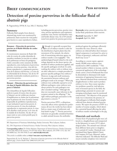
<strong>Detection</strong> <strong>of</strong> <strong>porc<strong>in</strong>e</strong> <strong>parvovirus</strong> <strong>in</strong> <strong>the</strong> <strong>follicular</strong> <strong>fluid</strong> <strong>of</strong><br />
<strong>abattoir</strong> <strong>pigs</strong><br />
R. Pogranichniy, DVM; K. Lee, MS; Z. Machaty, PhD<br />
Summary<br />
Follicular <strong>fluid</strong> samples from <strong>abattoir</strong>obta<strong>in</strong>ed<br />
pig ovaries were monitored by<br />
polymerase cha<strong>in</strong> reaction for viral pathogens<br />
known to cause reproductive failure,<br />
<strong>in</strong>clud<strong>in</strong>g <strong>porc<strong>in</strong>e</strong> <strong>parvovirus</strong>, <strong>porc<strong>in</strong>e</strong> circovirus,<br />
<strong>porc<strong>in</strong>e</strong> reproductive and respiratory<br />
syndrome virus, bov<strong>in</strong>e viral diarrhea virus,<br />
and border disease virus. Six <strong>of</strong> 49 animals<br />
tested were positive for <strong>porc<strong>in</strong>e</strong> <strong>parvovirus</strong>.<br />
Keywords: sw<strong>in</strong>e, <strong>porc<strong>in</strong>e</strong> <strong>parvovirus</strong>, <strong>follicular</strong><br />
<strong>fluid</strong>, polymerase cha<strong>in</strong> reaction<br />
Received: September 9, 2007<br />
Accepted: April 24, 2008<br />
Resumen – Detección de <strong>parvovirus</strong><br />
porc<strong>in</strong>o en el <strong>fluid</strong>o folicular de cerdos<br />
de matadero<br />
Se monitorearon muestras de <strong>fluid</strong>o folicular<br />
de ovarios de cerdas obtenidas de<br />
matadero mediante la reacción en cadena<br />
de la polimerasa en busca de patógenos<br />
virales conocidos como causantes de falla<br />
reproductiva, estos <strong>in</strong>cluyeron al <strong>parvovirus</strong><br />
porc<strong>in</strong>o, circovirus porc<strong>in</strong>o, virus del síndrome<br />
reproductivo y respiratorio porc<strong>in</strong>o,<br />
virus de la diarrea viral bov<strong>in</strong>a, y el virus de<br />
la enfermedad de la frontera. Seis de los 49<br />
animales exam<strong>in</strong>ados resultaron positivos a<br />
<strong>parvovirus</strong> porc<strong>in</strong>o.<br />
Résumé – Détection du <strong>parvovirus</strong> porc<strong>in</strong><br />
dans le liquide folliculaire chez des<br />
porcs à l’<strong>abattoir</strong><br />
Des échantillons de liquide folliculaire<br />
prélevés à partir d’ovaires chez des porcs<br />
à l’<strong>abattoir</strong> ont été vérifiés par réaction<br />
d’amplification en chaîne par la polymérase<br />
pour la présence de virus reconnus comme<br />
responsable de problèmes reproducteurs,<br />
et qui <strong>in</strong>cluaient le <strong>parvovirus</strong> porc<strong>in</strong>, le<br />
circovirus porc<strong>in</strong>, le virus du syndrome<br />
reproducteur et respiratoire porc<strong>in</strong>, le virus<br />
de la diarrhée virale <strong>porc<strong>in</strong>e</strong>, et le virus de<br />
la maladie de la frontière. Six des 49 animaux<br />
testés se sont avéréé positifs pour le<br />
<strong>parvovirus</strong> porc<strong>in</strong>.<br />
Although it is generally accepted that<br />
<strong>the</strong> use <strong>of</strong> embryo transfer is safer for<br />
<strong>the</strong> distribution <strong>of</strong> germ plasm than <strong>the</strong><br />
movement <strong>of</strong> live animals, <strong>the</strong> relative<br />
risk <strong>of</strong> transmitt<strong>in</strong>g <strong>in</strong>fectious agents via<br />
embryo transfer is not negligible. 1 The<br />
epidemiological hazard related to <strong>the</strong> technology<br />
depends on <strong>the</strong> donor species, <strong>the</strong><br />
method <strong>of</strong> embryo production applied, and<br />
<strong>the</strong> specific pathogens <strong>in</strong>volved. In cattle,<br />
wash<strong>in</strong>g <strong>the</strong> embryo with or without tryps<strong>in</strong><br />
after collection is a simple procedure to<br />
generate specific-pathogen-free embryos. 2<br />
However, <strong>in</strong> <strong>pigs</strong> and small rum<strong>in</strong>ants,<br />
<strong>the</strong> association between embryos and<br />
pathogens tends to be stronger. 3 When pig<br />
embryos were artificially exposed to seven<br />
different pathogens, some embryos were<br />
always positive for each pathogen even<br />
after extended wash<strong>in</strong>g. 4 Although this<br />
does not necessarily cause <strong>in</strong>fection <strong>in</strong> <strong>the</strong><br />
recipient animal or <strong>the</strong> <strong>of</strong>fspr<strong>in</strong>g, 5,6 <strong>the</strong><br />
tight embryo-pathogen association <strong>in</strong> <strong>the</strong><br />
pig rema<strong>in</strong>s a concern. 7,8<br />
Porc<strong>in</strong>e <strong>parvovirus</strong> (PPV), a ubiquitous<br />
virus that affects sw<strong>in</strong>e reproductive performance<br />
all over <strong>the</strong> world, has been identified<br />
as a major cause <strong>of</strong> embryonic and<br />
fetal death <strong>in</strong> <strong>pigs</strong>. 9-11 Porc<strong>in</strong>e <strong>parvovirus</strong><br />
<strong>in</strong>fections are generally considered harmless<br />
to adult animals because <strong>the</strong> antibodies<br />
RP: Department <strong>of</strong> Veter<strong>in</strong>ary Pathobiology, Purdue University, West Lafayette, Indiana.<br />
KL, ZM: Department <strong>of</strong> Animal Sciences, Purdue University, West Lafayette, Indiana.<br />
Correspond<strong>in</strong>g author: Dr Zoltan Machaty, Department <strong>of</strong> Animal Sciences, Purdue University,<br />
3-233 Lilly Hall <strong>of</strong> Life Sciences, 915 W State Street, West Lafayette, IN 47907-1151; Tel: 765-<br />
494-8008; Fax: 765-494-9346; E-mail: zmachaty@purdue.edu<br />
This article is available onl<strong>in</strong>e at http://www.aasv.org/shap.html.<br />
Pogranichniy R, Lee K, Machaty Z. <strong>Detection</strong> <strong>of</strong> <strong>porc<strong>in</strong>e</strong> <strong>parvovirus</strong> <strong>in</strong> <strong>the</strong> <strong>follicular</strong> <strong>fluid</strong> <strong>of</strong><br />
<strong>abattoir</strong> <strong>pigs</strong>. J Sw<strong>in</strong>e Health Prod. 2008;16(5):244–246.<br />
produced aga<strong>in</strong>st <strong>the</strong> pathogen efficiently<br />
neutralize <strong>the</strong> virus. However, when<br />
embryos are <strong>in</strong>fected before <strong>the</strong>ir immunological<br />
competency is activated (ie, prior to<br />
70 days <strong>of</strong> gestation), embryonic and fetal<br />
death may occur. 12<br />
Accord<strong>in</strong>g to a recent report, approximately<br />
30,000 sw<strong>in</strong>e embryos were<br />
transferred <strong>in</strong> 2005 worldwide. 13 This<br />
annual number is likely to <strong>in</strong>crease, and <strong>the</strong><br />
commercial application <strong>of</strong> <strong>the</strong> technology<br />
clearly carries a level <strong>of</strong> biohazard that must<br />
be dim<strong>in</strong>ished or elim<strong>in</strong>ated with implementation<br />
<strong>of</strong> appropriate biosecurity measures.<br />
Material obta<strong>in</strong>ed from commercial<br />
slaughterhouses is <strong>of</strong>ten used to produce<br />
embryos <strong>in</strong> vitro. S<strong>in</strong>ce many reproductive<br />
problems caused by viral <strong>in</strong>fection<br />
exist <strong>in</strong> livestock species <strong>in</strong> <strong>the</strong> field, <strong>the</strong><br />
epidemiologic potential <strong>of</strong> current and<br />
emerg<strong>in</strong>g technologies must be monitored<br />
cont<strong>in</strong>uously. 1 The objective <strong>of</strong> <strong>the</strong> present<br />
study was to determ<strong>in</strong>e <strong>the</strong> presence <strong>of</strong><br />
viral pathogens <strong>in</strong> <strong>follicular</strong> <strong>fluid</strong> samples<br />
collected from <strong>abattoir</strong>-obta<strong>in</strong>ed <strong>porc<strong>in</strong>e</strong><br />
ovaries. We report <strong>the</strong> presence <strong>of</strong> PPV <strong>in</strong> a<br />
number <strong>of</strong> ovarian samples, which identifies<br />
a potential hazard affect<strong>in</strong>g <strong>the</strong> safety<br />
<strong>of</strong> embryo transfer technologies <strong>in</strong> <strong>the</strong> pig.<br />
Materials and methods<br />
Ovaries from 49 prepubertal gilts were collected<br />
at a slaughterhouse and transported<br />
to <strong>the</strong> laboratory <strong>in</strong> warm (35˚C) physiological<br />
sal<strong>in</strong>e. The animals were selected<br />
from different farms prior to slaughter,<br />
over a period <strong>of</strong> 4 weeks. Chemicals were<br />
purchased from Sigma-Aldrich Chemical<br />
Company (St Louis, Missouri), unless<br />
o<strong>the</strong>rwise <strong>in</strong>dicated. The <strong>follicular</strong> <strong>fluid</strong> and<br />
oocyte-cumulus complexes were removed<br />
244<br />
Journal <strong>of</strong> Sw<strong>in</strong>e Health and Production — September and October 2008
from <strong>the</strong> <strong>in</strong>dividual ovaries us<strong>in</strong>g a disposable<br />
syr<strong>in</strong>ge and a 20-gauge hypodermic<br />
needle, transferred to separate microcentrifuge<br />
tubes, and frozen immediately at<br />
-80˚C. They were subsequently thawed and<br />
tested <strong>in</strong> duplicate by polymerase cha<strong>in</strong> reaction<br />
(PCR) for <strong>porc<strong>in</strong>e</strong> <strong>parvovirus</strong> (PPV),<br />
<strong>porc<strong>in</strong>e</strong> circovirus types 1 and 2 (PCV1 and<br />
PCV2, respectively), <strong>porc<strong>in</strong>e</strong> reproductive<br />
and respiratory syndrome virus (PRRSV),<br />
and pestiviruses, namely, bov<strong>in</strong>e viral diarrhea<br />
virus (BVDV) and border disease virus<br />
(BDV). Viral DNA or RNA was extracted<br />
from 0.15 to 0.2 mL <strong>of</strong> each sample us<strong>in</strong>g a<br />
commercial kit (Qiagen Inc, Valencia, California)<br />
accord<strong>in</strong>g to <strong>the</strong> <strong>in</strong>structions <strong>of</strong> <strong>the</strong><br />
manufacturer. Two µL <strong>of</strong> each nucleic acid<br />
extract was used for amplification under<br />
previously described PCR conditions. 14-18<br />
The target sequence for each virus was visualized<br />
after agarose gel electrophoresis <strong>of</strong> <strong>the</strong><br />
PCR products, us<strong>in</strong>g 100-bp DNA ladders<br />
(Roche; Indianapolis, Indiana) and molecular<br />
size determ<strong>in</strong>ation.<br />
Results<br />
A total <strong>of</strong> 49 samples was tested, with<br />
similar PCR results for each pair <strong>of</strong> duplicate<br />
samples. Six samples (12.2%) tested<br />
positive for PPV, with <strong>the</strong> amplified DNA<br />
fragment <strong>of</strong> <strong>the</strong> expected size clearly detectable<br />
after gel electrophoresis <strong>in</strong> each case.<br />
Nucleic acids <strong>of</strong> PRRSV, PCV1, PCV2,<br />
BVDV, and BDV were not identified <strong>in</strong> <strong>the</strong><br />
<strong>follicular</strong> <strong>fluid</strong> <strong>of</strong> <strong>the</strong> slaughtered animals.<br />
Discussion<br />
In this study, <strong>porc<strong>in</strong>e</strong> <strong>follicular</strong> <strong>fluid</strong> from<br />
pig ovaries collected at a slaughterhouse<br />
was monitored for a number <strong>of</strong> viral<br />
pathogens, <strong>in</strong>clud<strong>in</strong>g PPV, 9 that can cause<br />
reproductive failure <strong>in</strong> sows and gilts. The<br />
syndrome <strong>of</strong> stillbirth, mummification,<br />
embryonic death, and <strong>in</strong>fertility (SMEDI)<br />
was described several decades ago, 19 and<br />
subsequent research identified <strong>the</strong> pathogen<br />
associated with <strong>the</strong> syndrome as a<br />
DNA virus belong<strong>in</strong>g to <strong>the</strong> Parvoviridae<br />
family. 20 Later, <strong>the</strong> virus was isolated from<br />
<strong>pigs</strong> with various manifestations <strong>of</strong> reproductive<br />
failure, 21,22 and results <strong>of</strong> experiments<br />
<strong>in</strong>fect<strong>in</strong>g pregnant animals with<br />
<strong>the</strong> virus established that isolated PPV can<br />
<strong>in</strong>fect not only <strong>the</strong> dam but also <strong>the</strong> develop<strong>in</strong>g<br />
fetus. 10,23 Penetration <strong>of</strong> <strong>the</strong> placental<br />
barrier is believed to occur via maternal<br />
macrophages as vehicles. 24 The pathological<br />
outcome depends on <strong>the</strong> time <strong>of</strong> exposure<br />
dur<strong>in</strong>g gestation. Infection <strong>of</strong> <strong>the</strong> embryo<br />
before 35 days <strong>of</strong> gestation causes death<br />
and resorption, while fetuses <strong>in</strong>fected <strong>in</strong> late<br />
gestation survive <strong>in</strong>fection due to <strong>the</strong> immunocompetency<br />
acquired approximately day<br />
70 post conception. 9,25 The virus replicates<br />
<strong>in</strong> <strong>the</strong> proliferat<strong>in</strong>g cells <strong>of</strong> <strong>the</strong> fetus, and<br />
reproductive failure is caused by <strong>the</strong> direct<br />
effect <strong>of</strong> PPV on a variety <strong>of</strong> vital tissues and<br />
organs, <strong>in</strong>clud<strong>in</strong>g <strong>the</strong> placenta. 26,27<br />
An earlier study reported PPV antibody <strong>in</strong><br />
<strong>the</strong> <strong>follicular</strong> <strong>fluid</strong> <strong>of</strong> 94.3% <strong>of</strong> sows and<br />
78.1% <strong>of</strong> gilts exam<strong>in</strong>ed at an <strong>abattoir</strong>. 28<br />
In <strong>the</strong> present study, PPV was detected <strong>in</strong><br />
<strong>the</strong> antral follicles <strong>of</strong> 12.2% <strong>of</strong> animals.<br />
The presence <strong>of</strong> <strong>the</strong> virus <strong>in</strong> <strong>the</strong> oocyte<br />
cytoplasm was not monitored. However,<br />
even without viral contam<strong>in</strong>ation <strong>of</strong> <strong>the</strong><br />
oocyte cytoplasm, <strong>the</strong> presence <strong>of</strong> <strong>the</strong> virus<br />
on <strong>the</strong> surface <strong>of</strong> <strong>the</strong> zona pellucida poses<br />
a risk to <strong>the</strong> embryo after hatch<strong>in</strong>g. Several<br />
pathogens have been shown to adhere<br />
strongly to <strong>the</strong> zona <strong>of</strong> embryos produced<br />
<strong>in</strong> vitro, and mechanical wash<strong>in</strong>g or tryps<strong>in</strong><br />
treatment <strong>of</strong> <strong>in</strong> vitro-derived embryos do<br />
not provide <strong>the</strong> same benefit that results<br />
from cleans<strong>in</strong>g <strong>in</strong> vivo embryos. 4 Culture<br />
conditions <strong>of</strong> <strong>in</strong> vitro embryo production<br />
provide an ideal environment for replication<br />
<strong>of</strong> viruses through adequate time,<br />
substrate, and permissive cells. This can<br />
effectively amplify <strong>the</strong> number <strong>of</strong> viruses<br />
to high levels by <strong>the</strong> end <strong>of</strong> <strong>the</strong> culture<br />
period. 29 Failure to cleanse viruses from<br />
<strong>in</strong> vitro embryos and amplification <strong>of</strong> viral<br />
<strong>in</strong>fections dur<strong>in</strong>g cultur<strong>in</strong>g may be associated<br />
with higher embryonic death rates<br />
and lower pregnancy rates after embryo<br />
transfer. Therefore, test<strong>in</strong>g all <strong>abattoir</strong><br />
material for contam<strong>in</strong>at<strong>in</strong>g agents is a critical<br />
control for production and transfer <strong>of</strong><br />
<strong>in</strong> vitro embryos. 1 It rema<strong>in</strong>s to be determ<strong>in</strong>ed<br />
whe<strong>the</strong>r <strong>the</strong> quantity <strong>of</strong> PPV that is<br />
associated with <strong>the</strong> embryos is sufficient to<br />
constitute an <strong>in</strong>fectious dose for <strong>the</strong> recipients.<br />
Accord<strong>in</strong>g to one report, 30 PPV was<br />
able to cross <strong>the</strong> <strong>in</strong>tact zona pellucida and<br />
<strong>in</strong>fect embryonic cells. If this is verified, it<br />
would fur<strong>the</strong>r emphasize <strong>the</strong> potential biohazard<br />
PPV poses dur<strong>in</strong>g <strong>the</strong> application <strong>of</strong><br />
embryo-associated technologies.<br />
On <strong>the</strong> basis <strong>of</strong> our data, we conclude that<br />
PPV is a cl<strong>in</strong>ically significant pathogen<br />
that is present <strong>in</strong> <strong>the</strong> reproductive organs<br />
<strong>of</strong> females from a representative sw<strong>in</strong>e<br />
population. O<strong>the</strong>r viruses known to cause<br />
abortion and embryonic losses <strong>in</strong> <strong>pigs</strong><br />
(PCV1, PCV2, PRRSV, BVDV, and BDV)<br />
were not identified <strong>in</strong> <strong>the</strong> <strong>follicular</strong> <strong>fluid</strong><br />
analyzed <strong>in</strong> this study. The results emphasize<br />
<strong>the</strong> importance <strong>of</strong> us<strong>in</strong>g pathogen-free<br />
biomaterial for embryo production, especially<br />
with <strong>the</strong> development <strong>of</strong> new and<br />
<strong>in</strong>novative reproductive technologies that<br />
present a level <strong>of</strong> risk to biosecurity.<br />
Implications<br />
• Porc<strong>in</strong>e <strong>parvovirus</strong> is present <strong>in</strong> <strong>the</strong><br />
<strong>follicular</strong> <strong>fluid</strong> <strong>of</strong> some gilts and sows<br />
sent to slaughter.<br />
• When embryos produced by <strong>in</strong> vitro<br />
fertilization <strong>of</strong> <strong>abattoir</strong>-obta<strong>in</strong>ed<br />
oocytes are used for embryo transfer, it<br />
is advisable to perform an embryonic<br />
wash <strong>in</strong> an appropriate solution before<br />
transfer to elim<strong>in</strong>ate or dim<strong>in</strong>ish <strong>the</strong><br />
transmission <strong>of</strong> viral pathogens.<br />
• Ideally, oocytes used for <strong>in</strong> vitro<br />
fertilization and subsequent embryo<br />
transfer should be collected from<br />
females free <strong>of</strong> known reproductive<br />
pathogens.<br />
Acknowledgement<br />
The authors would like to thank Olivia<br />
Anstaett for technical assistance dur<strong>in</strong>g <strong>the</strong><br />
study.<br />
References<br />
1. Givens MD, Gard JA, Str<strong>in</strong>gfellow DA. Relative<br />
risks and approaches to biosecurity <strong>in</strong> <strong>the</strong> use <strong>of</strong><br />
embryo technologies <strong>in</strong> livestock. Theriogenology.<br />
2007;68:298–307.<br />
2. Str<strong>in</strong>gfellow DA. Recommendations for <strong>the</strong><br />
sanitary handl<strong>in</strong>g <strong>of</strong> <strong>in</strong> vivo-derived embryos.<br />
In: Str<strong>in</strong>gfellow DA, Seidel SM, eds. Manual <strong>of</strong><br />
<strong>the</strong> International Embryo Transfer Society. Savoy,<br />
Ill<strong>in</strong>ois: International Embryo Transfer Society;<br />
1998:79–84.<br />
3. Guer<strong>in</strong> B, Nibart M, Marquant-Leguienne<br />
B, Humblot P. Sanitary risks related to embryo<br />
transfer <strong>in</strong> domestic species. Theriogenology.<br />
1997;47:33–42.<br />
4. Str<strong>in</strong>gfellow DA, Riddell KP, Zurovac O. The<br />
potential <strong>of</strong> embryo transfer for <strong>in</strong>fectious disease<br />
control <strong>in</strong> livestock. NZ Vet J. 1991;39:8–17.<br />
5. Wrathall AE, Mengel<strong>in</strong>g WL. Effect <strong>of</strong> <strong>porc<strong>in</strong>e</strong><br />
<strong>parvovirus</strong> on development <strong>of</strong> fertilized pig eggs <strong>in</strong><br />
vitro. Br Vet J. 1979;135:249–254.<br />
6. Wrathall AE, Mengel<strong>in</strong>g WL. Effect <strong>of</strong> transferr<strong>in</strong>g<br />
<strong>parvovirus</strong>-<strong>in</strong>fected fertilized pig eggs <strong>in</strong>to<br />
seronegative gilts. Br Vet J. 1979;135:255-261.<br />
*7. Randall AE, Pettit MJ, Plante C, Buckrell<br />
BC, Randall GCB, Henderson JM, Larochelle<br />
R, Magar R, Pollard J. Elim<strong>in</strong>ation <strong>of</strong> <strong>porc<strong>in</strong>e</strong><br />
reproductive and respiratory syndrome virus<br />
through embryo transfer [Abstract]. Theriogenology.<br />
1999;51:274.<br />
8. S<strong>in</strong>gh EL, Thomas FC, Hare WC, Eaglesome<br />
MD. Embryo transfer as a means <strong>of</strong> controll<strong>in</strong>g<br />
<strong>the</strong> transmission <strong>of</strong> viral <strong>in</strong>fections: X. The <strong>in</strong> vivo<br />
exposure <strong>of</strong> zona pellucida-<strong>in</strong>tact <strong>porc<strong>in</strong>e</strong> embryos<br />
to sw<strong>in</strong>e vesicular disease virus. Theriogenology<br />
1987;27:451–457.<br />
Journal <strong>of</strong> Sw<strong>in</strong>e Health and Production — Volume 16, Number 5<br />
245
9. Mengel<strong>in</strong>g WL, Cutlip RC. Pathogenesis <strong>of</strong><br />
<strong>in</strong> utero <strong>in</strong>fection: experimental <strong>in</strong>fection <strong>of</strong> fiveweek-old<br />
<strong>porc<strong>in</strong>e</strong> fetuses with <strong>porc<strong>in</strong>e</strong> <strong>parvovirus</strong>.<br />
Am J Vet Res. 1975;36:1173–1177.<br />
10. Mengel<strong>in</strong>g WL, Cutlip RC. Reproductive<br />
disease experimentally <strong>in</strong>duced by expos<strong>in</strong>g<br />
pregnant gilts to <strong>porc<strong>in</strong>e</strong> <strong>parvovirus</strong>. Am J Vet Res.<br />
1976;37:1393–1400.<br />
11. Str<strong>in</strong>gfellow DA, Givens MD. Epidemiologic<br />
concerns relative to <strong>in</strong> vivo and <strong>in</strong> vitro production<br />
<strong>of</strong> livestock embryos. Anim Reprod Sci. 2000;60-<br />
61:629–642.<br />
12. Szelei J, Zadori Z, Tijssen P. Porc<strong>in</strong>e <strong>parvovirus</strong>.<br />
In: Kerr JR, Cotmore SF, Bloom ME, L<strong>in</strong>den<br />
RM, Parrish CR, eds. Parvoviruses. 1 st ed. London:<br />
Hodder Arnold Publication; 2006:435–446.<br />
*13. Tibier M. Data retrieval committee annual<br />
report. IETS Newsletter. 2006;24:12–18.<br />
14. Molitor TW, Oraveerakul K, Zhang QQ, Choi<br />
CS, Ludemann LR. Polymerase cha<strong>in</strong> reaction<br />
(PCR) amplification for <strong>the</strong> detection <strong>of</strong> <strong>porc<strong>in</strong>e</strong><br />
<strong>parvovirus</strong>. J Virol Methods. 1991;32:201–211.<br />
15. Pogranichniy RM, Yoon KJ, Harms PA,<br />
Swenson SL, Zimmerman JJ, Sorden SD. Characterization<br />
<strong>of</strong> immune response <strong>of</strong> young <strong>pigs</strong> to<br />
<strong>porc<strong>in</strong>e</strong> circovirus type 2 <strong>in</strong>fection. Viral Immunol.<br />
2000;13:143–153.<br />
16. Christopher-Henn<strong>in</strong>gs J, Nelson EA, Nelson<br />
JK, H<strong>in</strong>es RJ, Swenson SL, Hill HT, Zimmerman<br />
JJ, Katz JB, Yaeger MJ, Chase CC. <strong>Detection</strong> <strong>of</strong><br />
<strong>porc<strong>in</strong>e</strong> reproductive and respiratory syndrome<br />
virus <strong>in</strong> boar semen by PCR. J Cl<strong>in</strong> Microbiol.<br />
1995;33:1730–1734.<br />
17. Vilcek S, Herr<strong>in</strong>g AJ, Herr<strong>in</strong>g JA, Nettleton<br />
PF, Low<strong>in</strong>gs JP, Paton DJ. Pestiviruses isolated<br />
from <strong>pigs</strong>, cattle and sheep can be allocated <strong>in</strong>to<br />
at least three genogroups us<strong>in</strong>g polymerase cha<strong>in</strong><br />
reaction and restriction endonuclease analysis. Arch<br />
Virol. 1994;136:309–323.<br />
18. Sandvik T, Paton DJ, Low<strong>in</strong>gs PJ. <strong>Detection</strong><br />
and identification <strong>of</strong> rum<strong>in</strong>ant and <strong>porc<strong>in</strong>e</strong> pestiviruses<br />
by nested amplification <strong>of</strong> 5’ untranslated<br />
cDNA regions. J Virol Methods. 1997;64:43–56.<br />
19. Dunne HW, Gobble JL, Hokanson JF, Kradel<br />
DC, Bubash GR. Porc<strong>in</strong>e reproductive failure associated<br />
with a newly identified “SMEDI” group <strong>of</strong><br />
picorna viruses. Am J Vet Res. 1965;26:1284–1297.<br />
20. Cartwright SF, Huck RA. Viruses isolated <strong>in</strong><br />
association with herd <strong>in</strong>fertility, abortions and<br />
stillbirths <strong>in</strong> <strong>pigs</strong>. Vet Rec. 1967;81:196–197.<br />
21. Narita M, Inui S, Kawakami Y, Kitamura K,<br />
Maeda A. Histopathological changes <strong>of</strong> <strong>the</strong> bra<strong>in</strong><br />
<strong>in</strong> sw<strong>in</strong>e fetuses naturally <strong>in</strong>fected with <strong>porc<strong>in</strong>e</strong><br />
<strong>parvovirus</strong>. Natl Inst Anim Health Q (Tokyo).<br />
1975;15:24–28.<br />
22. Forman AJ, Lenghaus C, Hogg GG, Hale CJ.<br />
Association <strong>of</strong> a <strong>parvovirus</strong> with an outbreak <strong>of</strong><br />
foetal death and mummification <strong>in</strong> <strong>pigs</strong>. Aust Vet J.<br />
1977;53:326–329.<br />
23. Joo HS, Donaldson-Wood CR, Johnson RH.<br />
Observations on <strong>the</strong> pathogenesis <strong>of</strong> <strong>porc<strong>in</strong>e</strong><br />
<strong>parvovirus</strong> <strong>in</strong>fection. Arch Virol. 1976;51:123–129.<br />
24. Paul PS, Mengel<strong>in</strong>g WL, Brown TT Jr. Replication<br />
<strong>of</strong> <strong>porc<strong>in</strong>e</strong> <strong>parvovirus</strong> <strong>in</strong> peripheral blood<br />
lymphocytes, monocytes, and peritoneal macrophages.<br />
Infect Immun. 1979;25:1003–1007.<br />
25. Mengel<strong>in</strong>g WL, Lager KM, Vorwald AC. The<br />
effect <strong>of</strong> <strong>porc<strong>in</strong>e</strong> <strong>parvovirus</strong> and <strong>porc<strong>in</strong>e</strong> reproductive<br />
and respiratory syndrome virus on <strong>porc<strong>in</strong>e</strong><br />
reproductive performance. Anim Reprod Sci.<br />
2000;60-61:199–210.<br />
26. Mengel<strong>in</strong>g WL, Paul PS, Brown TT. Transplacental<br />
<strong>in</strong>fection and embryonic death follow<strong>in</strong>g<br />
maternal exposure to <strong>porc<strong>in</strong>e</strong> <strong>parvovirus</strong> near <strong>the</strong><br />
time <strong>of</strong> conception. Arch Virol. 1980;65:55–62.<br />
27. Mengel<strong>in</strong>g WL. Porc<strong>in</strong>e <strong>parvovirus</strong>. In: Leman<br />
AD, Straw BE, Mengel<strong>in</strong>g WL, D’Allaire S, Taylor<br />
DJ, eds. Diseases <strong>of</strong> Sw<strong>in</strong>e. 7 th ed. Ames, Iowa:<br />
Iowa State University Press; 1992:299–311.<br />
28. Thacker BJ, Leman AD, Hurtgen JP, Sauber<br />
TE, Joo HS. Survey <strong>of</strong> <strong>porc<strong>in</strong>e</strong> <strong>parvovirus</strong> <strong>in</strong>fection<br />
<strong>in</strong> sw<strong>in</strong>e fetuses and <strong>the</strong>ir dams at a M<strong>in</strong>nesota<br />
<strong>abattoir</strong>. Am J Vet Res. 1981;42:865–867.<br />
29. Str<strong>in</strong>gfellow DA, Givens MD, Waldrop JG.<br />
Biosecurity issues associated with current and<br />
emerg<strong>in</strong>g embryo techniques. Reprod Fertil Dev.<br />
2004;16:93–102.<br />
30. Bane DP, James JE, Gradil CM, Molitor TW.<br />
In vitro exposure <strong>of</strong> preimplantation <strong>porc<strong>in</strong>e</strong><br />
embryos to <strong>porc<strong>in</strong>e</strong> <strong>parvovirus</strong>. Theriogenology.<br />
1990;33:553–561.<br />
* Non-refereed references.<br />
246<br />
Journal <strong>of</strong> Sw<strong>in</strong>e Health and Production — September and October 2008