structure of the ribosome
structure of the ribosome
structure of the ribosome

Create successful ePaper yourself
Turn your PDF publications into a flip-book with our unique Google optimized e-Paper software.
RNA<br />
S PECIAL S ECTION<br />
REVIEW<br />
RNA Structure: Reading <strong>the</strong> Ribosome<br />
Harry F. Noller<br />
The crystal <strong>structure</strong>s <strong>of</strong> <strong>the</strong> <strong>ribosome</strong> and its subunits have increased <strong>the</strong> amount<br />
<strong>of</strong> information about RNA <strong>structure</strong> by about two orders <strong>of</strong> magnitude. This is<br />
leading to an understanding <strong>of</strong> <strong>the</strong> principles <strong>of</strong> RNA folding and <strong>of</strong> <strong>the</strong> molecular<br />
interactions that underlie <strong>the</strong> functional capabilities <strong>of</strong> <strong>the</strong> <strong>ribosome</strong> and o<strong>the</strong>r RNA<br />
systems. Nearly all <strong>of</strong> <strong>the</strong> possible types <strong>of</strong> RNA tertiary interactions have been<br />
found in ribosomal RNA. One <strong>of</strong> <strong>the</strong>se, an abundant tertiary structural motif called<br />
<strong>the</strong> A-minor interaction, has been shown to participate in both aminoacyl-transfer<br />
RNA selection and in peptidyl transferase; it may also play an important role in <strong>the</strong><br />
structural dynamics <strong>of</strong> <strong>the</strong> <strong>ribosome</strong>.<br />
As awareness <strong>of</strong> <strong>the</strong> biological importance <strong>of</strong><br />
RNA continues to unfold, <strong>the</strong> ways in which <strong>the</strong><br />
structural properties <strong>of</strong> RNA enable its functional<br />
capabilities are becoming all <strong>the</strong> more interesting.<br />
For more than 20 years, our understanding <strong>of</strong><br />
RNA <strong>structure</strong> was based almost entirely on <strong>the</strong><br />
x-ray crystal <strong>structure</strong> <strong>of</strong> <strong>the</strong> 25-kD transfer RNA<br />
(tRNA), which appeared in 1974 (1, 2). The<br />
widespread lack <strong>of</strong> success in obtaining useful<br />
crystals <strong>of</strong> o<strong>the</strong>r RNA molecules discouraged<br />
efforts to solve new <strong>structure</strong>s <strong>of</strong> more complex<br />
RNA molecules. Except for x-ray <strong>structure</strong>s <strong>of</strong><br />
<strong>the</strong> smaller hammerhead ribozyme (3, 4)nonew<br />
RNA <strong>structure</strong>s <strong>of</strong> comparable size appeared<br />
until <strong>the</strong> 160-nucleotide (nt) P4-P6 domain <strong>of</strong><br />
<strong>the</strong> group I ribozyme, in 1996 (5).Only4years<br />
later, <strong>the</strong> first high-resolution x-ray crystal<br />
<strong>structure</strong>s <strong>of</strong> <strong>the</strong> ribosomal subunits emerged<br />
(6–8), suddenly increasing information on RNA<br />
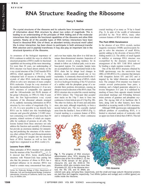
<strong>structure</strong> by two orders <strong>of</strong> magnitude (Fig. 1)<br />
(9, 10). It is now possible to see directly how<br />
RNA can be folded into this breathtakingly<br />
intricate and graceful globular 2.5-MD <strong>structure</strong><br />
containing over 4500 nt and more than 50<br />
proteins, related versions <strong>of</strong> which are responsible<br />
for syn<strong>the</strong>sis <strong>of</strong> proteins in all cells. The<br />
lessons learned from <strong>the</strong>se <strong>structure</strong>s not only<br />
address <strong>the</strong> function and assembly <strong>of</strong> <strong>ribosome</strong>s<br />
but provide an enormous database for interpreting<br />
and predicting <strong>the</strong> <strong>structure</strong>s <strong>of</strong> <strong>the</strong> numerous<br />
o<strong>the</strong>r cellular RNAs and ribonucleoproteins<br />
(RNPs), giving new insights into <strong>the</strong> structural<br />
basis <strong>of</strong> RNA function as well as how life<br />
might have originated in an RNA world (11).<br />
and even base triples, that allow it to fold into its<br />
unique three-dimensional <strong>structure</strong>. Inspection <strong>of</strong><br />
its <strong>structure</strong> reveals a strong tendency for its<br />
strands to follow an A-helical path, even in nonbase-paired<br />
regions. For example, hairpin turns<br />
are accomplished not by incremental bends in <strong>the</strong><br />
RNA chain but by abrupt local changes in<br />
direction, usually centered around one or two<br />
nucleotides. A commonly observed motif is <strong>the</strong> U<br />
turn, seen in <strong>the</strong> anticodon loop <strong>of</strong> tRNA, which<br />
involves hydrogen bonding <strong>of</strong> <strong>the</strong> N3 position<br />
<strong>of</strong> a uridine with <strong>the</strong> phosphate group <strong>of</strong> a nucleotide<br />
three positions downstream, causing an<br />
abrupt reversal in direction <strong>of</strong> <strong>the</strong> RNA chain. The<br />
tRNA <strong>structure</strong> also revealed <strong>the</strong> coaxial stacking<br />
<strong>of</strong> RNA helices: The 7-base-pair (bp) acceptor<br />
stem stacks on <strong>the</strong> 5-bp T stem to form one<br />
continuous A-form helical arm <strong>of</strong> 12 bp (Fig. 2B).<br />
The o<strong>the</strong>r two helices, <strong>the</strong> D stem and anticodon<br />
stem, also stack, although imperfectly, to form a<br />
second helical arm. The two coaxially stacked<br />
arms form <strong>the</strong> familiar L form <strong>of</strong> tRNA (Fig. 1).<br />
Coaxial stacking is a common feature <strong>of</strong> RNA<br />
and is widespread in rRNA, where continuous<br />
coaxial stacking <strong>of</strong> as many as 70 bp is found<br />
(Fig. 2). In spite <strong>of</strong> <strong>the</strong> wealth <strong>of</strong> information<br />
provided by <strong>the</strong> 76-nt tRNA, many o<strong>the</strong>r<br />
common features <strong>of</strong> RNA <strong>structure</strong> were absent.<br />
The Post-tRNA Renaissance<br />
In <strong>the</strong> absence <strong>of</strong> new RNA crystals, nuclear<br />
magnetic resonance (NMR) spectroscopists began<br />
to solve <strong>the</strong> <strong>structure</strong>s <strong>of</strong> small RNAs,<br />
quickly adding to <strong>the</strong> diversity <strong>of</strong> known RNA<br />
folding motifs (12). The ability <strong>of</strong> small ligands<br />
to stabilize or rearrange RNA <strong>structure</strong> was<br />
exemplified by <strong>the</strong> dramatic structural rearrangement<br />
<strong>of</strong> <strong>the</strong> HIV TAR RNA induced<br />
by binding a single arginine residue (13).<br />
One <strong>of</strong> <strong>the</strong> first rRNA <strong>structure</strong>s obtained by<br />
NMR spectroscopy was <strong>the</strong> sarcin-ricin loop<br />
(SRL)<strong>of</strong>28SrRNA(14), a <strong>structure</strong> that interacts<br />
with elongation factors EF1 and EF2 and is<br />
targeted by <strong>the</strong> lethal ribotoxins a-sarcin and<br />
ricin. The compact 29-nt <strong>structure</strong> was found to<br />
contain several purine-purine base pairs, a<br />
tetraloop, and a bulged guanosine adjacent to a<br />
reverse Hoogsteen A-U pair. It is stabilized by<br />
stacking <strong>of</strong> bases from opposite strands (termed<br />
cross-strand stacking) and H-bonding between<br />
imino protons <strong>of</strong> guanines and phosphate oxygens.<br />
The zig-zag fold <strong>of</strong> its backbone (<strong>the</strong> S<br />
turn), along with its o<strong>the</strong>r features, have been<br />
identified as recurring motifs in RNA <strong>structure</strong>s.<br />
Although NMR spectroscopy sidestepped <strong>the</strong><br />
difficult problem <strong>of</strong> crystallizing RNA, it is<br />
limited to structural analysis <strong>of</strong> molecules with<br />
an upper size limit <strong>of</strong> about that <strong>of</strong> tRNA. This led<br />
Lessons from tRNA<br />
Many principles <strong>of</strong> RNA <strong>structure</strong> were gleaned<br />
from <strong>the</strong> <strong>structure</strong> <strong>of</strong> <strong>the</strong> 76-nt tRNA Phe yeast<br />
(1, 2).<br />
It showed that RNA forms double-helical<br />
<strong>structure</strong>s with Watson-Crick base pairing but<br />
alsothat<strong>the</strong>presence<strong>of</strong>riboseinRNAhasa<br />
pr<strong>of</strong>ound influence on its <strong>structure</strong>. tRNA was<br />
found to contain many noncanonical base pairs,<br />
Center for Molecular Biology <strong>of</strong> RNA, Department <strong>of</strong><br />
Molecular, Cell, and Developmental Biology, Sinsheimer<br />
Laboratories, University <strong>of</strong> California, Santa Cruz, Santa<br />
Cruz, CA 95064, USA.<br />
Fig. 1. The progress <strong>of</strong> RNA <strong>structure</strong><br />
determination since 1974, showing <strong>the</strong><br />
relative sizes <strong>of</strong> <strong>the</strong> 76-nt tRNA (1, 2), <strong>the</strong><br />
160-nt P4-P6 domain <strong>of</strong> <strong>the</strong> group I<br />
ribozyme (5), and <strong>the</strong> 4530-nt 70S<br />
<strong>ribosome</strong>, which also contains more than<br />
50 proteins (31, 32). In <strong>the</strong> <strong>ribosome</strong> <strong>structure</strong>,<br />
<strong>the</strong> 16S, 23S, and 5S rRNAs are<br />
colored cyan, gray, and gray-blue, respectively, and <strong>the</strong> small and large subunit ribosomal proteins<br />
are dark blue and magenta, respectively. Two tRNAs (yellow and orange) and a mRNA (green) are<br />
visible inside <strong>the</strong> <strong>ribosome</strong>.<br />
1508<br />
2 SEPTEMBER 2005 VOL 309 SCIENCE www.sciencemag.org
to an increased effort to improve methods for<br />
RNA crystallization (15). An encouraging sign<br />
was <strong>the</strong> appearance <strong>of</strong> <strong>the</strong> first crystal <strong>structure</strong>s <strong>of</strong> a<br />
catalytic RNA, <strong>the</strong> hammerhead ribozyme, solved<br />
first as an RNA-DNA chimera and subsequently<br />
as an all-RNA <strong>structure</strong> (3, 4). Both <strong>structure</strong>s<br />
revealed essentially <strong>the</strong> same fold, with three<br />
helices arranged in a Y configuration containing a<br />
U turn at <strong>the</strong> three-helix junction. Scott and coworkershavegoneontosolve<strong>the</strong><strong>structure</strong>s<strong>of</strong><br />
four additional constructs by using strategies that<br />
trap <strong>the</strong> hammerhead ribozyme in different states<br />
<strong>of</strong> its catalytic cycle, revealing for <strong>the</strong> first time a<br />
detailed high-resolution ‘‘movie’’ <strong>of</strong> <strong>the</strong> mechanism<br />
<strong>of</strong> action <strong>of</strong> a catalytic RNA<br />
(16). Since <strong>the</strong> hammerhead <strong>structure</strong>,<br />
crystal <strong>structure</strong>s <strong>of</strong> three more<br />
ribozymes have been solved, including<br />
<strong>the</strong> hepatitis delta virus ribozyme<br />
(17), <strong>the</strong> hairpin ribozyme (18), and<br />
<strong>the</strong> group I self-splicing intron<br />
(19–21), providing <strong>the</strong> structural<br />
basis for understanding <strong>the</strong>ir respective<br />
catalytic mechanisms.<br />
The first RNA <strong>structure</strong> to be<br />
solved that exceeded <strong>the</strong> size <strong>of</strong><br />
tRNA was <strong>the</strong> 160-nt P4-P6 domain<br />
<strong>of</strong> <strong>the</strong> Tetrahymena group I intron<br />
at 2.8 ) resolution (5). It consists <strong>of</strong><br />
two extended coaxial helical elements<br />
connected at one end by an<br />
internal loop containing a 150- bend<br />
(Fig. 1). For <strong>the</strong> first time, examples<br />
could be seen <strong>of</strong> <strong>the</strong> kinds <strong>of</strong> RNA-<br />
RNA interactions that are used to<br />
stabilize <strong>the</strong> packing <strong>of</strong> RNA helices<br />
into larger, more complex globular<br />
<strong>structure</strong>s. One <strong>of</strong> <strong>the</strong>se has been<br />
named <strong>the</strong> A-minor motif (22), one<br />
<strong>of</strong> <strong>the</strong> most abundant long-range<br />
interactions in rRNA, in which<br />
single-stranded adenosines make<br />
tertiary contacts with <strong>the</strong> minor<br />
grooves <strong>of</strong> double helices. A-minor<br />
interactions also play important<br />
functional roles. Helix-helix interactions<br />
were also formed by ribose<br />
zippers involving H bonding between<br />
<strong>the</strong> 2-hydroxyl group <strong>of</strong> a<br />
ribose in one helix and <strong>the</strong> 2-<br />
hydroxyl and <strong>the</strong> 2-oxygen <strong>of</strong> a<br />
pyrimidine base (or <strong>the</strong> 3-nitrogen <strong>of</strong> a purine<br />
base) <strong>of</strong> <strong>the</strong> o<strong>the</strong>r helix between <strong>the</strong>ir respective<br />
minor groove surfaces. In addition, close approach<br />
<strong>of</strong> phosphates was <strong>of</strong>ten mediated by<br />
bound hydrated magnesium ions. A recurring<br />
motif in <strong>the</strong> P4-P6 <strong>structure</strong>, called <strong>the</strong> A<br />
platform, positions adenines side by side in a<br />
pseudo–base pair within a helix, opening <strong>the</strong><br />
minor groove for interactions with nucleotides<br />
from noncontiguous RNA strands.<br />
rRNA Secondary Structure Prediction<br />
Long before <strong>the</strong> first <strong>ribosome</strong> crystal <strong>structure</strong>s<br />
appeared, <strong>the</strong> essential features <strong>of</strong> rRNA secondary<br />
<strong>structure</strong>s were correctly predicted by<br />
using comparative sequence analysis (23–25).<br />
At about this same time, Michel and colleagues<br />
used a similar approach to establish <strong>the</strong><br />
secondary <strong>structure</strong>s <strong>of</strong> group I introns (26).<br />
Comparative analysis establishes base pairing<br />
by identification <strong>of</strong> compensating base changes<br />
in complementary nucleotides between two or<br />
more sequences. This approach was first explicitly<br />
applied by Fox and Woese (23), who,<br />
studying 5S rRNA sequences as phylogenetic<br />
markers, realized <strong>the</strong>re was a common secondary<br />
<strong>structure</strong> that was compatible with several<br />
different sequences. Comparative analysis<br />
Fig. 2. Secondary <strong>structure</strong>s <strong>of</strong> (A) 16S rRNA<br />
and (B) tRNA. Coaxial stacking <strong>of</strong> individual<br />
helices is indicated by <strong>the</strong> colored bars.<br />
wasusedon<strong>the</strong>large16Sand23SrRNAs<br />
from <strong>the</strong> outset; consequently, <strong>the</strong>ir main<br />
secondary <strong>structure</strong> features were deduced<br />
ra<strong>the</strong>r quickly (24, 25), to be confirmed<br />
crystallographically some 20 years later. Even<br />
some rRNA tertiary interactions were discovered<br />
by comparative analysis (27, 28), as<br />
hadbeen<strong>the</strong>caseearlierfortRNA(29). The<br />
secondary <strong>structure</strong>s <strong>of</strong> most globular RNAs<br />
have been determined by comparative analysis,<br />
including ribonuclease (RNase) P RNA, <strong>the</strong><br />
group I and group II self-splicing introns,<br />
snRNAs, and telomerase RNA. For some<br />
RNAs, such as in vitro–selected RNA aptamers<br />
RNA<br />
and ribozymes, a lack <strong>of</strong> phylogenetic sequence<br />
information has been overcome by introducing<br />
base variation with <strong>the</strong> use <strong>of</strong> ei<strong>the</strong>r<br />
site-directed or random mutagenesis (30).<br />
About 60% <strong>of</strong> <strong>the</strong> nucleotides in <strong>the</strong> large<br />
rRNAs are involved in Watson-Crick base<br />
pairing. However, <strong>the</strong> unpaired bases are not<br />
distributed evenly among <strong>the</strong> four bases. In<br />
Escherichia coli 16S rRNA, for example, <strong>the</strong><br />
proportions <strong>of</strong> unpaired bases for G, C, and U<br />
are 31%, 29%, and 33%, respectively, whereas<br />
62% <strong>of</strong> As are unpaired (27), a tendency that<br />
extends to o<strong>the</strong>r functional RNAs. The preponderance<br />
<strong>of</strong> unpaired adenosines reflects <strong>the</strong>ir<br />
participation in special tertiary<br />
interactions.<br />
Implications for RNA<br />
Tertiary Structure<br />
The <strong>ribosome</strong> and its subunits<br />
are <strong>the</strong> largest asymmetric <strong>structure</strong>s<br />
that have been solved so far<br />
by crystallography. The 2.4 )<br />
Halocarcula marismortui 50S subunit<br />
<strong>structure</strong> (8) and<strong>the</strong>È3 )<br />
Thermus <strong>the</strong>rmophilus 30S subunit<br />
<strong>structure</strong> (6, 7) provided <strong>the</strong> first<br />
detailed views <strong>of</strong> <strong>the</strong> molecular<br />
interactions that are responsible<br />
for <strong>the</strong> <strong>structure</strong>s <strong>of</strong> both ribosomal<br />
subunits. A 5.5 ) <strong>structure</strong><br />
<strong>of</strong> a functional complex <strong>of</strong> <strong>the</strong> T.<br />
<strong>the</strong>rmophilus 70S <strong>ribosome</strong> revealed<br />
<strong>the</strong> positions <strong>of</strong> <strong>the</strong> tRNAs<br />
and mRNA and <strong>the</strong>ir interactions<br />
with <strong>the</strong> <strong>ribosome</strong>, as well as <strong>the</strong><br />
features <strong>of</strong> <strong>the</strong> intersubunit bridges<br />
(31, 32). Many co-crystals <strong>of</strong> <strong>ribosome</strong>s<br />
and subunits containing<br />
tRNA and mRNA fragments, protein<br />
factors, and antibiotics have<br />
nowbeensolvedinanefforttounderstand<br />
<strong>the</strong> mechanism <strong>of</strong> translation<br />
(33). These analyses have<br />
been complemented by extensive<br />
cryogenic electron microscopy<br />
(cryo-EM) reconstruction<br />
studies, which have led to lowerresolution<br />
<strong>structure</strong>s for many<br />
functional complexes <strong>of</strong> <strong>the</strong> <strong>ribosome</strong><br />
that have so far defied<br />
crystallization (34).<br />
Many long-standing questions were immediately<br />
resolved by <strong>the</strong> crystal <strong>structure</strong>s. A critical<br />
issue was whe<strong>the</strong>r <strong>the</strong> rRNA merely serves as a<br />
structural scaffold, or whe<strong>the</strong>r it is directly<br />
involved in ribosomal function. The <strong>structure</strong>s<br />
showed that rRNA in fact does both <strong>of</strong> <strong>the</strong>se<br />
things, creating <strong>the</strong> structural framework for <strong>the</strong><br />
<strong>ribosome</strong>,andat<strong>the</strong>sametimeforming<strong>the</strong>main<br />
features <strong>of</strong> its functional sites, confirming that<br />
<strong>the</strong> <strong>ribosome</strong> is indeed a ribozyme (35).<br />
It was already clear from <strong>the</strong> secondary<br />
<strong>structure</strong>s <strong>of</strong> 16S and 23S rRNA that <strong>the</strong>y are<br />
organized into domains <strong>of</strong> a few hundred nu-<br />
S PECIAL S ECTION<br />
www.sciencemag.org SCIENCE VOL 309 2 SEPTEMBER 2005 1509
S PECIAL S ECTION<br />
RNA<br />
cleotides each, four for 16S rRNA and six for<br />
23S rRNA (24, 25). The three major domains <strong>of</strong><br />
16S rRNA were assigned to <strong>the</strong> head, body,<br />
and platform features <strong>of</strong> <strong>the</strong> low-resolution EM<br />
<strong>structure</strong> for <strong>the</strong> 30S subunit (36, 37), and this<br />
has been confirmed by crystallography (6, 7).<br />
Their structural autonomy appears to facilitate<br />
<strong>the</strong>ir independent movement during translation.<br />
The six domains <strong>of</strong> 23S rRNA are more<br />
closely packed against one ano<strong>the</strong>r (8) and<br />
were not distinguishable as separate domains<br />
<strong>of</strong> <strong>the</strong> 50S subunit at low resolution.<br />
Comparative analysis <strong>of</strong> 16S and 23S rRNA<br />
secondary <strong>structure</strong> also provided a sense <strong>of</strong> <strong>the</strong><br />
allowed variation in <strong>the</strong> sizes<br />
<strong>of</strong> <strong>the</strong> different helical elements<br />
(24, 25, 27). Some<br />
helices are strictly conserved<br />
in length, showing no phylogenetic<br />
variation. O<strong>the</strong>rs vary,<br />
showing both shorter and<br />
longer versions relative to E.<br />
coli in different phylogenetic<br />
branches. In some cases,<br />
shortening but not leng<strong>the</strong>ning<br />
is permitted. These observations<br />
can now be interpreted in<br />
view <strong>of</strong> <strong>the</strong> three-dimensional<br />
<strong>structure</strong>s. Variable-length helices<br />
are always found on <strong>the</strong><br />
surface, distant from <strong>the</strong> functional<br />
center <strong>of</strong> <strong>the</strong> <strong>ribosome</strong>,<br />
with <strong>the</strong>ir extensible ends<br />
pointing into <strong>the</strong> solvent. Ones<br />
that can be shortened, but not<br />
leng<strong>the</strong>ned, have ends whose<br />
maximum lengths are restricted<br />
by potential clash with<br />
o<strong>the</strong>r structural elements.<br />
The hundreds <strong>of</strong> individual<br />
rRNA helices in <strong>the</strong> <strong>ribosome</strong><br />
allowustodrawnewgeneralities<br />
about RNA secondary<br />
<strong>structure</strong>. Most rRNA helices<br />
terminate at both ends in G-C<br />
pairs. As predicted from sequence<br />
analysis and chemical<br />
probing studies, noncanonical<br />
A-G pairs <strong>of</strong>ten flank <strong>the</strong> ends<br />
<strong>of</strong> helices (38, 39). The crystal<br />
<strong>structure</strong>s show that <strong>the</strong>y are most commonly<br />
sheared A-G pairs, as well as Watson-<br />
Crick-like A-G imino pairs (40, 41). As first<br />
observed for tRNA, bases that fall into nonhelical<br />
(so-called single-stranded) regions <strong>of</strong><br />
<strong>the</strong> secondary <strong>structure</strong> are typically found to<br />
be highly <strong>structure</strong>d, participating in H bonding<br />
and stacking interactions with o<strong>the</strong>r elements<br />
<strong>of</strong> <strong>the</strong> RNA. Of <strong>the</strong> 25 possible kinds <strong>of</strong><br />
noncanonical base pairs involving two or<br />
more hydrogen bonds (40, 41), 20 are found<br />
in <strong>the</strong> <strong>ribosome</strong>. For example, <strong>the</strong> sheared<br />
A-G pair is represented 20 times in 16S rRNA<br />
and 46 times in 23S rRNA, and <strong>the</strong>re are 7<br />
and 22 examples, respectively, <strong>of</strong> <strong>the</strong> reverse<br />
Hoogsteen A-U pair. Westh<strong>of</strong> and co-workers<br />
have made a comprehensive study <strong>of</strong> <strong>the</strong><br />
kinds <strong>of</strong> noncanonical interactions that appear<br />
in RNA and <strong>the</strong>ir geometric and stereochemical<br />
classification (42, 43).<br />
Among <strong>the</strong> most interesting structural<br />
motifs are <strong>the</strong> A-minor interactions, <strong>of</strong> which<br />
hundreds <strong>of</strong> examples are found in rRNA (22).<br />
In <strong>the</strong>se motifs, single-stranded adenosines<br />
reach into <strong>the</strong> minor groove <strong>of</strong> a helix, making<br />
both H bonding and van der Waals contacts.<br />
They are not simply base-base interactions, but<br />
nucleoside-nucleoside interactions, because<br />
crucial contacts are also made with <strong>the</strong> riboses<br />
Fig. 3. (A) TypeIand(B) typeIIA-minor<br />
nucleoside interactions (22). These precise<br />
lock-and-key minor-groove interactions between<br />
(usually) an adenosine and a Watson-<br />
Crick base pair are found extensively in 16S<br />
and 23S rRNA (6, 8). They were first observed<br />
in crystal packing <strong>of</strong> <strong>the</strong> hammerhead<br />
ribozyme (3, 4) and in <strong>the</strong> P4-P6 domain <strong>of</strong><br />
<strong>the</strong> group I ribozyme (5). (C to E) A-minor<br />
interactions play an important functional role<br />
in monitoring codon-anticodon interaction<br />
by <strong>the</strong> <strong>ribosome</strong> via <strong>the</strong>ir unique stereochemical<br />
fit to Watson-Crick base pairs (44).<br />
as well as <strong>the</strong> bases (Fig. 3). Pairs <strong>of</strong> consecutive<br />
A-minor interactions are <strong>of</strong>ten found, in which<br />
two adjacent adenosines sequentially form type<br />
II and type I interactions (although some type II<br />
interactions are also made by guanosines) with<br />
adjacent base pairs (Fig. 3, A and B), which are<br />
typically G-C pairs. Although <strong>the</strong>y form many<br />
important structural contacts, <strong>the</strong>y are also intimately<br />
involved in <strong>ribosome</strong> function. For<br />
example, <strong>the</strong> 3-terminal adenosines <strong>of</strong> both<br />
<strong>the</strong> A- and P-site tRNAs are positioned in <strong>the</strong><br />
peptidyl transferase site by A-minor interactions<br />
with 23S rRNA (35). An elegant<br />
RNA-based mechanism using <strong>the</strong> A-minor<br />
motif occurs in <strong>the</strong> decoding site <strong>of</strong> <strong>the</strong> 30S<br />
subunit (44), where <strong>the</strong> stereochemical fit <strong>of</strong><br />
codon-anticodon pairing is monitored by A-<br />
minor interactions between A1492 and A1493<br />
<strong>of</strong> 16S rRNA (supported by additional interactions<br />
from G530) and <strong>the</strong> minor groove<br />
surface <strong>of</strong> <strong>the</strong> codon-anticodon helix (Fig. 3,<br />
C to E). The prevalence <strong>of</strong> A-minor interactions<br />
in rRNA helps to account for <strong>the</strong> overrepresentation<br />
<strong>of</strong> single-stranded adenosines in<br />
rRNA secondary <strong>structure</strong>s.<br />
About half <strong>of</strong> <strong>the</strong> helices in rRNA<br />
terminate in hairpin loops. In T. <strong>the</strong>rmophilus<br />
16S rRNA, 17 <strong>of</strong> its 32 hairpin loops are<br />
tetraloops (Fig. 2), first identified as <strong>the</strong> most<br />
common type <strong>of</strong> hairpin loop<br />
in rRNA by inspection <strong>of</strong> <strong>the</strong>ir<br />
phylogenetically derived secondary<br />
<strong>structure</strong>s (45). As<br />
found for many RNAs, <strong>the</strong><br />
GNRA tetraloop is most common<br />
in rRNA, representing<br />
about half <strong>of</strong> <strong>the</strong> observed<br />
tetraloops. The o<strong>the</strong>r hairpin<br />
loops use a variety <strong>of</strong> strategies<br />
to execute <strong>the</strong>ir turns.<br />
In 16S rRNA, <strong>the</strong>re are five<br />
examples <strong>of</strong> U turns, and G<br />
turns are also found, in<br />
which <strong>the</strong> stabilizing hydrogen<br />
bond to <strong>the</strong> backbone<br />
phosphate is made from <strong>the</strong><br />
N1 position <strong>of</strong> a guanine<br />
base; <strong>the</strong>se include <strong>the</strong> G<br />
turns that are an intrinsic<br />
feature <strong>of</strong> GNRA tetraloop<br />
<strong>structure</strong>s. Indeed, G(N1)-<br />
phosphate H bonds are widespread,<br />
making many kinds<br />
<strong>of</strong> base-backbone interactions<br />
in addition to G turns,<br />
<strong>of</strong> which <strong>the</strong>re are dozens <strong>of</strong><br />
examples in both 16S and<br />
23S rRNA.<br />
It has been said that<br />
‘‘tRNA looks like Nature’s<br />
attempt to make RNA do<br />
<strong>the</strong> job <strong>of</strong> a protein’’ (46).<br />
rRNA takes this notion to<br />
<strong>the</strong> extreme, representing <strong>the</strong><br />
limit <strong>of</strong> what can be done to<br />
make a globular, functional molecule out <strong>of</strong><br />
RNA, beyond which nature has resorted to<br />
proteins. The basic building block <strong>of</strong> RNA<br />
<strong>structure</strong>, <strong>the</strong> double helix, greatly restricts <strong>the</strong><br />
ability <strong>of</strong> RNA to form globular <strong>structure</strong>s<br />
because <strong>of</strong> its rigidity and limited geometry.<br />
How <strong>the</strong>n, does RNA manage to form a <strong>structure</strong><br />
such as <strong>the</strong> <strong>ribosome</strong>, with its complex,<br />
curving three-dimensional surfaces, stereospecific<br />
binding pockets, and o<strong>the</strong>r intricate<br />
molecular features? Almost all rRNA helices<br />
contain seven or fewer contiguous Watson-<br />
Crick base pairs, in spite <strong>of</strong> <strong>the</strong> fact that <strong>the</strong><br />
overall dimensions <strong>of</strong> <strong>the</strong> <strong>ribosome</strong> (È250 ))<br />
would in principle allow for continuous heli-<br />
1510<br />
2 SEPTEMBER 2005 VOL 309 SCIENCE www.sciencemag.org
ces <strong>of</strong> as many as 80 bp. A general strategy<br />
found throughout <strong>the</strong> <strong>ribosome</strong> is to connect<br />
<strong>the</strong>se short helices by bulge loops or internal<br />
loops <strong>of</strong> unequal length, introducing bends<br />
that allow a high degree <strong>of</strong> structural curvature.<br />
The connecting loops <strong>the</strong>mselves are<br />
highly <strong>structure</strong>d, rich in noncanonical base<br />
pairs as well as base-phosphate and baseribose<br />
interactions that constrain <strong>the</strong> geometries<br />
<strong>of</strong> <strong>the</strong> individual bends. Indeed, bases that<br />
are not involved in ei<strong>the</strong>r Watson-Crick or some<br />
kind <strong>of</strong> noncanonical interaction are very rare,<br />
explaining why so few bases are reactive toward<br />
chemical probes and <strong>the</strong>ir<br />
inability to hybridize with<br />
oligonucleotide probes. Some<br />
<strong>of</strong> <strong>the</strong> connecting loop features<br />
have been recognized<br />
as recurring motifs in RNA<br />
<strong>structure</strong>: for example, <strong>the</strong><br />
S turn motif and <strong>the</strong> kink<br />
turn that creates a sharp 120-<br />
angle between two adjacent<br />
helices (47).<br />
These irregular compound<br />
helices are packed<br />
against one ano<strong>the</strong>r to form<br />
<strong>the</strong> final globular <strong>structure</strong>.<br />
Earlier, it was thought that<br />
RNA-RNA packing would<br />
be mediated by <strong>the</strong> basic<br />
ribosomal proteins to alleviate<br />
charge repulsion between<br />
<strong>the</strong> high density <strong>of</strong> negatively<br />
charged phosphate groups<br />
lining <strong>the</strong> RNA backbone. It<br />
was <strong>the</strong>refore surprising to<br />
find extensive regions <strong>of</strong><br />
closely packed RNA helices<br />
containing little or no protein.<br />
Packing <strong>of</strong> RNA structural<br />
elements is <strong>of</strong> special<br />
interest in <strong>the</strong> functional sites,<br />
which are mostly devoid <strong>of</strong><br />
proteins. In fact, ribosomal<br />
proteins are found mainly on<br />
<strong>the</strong> outer surface <strong>of</strong> <strong>the</strong> <strong>ribosome</strong>, although many<br />
<strong>of</strong> <strong>the</strong>m contain long, un<strong>structure</strong>d tails that<br />
penetrate <strong>the</strong> RNA (48, 49). Not surprisingly,<br />
both divalent and monovalent cations as well<br />
as polyamines, which have long been known<br />
to be essential for <strong>the</strong> structural and functional<br />
integrity <strong>of</strong> <strong>ribosome</strong>s, mediate RNA-RNA<br />
packing interactions in <strong>the</strong> <strong>ribosome</strong>, helping to<br />
neutralize phosphate-phosphate repulsion (50).<br />
The ribose zipper (5) is yet ano<strong>the</strong>r strategy<br />
that is used for packing <strong>the</strong> minor grooves <strong>of</strong><br />
rRNA helices against each o<strong>the</strong>r.<br />
Folding<strong>of</strong>RNAdiffersinmanywaysfrom<br />
that <strong>of</strong> proteins. There are only four types <strong>of</strong><br />
nucleotide monomers; <strong>the</strong>re are six backbone<br />
torsion angles, instead <strong>of</strong> two; and RNA<br />
<strong>structure</strong> is not nucleated by a hydrophobic<br />
core, as are most proteins. Instead, RNA<br />
folding uses <strong>the</strong> two principle devices that<br />
were first seen in <strong>the</strong> double-helical <strong>structure</strong>s<br />
<strong>of</strong> DNA and RNA: hydrogen bonding and<br />
base stacking.<br />
An example <strong>of</strong> how noncanonical H-bonded<br />
interactions can direct <strong>the</strong> packing <strong>of</strong> RNA<br />
helices is <strong>the</strong> helix 6–helix 8 interaction in 16S<br />
rRNA (6) (Fig. 4). These two helices pack<br />
against each o<strong>the</strong>r at a 90- angle, via <strong>the</strong>ir<br />
respective minor-groove surfaces. They are positioned<br />
by two layers <strong>of</strong> coplanar bases that<br />
form two exquisitely stereospecific H-bonded<br />
networks. Both layers contain central A-minor<br />
interactions in which adenosines in helix 8 bind<br />
Fig. 4. An example <strong>of</strong> how <strong>the</strong> <strong>ribosome</strong> packs two helices (h6 and h8) in 16S rRNA<br />
toge<strong>the</strong>r at right angles to each o<strong>the</strong>r (6). Two layers <strong>of</strong> nucleotides (yellow and red)<br />
form extensive hydrogen-bonded networks (dotted lines) that precisely locate <strong>the</strong> two<br />
helices. In <strong>the</strong> top (yellow) layer, nucleotide A151 in h8 makes a type II A-minor<br />
interaction with <strong>the</strong> G102-C67 base pair in h6, itself bolstered by a Hoogsteen pair with<br />
U170. In <strong>the</strong> bottom (red) layer, A152 <strong>of</strong> h8 makes a type I A-minor interaction with a<br />
Watson-Crick-like pair between G68 and A101 <strong>of</strong> h6. Both A152 and its A-G receptor<br />
are bolstered by additional noncanonical base pairings with C169 and G64.<br />
to receptors in helix 6, forming <strong>the</strong> heart <strong>of</strong> <strong>the</strong><br />
interhelical connection. The upper (yellow) layer<br />
is formed by interaction <strong>of</strong> <strong>the</strong> minor-groove<br />
side <strong>of</strong> a Watson-Crick G-C pair in helix 6<br />
through a type II A-minor interaction with <strong>the</strong><br />
adenosine <strong>of</strong> a noncanonical Hoogsteen base<br />
pair in helix 8. The lower (red) layer is formed<br />
from a noncanonical A-G-G base triple, <strong>of</strong><br />
which one <strong>of</strong> <strong>the</strong> guanosines forms <strong>the</strong> receptor<br />
for a Type I A-minor interaction from an<br />
adenosine involved in a noncanonical A-C<br />
pair in helix 8. The positions <strong>of</strong> both <strong>of</strong> <strong>the</strong><br />
A-minor adenosines are constrained by <strong>the</strong>ir<br />
additional base-base interactions, tightly<br />
restricting <strong>the</strong> overall geometry. It seems<br />
counterintuitive that this apparently coplanar<br />
arrangement <strong>of</strong> bases results in a 90- packing<br />
angle between <strong>the</strong> two helices. This is <strong>the</strong> result<br />
<strong>of</strong> three effects: first, <strong>the</strong> bases <strong>of</strong> RNA<br />
RNA<br />
helices are tilted at an angle to <strong>the</strong> helical axis;<br />
second, <strong>the</strong> adenine bases in A-minor interactions<br />
typically form È30- angles with <strong>the</strong><br />
planes <strong>of</strong> <strong>the</strong>ir receptor bases; and third, <strong>the</strong><br />
adenines are held at an angle to helix 8 by additional<br />
noncanonical base-base interactions.<br />
In addition to coaxial stacking <strong>of</strong> helices, <strong>the</strong><br />
<strong>ribosome</strong> contains some remarkable examples<br />
<strong>of</strong> base stacking <strong>of</strong> unpaired bases, such as in<br />
<strong>the</strong> noncanonical <strong>structure</strong> known as helix 70 in<br />
23S rRNA (Fig. 5). Helix 70 is located at <strong>the</strong><br />
subunit interface <strong>of</strong> <strong>the</strong> 50S subunit near <strong>the</strong><br />
geometric center <strong>of</strong> <strong>the</strong> <strong>ribosome</strong> (8). It forms<br />
<strong>the</strong> attachment point for helix<br />
69, which interacts with both<br />
<strong>the</strong> A- and P-site tRNAs, as<br />
well as forming a bridge<br />
to <strong>the</strong> decoding site <strong>of</strong> 16S<br />
rRNA (31, 32). Its compact,<br />
23-nucleotide <strong>structure</strong><br />
is a tour de force <strong>of</strong><br />
noncanonical complexity<br />
and is one <strong>of</strong> <strong>the</strong> most conserved<br />
features <strong>of</strong> rRNA. It<br />
contains no fewer than four<br />
systems <strong>of</strong> stacked bases,<br />
one <strong>of</strong> which is bifurcated to<br />
form a short fifth stack. Although<br />
helix 70 superficially<br />
resembles a normal RNA<br />
helix, it in fact contains only<br />
a single canonical Watson-<br />
Crick base pair (G1964-<br />
C1934). Although its role in<br />
translation is not known, <strong>the</strong><br />
projection <strong>of</strong> bases A1966<br />
and U1944 into <strong>the</strong> minor<br />
grooves <strong>of</strong> <strong>the</strong> functionally<br />
important helices 93 and 92,<br />
respectively, are suggestive<br />
<strong>of</strong> some relationship to <strong>the</strong><br />
peptidyl transferase activity<br />
<strong>of</strong> <strong>the</strong> <strong>ribosome</strong>.<br />
rRNA folds correctly only<br />
by assembling with ribosomal<br />
proteins, which appear to<br />
stage <strong>the</strong> order <strong>of</strong> folding <strong>of</strong> rRNA during<br />
<strong>ribosome</strong> assembly to avoid losing improperly<br />
folded <strong>ribosome</strong>s in kinetic traps. Their role in<br />
translational function appears to be subordinate<br />
to that <strong>of</strong> rRNA, helping to improve <strong>the</strong><br />
efficiency and accuracy <strong>of</strong> mechanisms that<br />
are based on RNA. This view is supported by<br />
<strong>the</strong>ir location mainly on <strong>the</strong> exterior <strong>of</strong> <strong>the</strong><br />
<strong>ribosome</strong>, away from <strong>the</strong> functional subunit<br />
interface region (6–8, 31, 32). Fur<strong>the</strong>r evidence<br />
comes from <strong>the</strong> observation that at least<br />
one-third <strong>of</strong> <strong>the</strong> ribosomal proteins can be<br />
deleted singly without conferring a lethal<br />
phenotype (51). Nearly all ribosomal proteins<br />
interact directly with rRNA, and few have<br />
contact with o<strong>the</strong>r ribosomal proteins. They<br />
are typically small and basic, representing a<br />
diverse collection <strong>of</strong> structural types that span<br />
<strong>the</strong> range <strong>of</strong> known protein folds, giving <strong>the</strong><br />
S PECIAL S ECTION<br />
www.sciencemag.org SCIENCE VOL 309 2 SEPTEMBER 2005 1511
RNA<br />
S PECIAL S ECTION<br />
impression that <strong>the</strong>y were recruited to <strong>the</strong><br />
<strong>ribosome</strong> in many independent evolutionary<br />
events. As mentioned above, some ribosomal<br />
proteins have long, un<strong>structure</strong>d tails that penetrate,<br />
and co-assemble with, <strong>the</strong> rRNA (48, 49).<br />
The C-terminal tails <strong>of</strong> proteins S9 and S13<br />
contact <strong>the</strong> anticodon stem loop <strong>of</strong> tRNA in <strong>the</strong><br />
30S P site; cells in which <strong>the</strong> S9 and S13 tails<br />
have been deleted are viable, showing that<br />
<strong>the</strong>se interactions are not essential for <strong>ribosome</strong><br />
function (52). In keeping with <strong>the</strong>ir<br />
diverse <strong>structure</strong>s, <strong>the</strong>ir rRNA binding sites<br />
are comparably diverse,<br />
comprising both helical<br />
and loop features; unlike<br />
DNA-binding proteins, ribosomal<br />
proteins mainly<br />
recognize higher-order<br />
structural features <strong>of</strong><br />
rRNA, ra<strong>the</strong>r than base<br />
sequence (48, 49). Binding<br />
to rRNA helices occurs<br />
preferentially on <strong>the</strong>ir<br />
minor-groove surfaces.<br />
Apart from contributing<br />
to <strong>the</strong> neutralization <strong>of</strong><br />
negative charges on <strong>the</strong><br />
rRNA backbone, ribosomal<br />
proteins are known<br />
to stabilize certain tertiary<br />
folds (53) and to help fix<br />
<strong>the</strong> relative orientation <strong>of</strong><br />
helices at multihelix junctions<br />
(54). Indeed, proteins<br />
may have initially evolved<br />
to extend <strong>the</strong> structural<br />
repertoire <strong>of</strong> RNA in an<br />
RNA world (55).<br />
Ribosome<br />
Dynamics<br />
Ribosomes are molecular<br />
machines, whose moving<br />
parts enable <strong>the</strong> dynamic<br />
process <strong>of</strong> translation.<br />
Each tRNA traverses a<br />
distance <strong>of</strong> more than<br />
130 ) from <strong>the</strong> time it enters <strong>the</strong> <strong>ribosome</strong><br />
as an aminoacyl-tRNA until it is released as a<br />
deacylated tRNA (31, 32, 56); it was anticipated<br />
that such large-scale tRNA movement<br />
must be matched by corresponding<br />
movements in <strong>the</strong> <strong>ribosome</strong>. Evidence for this,<br />
ranging from local conformational changes<br />
to relative movement <strong>of</strong> <strong>the</strong> 30S and 50S subunits,<br />
comes from structural changes that are<br />
observed between different crystal <strong>structure</strong>s<br />
(31, 32, 44, 57–59) and from cryo-EM studies<br />
<strong>of</strong> <strong>ribosome</strong>s trapped in different functional<br />
states (34, 60, 61).<br />
An example <strong>of</strong> a local rearrangement is <strong>the</strong><br />
flipping <strong>of</strong> bases G530, A1492, and A1493 in<br />
<strong>the</strong> 30S decoding site to monitor <strong>the</strong> accuracy<br />
<strong>of</strong> codon-anticodon interaction (44) (Fig. 3, C<br />
to E). Accompanying this local change is a<br />
larger-scale movement, in which <strong>the</strong> 30S subunit<br />
goes from an open to a closed conformation<br />
that is induced by binding <strong>of</strong> a cognate tRNA<br />
(44, 62, 63). It is believed that <strong>the</strong> energy<br />
derived from binding <strong>the</strong> cognate tRNA compensates<br />
for <strong>the</strong> energetic costs <strong>of</strong> <strong>the</strong> transition<br />
to <strong>the</strong> closed form. The altered conformation <strong>of</strong><br />
<strong>the</strong> 30S subunit may affect <strong>the</strong> interactions<br />
between <strong>the</strong> aminoacyl-tRNAIEF-TuI guanosine<br />
triphosphate (GTP) ternary complex and<br />
<strong>the</strong> conserved sarcin-ricin loop <strong>of</strong> 23S rRNA in<br />
<strong>the</strong> 50S subunit, leading to acceleration <strong>of</strong> GTP<br />
Fig. 5. Helix 70 <strong>of</strong> 23S rRNA (8) contains four different systems <strong>of</strong> stacked bases and contains<br />
only a single canonical Watson-Crick base pair (G1964-C1934). Its <strong>structure</strong> positions U1944<br />
and A1966 to interact with <strong>the</strong> minor grooves <strong>of</strong> helices 92 and 93 in <strong>the</strong> peptidyl transferase<br />
region <strong>of</strong> <strong>the</strong> 50S subunit.<br />
hydrolysis and accommodation <strong>of</strong> aminoacyltRNA<br />
(62, 63).<br />
An example <strong>of</strong> a larger-scale movement is<br />
that <strong>of</strong> <strong>the</strong> L1 arm <strong>of</strong> <strong>the</strong> 50S subunit. Exit <strong>of</strong> <strong>the</strong><br />
E site–bound deacylated tRNA is obstructed by<br />
protein L1 and <strong>the</strong> extended arm <strong>of</strong> 23S rRNA to<br />
which it is bound (32). In addition, <strong>the</strong> observed<br />
contact with <strong>the</strong> elbow <strong>of</strong> tRNA bound in <strong>the</strong><br />
P/E state (64) requires movement <strong>of</strong> <strong>the</strong> L1 arm<br />
by about 20 ) (65). In <strong>the</strong> Dinococcus radiodurans<br />
50S crystal <strong>structure</strong> (59), <strong>the</strong> position <strong>of</strong><br />
<strong>the</strong> L1 arm is shifted downward by about 20 )<br />
relative to that seen in <strong>the</strong> T. <strong>the</strong>rmophilus<br />
crystal and <strong>the</strong> E. coli cryo-EM <strong>structure</strong>s,<br />
sufficient to allow release <strong>of</strong> <strong>the</strong> tRNA.<br />
Coupled movement <strong>of</strong> tRNA and mRNA<br />
occurs during <strong>the</strong> EF-G–catalyzed process <strong>of</strong><br />
translocation, <strong>the</strong> most dynamic <strong>of</strong> ribosomal<br />
functions. Translocation takes place in at least<br />
two steps, <strong>the</strong> first <strong>of</strong> which mainly involves<br />
movement <strong>of</strong> <strong>the</strong> acceptor arms <strong>of</strong> tRNA relative<br />
to <strong>the</strong> 50S subunit. This results in tRNAs bound<br />
in hybrid states, in which <strong>the</strong>ir anticodon ends<br />
remain in <strong>the</strong>ir original positions in <strong>the</strong> A and P<br />
sites <strong>of</strong> <strong>the</strong> 30S subunit while <strong>the</strong>ir acceptor ends<br />
move to <strong>the</strong> P and E sites <strong>of</strong> <strong>the</strong> 50S subunits<br />
(64). In <strong>the</strong> second step, <strong>the</strong> anticodon ends<br />
move to <strong>the</strong> P and E sites <strong>of</strong> <strong>the</strong> 30S subunit,<br />
coupled to movement <strong>of</strong> <strong>the</strong> mRNA, completing<br />
<strong>the</strong> translocation <strong>of</strong> tRNA from <strong>the</strong> A to P<br />
and P to E sites. Structural<br />
changes accompanying<br />
translocation<br />
have been analyzed by<br />
comparison <strong>of</strong> cryo-EM<br />
reconstructions in which<br />
<strong>ribosome</strong>s were trapped<br />
in <strong>the</strong> pre- and posttranslocation<br />
states<br />
(60, 61). Pretranslocation<br />
or posttranslocation<br />
<strong>ribosome</strong>s,<br />
containing peptidyltRNA<br />
bound to <strong>the</strong> A<br />
site or P site, respectively,<br />
were bound with<br />
EF-G and a nonhydrolyzable<br />
GTP analog or<br />
guanosine diphosphate<br />
(GDP). These experiments<br />
show structural<br />
differences between <strong>the</strong><br />
pre- and posttranslocation<br />
states <strong>of</strong> <strong>the</strong> <strong>ribosome</strong><br />
corresponding to<br />
a rotational movement<br />
<strong>of</strong> about 6- between <strong>the</strong><br />
30S and 50S subunits,<br />
causing relative displacements<br />
<strong>of</strong> as much<br />
as 20 ) at <strong>the</strong>ir extremities.<br />
This movement<br />
is accompanied by<br />
o<strong>the</strong>r structural changes,<br />
including rearrangement<br />
<strong>of</strong> intersubunit bridge contacts between <strong>the</strong><br />
head <strong>of</strong> <strong>the</strong> 30S subunit and <strong>the</strong> central protuberance<br />
<strong>of</strong> <strong>the</strong> 50S subunit, as well as a 20 )<br />
displacement <strong>of</strong> <strong>the</strong> L1 arm. On <strong>the</strong> basis <strong>of</strong><br />
<strong>the</strong>se observations, Frank and co-workers have<br />
proposed a ratchet model for translocation, in<br />
which rotational movement between <strong>the</strong> subunits<br />
and movement <strong>of</strong> <strong>the</strong> L1 arm, coupled<br />
with alternate binding and release <strong>of</strong> <strong>the</strong> two<br />
ends <strong>of</strong> <strong>the</strong> tRNA, is used to drive movement<br />
<strong>of</strong> tRNA and mRNA through <strong>the</strong> <strong>ribosome</strong><br />
(60, 61). GTP hydrolysis is coupled to translocation<br />
under normal cellular conditions,<br />
although <strong>the</strong> first step <strong>of</strong> translocation leading<br />
to formation <strong>of</strong> hybrid states can proceed<br />
spontaneously in vitro after peptide bond<br />
formation (64). Fur<strong>the</strong>rmore, <strong>the</strong> observation<br />
that a complete single round <strong>of</strong> highly accurate<br />
1512<br />
2 SEPTEMBER 2005 VOL 309 SCIENCE www.sciencemag.org
translocation can proceed in <strong>the</strong> absence <strong>of</strong><br />
EF-G and GTP, stimulated by <strong>the</strong> antibiotic<br />
sparsomycin (66), indicates that translocation<br />
is an inherent property <strong>of</strong> <strong>the</strong> <strong>ribosome</strong> itself.<br />
Although <strong>the</strong> resolution <strong>of</strong> cryo-EM reconstructions<br />
is insufficient to draw detailed conclusions<br />
about <strong>the</strong> mechanism <strong>of</strong> translocational dynamics,<br />
it is likely to be yet ano<strong>the</strong>r function <strong>of</strong> rRNA. A<br />
puzzle is how large-scale movements, such as those<br />
<strong>of</strong> translocation, which must occur at <strong>the</strong> rate <strong>of</strong><br />
about 20 per second, avoid<br />
<strong>the</strong> potential kinetic barriers<br />
that would be expected from<br />
making and breaking <strong>of</strong> <strong>the</strong><br />
many molecular interactions<br />
that maintain <strong>the</strong> precise geometry<br />
<strong>of</strong> <strong>the</strong> different conformational<br />
states needed for<br />
accurate translation. Helical<br />
switches, in which certain<br />
RNA sequences alternate between<br />
two different <strong>structure</strong>s<br />
by base pairing with different<br />
complementary strands, have<br />
two disadvantages. First,<br />
disruption<strong>of</strong>anRNAhelix<br />
has a high energy <strong>of</strong> activation,<br />
and second, it leads to<br />
single-stranded intermediates<br />
that lack <strong>the</strong> necessary rigidity<br />
to maintain precise geometry.<br />
Helical switches have not<br />
been found in <strong>ribosome</strong>s,<br />
perhaps for <strong>the</strong>se reasons.<br />
The ideal dynamic interactions<br />
would thus be ones<br />
whose disruption and formation<br />
have relatively low activation<br />
barriers, maintain<br />
<strong>the</strong>ir local conformations in<br />
<strong>the</strong> disrupted state, and form<br />
with precise stereochemistry.<br />
The abundant A-minor interactions<br />
fit this description<br />
well. We have already seen<br />
that <strong>the</strong>y participate in dynamic<br />
yet precise interactions<br />
in aminoacyl-tRNA<br />
selection and in <strong>the</strong> peptidyl<br />
transferase active site (Fig.<br />
3) (35, 44). The crystallographic<br />
evidence suggests that involvement <strong>of</strong><br />
A-minor interactions in ribosomal dynamics<br />
may be much more widespread.<br />
The 3.0 ) crystal <strong>structure</strong> <strong>of</strong> <strong>the</strong> isolated<br />
30S subunit shows that <strong>the</strong>re are about 55 A-<br />
minor interactions, or potential A-minor interactions,<br />
in 16S rRNA (Fig. 6). They are<br />
typically found in consecutive pairs consisting<br />
<strong>of</strong> a type II interaction followed by a type I<br />
interaction. The vast majority <strong>of</strong> <strong>the</strong>m are longrange<br />
interactions; i.e., <strong>the</strong>y connect parts <strong>of</strong> <strong>the</strong><br />
secondary <strong>structure</strong> that lie in different domains<br />
or subdomains <strong>of</strong> <strong>the</strong> RNA. In contrast, <strong>the</strong><br />
o<strong>the</strong>r base-base and base-backbone tertiary<br />
interactions are overwhelmingly local (67).<br />
Most intriguing is that eight <strong>of</strong> <strong>the</strong> sets <strong>of</strong> A-<br />
minor examples in Fig. 6 have optimal geometries<br />
except that <strong>the</strong> adenosines are out <strong>of</strong><br />
contact range from <strong>the</strong>ir putative helical<br />
receptors. This suggests that <strong>the</strong>se eight sets <strong>of</strong><br />
potential A-minor contacts could play a role in<br />
<strong>the</strong> conformational dynamics <strong>of</strong> <strong>the</strong> 30S subunit.<br />
Direct support for formation <strong>of</strong> one <strong>of</strong> <strong>the</strong>m<br />
comes from <strong>the</strong> electron density map <strong>of</strong> <strong>the</strong> T.<br />
Fig. 6. A-minor interactions in 16S rRNA, as<br />
found in <strong>the</strong> 3.0 Å crystal <strong>structure</strong> <strong>of</strong> <strong>the</strong> T.<br />
<strong>the</strong>rmophilus 30S ribosomal subunit (6).<br />
Potential A-minor interactions with adenosines<br />
that have optimal geometry but are out<br />
<strong>of</strong> contact range with <strong>the</strong>ir helical receptors<br />
are shown in red; interactions that form at<br />
proper contact distances are shown in black.<br />
Nucleotides that form intersubunit bridge<br />
contacts (32) are highlighted in blue.<br />
<strong>the</strong>rmophilus 70S <strong>ribosome</strong> (32), in which <strong>the</strong><br />
potential interaction between helix 13 and helix<br />
44 (Fig. 6) is clearly formed. Intriguingly, most<br />
<strong>of</strong> <strong>the</strong>se potentially dynamic interactions are<br />
positioned immediately adjacent to features<br />
<strong>of</strong> 16S rRNA that form intersubunit bridges<br />
(Fig. 6) (32); this observation is consistent<br />
with <strong>the</strong>ir possible involvement in translocation,<br />
in which molecular rearrangements at <strong>the</strong><br />
subunit interface are known to occur (60, 61).<br />
Conclusions<br />
We have now seen enough RNA <strong>structure</strong>s to<br />
infer some generalities about what we can expect<br />
RNA<br />
to find in o<strong>the</strong>r RNAs. We have probably seen<br />
most, if not all <strong>of</strong> <strong>the</strong> possible local RNA<br />
folding motifs (10); U turns, T loops, S turns,<br />
kink turns, hook turns, A minor interactions, A<br />
platforms, and tetraloops are all recurring<br />
features <strong>of</strong> <strong>the</strong> <strong>structure</strong>s <strong>of</strong> globular RNAs.<br />
Toge<strong>the</strong>r with <strong>the</strong> A-form double helix and<br />
<strong>the</strong> more than 20 types <strong>of</strong> noncanonical<br />
base pairs, we can now say that <strong>the</strong>se<br />
comprise <strong>the</strong> building blocks <strong>of</strong> RNA architecture.<br />
It has been shown that<br />
we can already predict with<br />
good accuracy <strong>the</strong> occurrence<br />
<strong>of</strong> many <strong>of</strong> <strong>the</strong>se structural<br />
features with <strong>the</strong> use <strong>of</strong> only<br />
sequence information. With <strong>the</strong><br />
availability <strong>of</strong> many thousands<br />
<strong>of</strong> rRNA sequences plus examples<br />
<strong>of</strong> <strong>the</strong>ir high-resolution<br />
crystal <strong>structure</strong>s, it may be<br />
possible to fur<strong>the</strong>r extend <strong>the</strong><br />
rules for prediction <strong>of</strong> RNA<br />
<strong>structure</strong> by using sophisticated<br />
bioinformatic approaches. Lastly,<br />
and most importantly, <strong>the</strong><br />
<strong>ribosome</strong> is a dynamic <strong>structure</strong>,<br />
no doubt facilitated by <strong>the</strong><br />
inherent flexibility <strong>of</strong> its RNA.<br />
The functional capabilities <strong>of</strong> a<br />
number <strong>of</strong> cellular RNAs, including<br />
<strong>the</strong> hammerhead ribozyme,<br />
group I intron, and<br />
spliceosomal RNAs also appear<br />
to depend on <strong>the</strong>ir structural<br />
dynamics (68). There is little<br />
doubt that <strong>the</strong> <strong>ribosome</strong> will<br />
continue to help us understand<br />
<strong>the</strong> strategies by which RNA<br />
<strong>structure</strong> enables movement<br />
and biological function.<br />
References and Notes<br />
1. J. D. Robertus et al., Nature 250,<br />
546 (1974).<br />
2. S. H. Kim et al., Science 185, 435<br />
(1974).<br />
3. H. W. Pley, K. M. Flaherty, D. B.<br />
McKay, Nature 372, 68 (1994).<br />
4. W. G. Scott, J. T. Finch, A. Klug,<br />
Cell 81, 991 (1995).<br />
5. J. H. Cate et al., Science 273, 1678<br />
(1996).<br />
6. B. T. Wimberly et al., Nature 407,<br />
327 (2000).<br />
7. F. Schluenzen et al., Cell 102, 615 (2000).<br />
8. N. Ban, P. Nissen, J. Hansen, P. B. Moore, T. A. Steitz,<br />
Science 289, 905 (2000).<br />
9. V. Ramakrishnan, Cell 108, 557 (2002).<br />
10. P. B. Moore, T. A. Steitz, Annu. Rev. Biochem. 72, 813<br />
(2003).<br />
11. R. F. Gesteland, T. R. Cech, J. F. Atkins, Eds., The RNA<br />
World (Cold Spring Harbor Laboratory Press, Cold<br />
Spring Harbor, NY, ed. 2, 1999).<br />
12. O. C. Uhlenbeck, A. Pardi, J. Feigon, Cell 90, 833 (1997).<br />
13. J. D. Puglisi, R. Tan, B. J. Calnan, A. D. Frankel, J. R.<br />
Williamson, Science 257, 76 (1992).<br />
14. A. A. Szewczak, P. B. Moore, Y. L. Chang, I. G. Wool,<br />
Proc. Natl. Acad. Sci. U.S.A. 90, 9581 (1993).<br />
15. J. A. Doudna, C. Grosshans, A. Gooding, C. E. Kundrot,<br />
Proc. Natl. Acad. Sci. U.S.A. 90, 7829 (1993).<br />
16. Movie is available online (http://chemistry.ucsc.edu/<br />
Èwgscott/pubs/movies2.html).<br />
S PECIAL S ECTION<br />
www.sciencemag.org SCIENCE VOL 309 2 SEPTEMBER 2005 1513
RNA<br />
S PECIAL S ECTION<br />
17. A. R. Ferre-D’Amare, K. Zhou, J. A. Doudna, Nature<br />
395, 567 (1998).<br />
18. P. B. Rupert, A. R. Ferre-D’Amare, Nature 410, 780<br />
(2001).<br />
19. P. L. Adams, M. R. Stahley, A. B. Kosek, J. Wang, S. A.<br />
Strobel, Nature 430, 45 (2004).<br />
20. F. Guo, A. R. Gooding, T. R. Cech, Mol. Cell 16, 351<br />
(2004).<br />
21. B. L. Golden, H. Kim, E. Chase, Nat. Struct. Mol. Biol.<br />
12, 82 (2005).<br />
22. P. Nissen, J. A. Ippolito, N. Ban, P. B. Moore, T. A.<br />
Steitz, Proc. Natl. Acad. Sci. U.S.A. 98, 4899 (2001).<br />
23. G. W. Fox, C. R. Woese, Nature 256, 505 (1975).<br />
24. C. R. Woese et al., Nucleic Acids Res. 8, 2275<br />
(1980).<br />
25. H. F. Noller et al., Nucleic Acids Res. 9, 6167 (1981).<br />
26. F. Michel, A. Jacquier, B. Dujon, Biochimie 64, 867 (1982).<br />
27. R. R. Gutell, B. Weiser, C. R. Woese, H. F. Noller, Prog.<br />
Nucleic Acid Res. Mol. Biol. 32, 155 (1985).<br />
28. R. R. Gutell, H. F. Noller, C. R. Woese, EMBO J. 5,<br />
1111 (1986).<br />
29. M. Levitt, Nature 224, 759 (1969).<br />
30. R. Green, A. D. Ellington, J. W. Szostak, Nature 347,<br />
406 (1990).<br />
31. J. H. Cate, M. M. Yusupov, G. Z. Yusupova, T. N.<br />
Earnest, H. F. Noller, Science 285, 2095 (1999).<br />
32. M. Yusupov et al., Science 292, 883 (2001).<br />
33. For example, (57, 69, 70).<br />
34. J. Frank, Biopolymers 68, 223 (2003).<br />
35. P. Nissen, J. Hansen, N. Ban, P. B. Moore, T. A. Steitz,<br />
Science 289, 920 (2000).<br />
36. H. F. Noller, C. R. Woese, Science 212, 403 (1981).<br />
37. R. Brimacombe, P. Maly, C. Zwieb, Prog. Nucleic Acid<br />
Res. Mol. Biol. 28, 1 (1983).<br />
38. D. Moazed, S. Stern, H. F. Noller, J. Mol. Biol. 187,<br />
399 (1986).<br />
39. T. Elgavish, J. J. Cannone, J. C. Lee, S. C. Harvey, R. R.<br />
Gutell, J. Mol. Biol. 310, 735 (2001).<br />
40. M. E. Burkard, D. H. Turner, I. Tinoco Jr., in (11), pp. 675–685.<br />
41. W. Saenger, Principles <strong>of</strong> Nucleic Acid Structure<br />
(Springer-Verlag, New York, 1984).<br />
42. N. B. Leontis, E. Westh<strong>of</strong>, Curr. Opin. Struct. Biol. 13,<br />
300 (2003).<br />
43. A. Lescoute, N. B. Leontis, C. Massire, E. Westh<strong>of</strong>,<br />
Nucleic Acids Res. 33, 2395 (2005).<br />
44. J. M. Ogle et al., Science 292, 897 (2001).<br />
45. C. R. Woese, S. Winker, R. R. Gutell, Proc. Natl. Acad.<br />
Sci. U.S.A. 87, 8467 (1990).<br />
46. F. H. C. Crick, Cold Spring Harb. Symp. Quant. Biol.<br />
31, 3 (1966).<br />
47. D. J. Klein, T. M. Schmeing, P. B. Moore, T. A. Steitz,<br />
EMBO J. 20, 4214 (2001).<br />
48. D. E. Brodersen, W. M. Clemons Jr., A. P. Carter, B. T.<br />
Wimberly, V. Ramakrishnan, J. Mol. Biol. 316, 725<br />
(2002).<br />
49. D. J. Klein, P. B. Moore, T. A. Steitz, J. Mol. Biol. 340,<br />
141 (2004).<br />
50. D. J. Klein, P. B. Moore, T. A. Steitz, RNA 10, 1366<br />
(2004).<br />
51. E. R. Dabbs, J. Bacteriol. 140, 734 (1979).<br />
52. L. Hoang, K. Fredrick, H. F. Noller, Proc. Natl. Acad.<br />
Sci. U.S.A. 101, 12439 (2004).<br />
53. S. Stern, T. Powers, L.-M. Changchien, H. F. Noller,<br />
Science 244, 783 (1989).<br />
54. J. W. Orr, P. J. Hagerman, J. R. Williamson, J. Mol.<br />
Biol. 275, 453 (1998).<br />
55. H. F. Noller, RNA 10, 1833 (2004).<br />
56. R. K. Agrawal et al., J. Cell Biol. 150, 447 (2000).<br />
57. A. P. Carter et al., Science 291, 498 (2001); published<br />
online 4 January 2001 (10.1126/science.1057766).<br />
58. H. F. Noller, A. Baucom, Biochem. Soc. Trans. 30,<br />
1159 (2002).<br />
59. J. Harms et al., Cell 107, 679 (2001).<br />
60. J. Frank, R. K. Agrawal, Nature 406, 318 (2000).<br />
61. M. Valle et al., Cell 114, 123 (2003).<br />
62. J. M. Ogle, F. V. Murphy, M. J. Tarry, V. Ramakrishnan,<br />
Cell 111, 721 (2002).<br />
63. J. M. Ogle, A. P. Carter, V. Ramakrishnan, Trends<br />
Biochem. Sci. 28, 259 (2003).<br />
64. D. Moazed, H. F. Noller, Nature 342, 142 (1989).<br />
65. H.F.Noller,M.M.Yusupov,G.Z.Yusupova,A.Baucom,<br />
J. H. Cate, FEBS Lett. 514, 11 (2002).<br />
66. K. Fredrick, H. F. Noller, Science 300, 1159 (2003).<br />
67. Global searches for RNA tertiary interactions, including<br />
A-minor interactions, in 16S (4) and 23S (6) rRNA<br />
were carried out by using CASTER (71).<br />
68. E. A. Doherty, J. A. Doudna, Annu. Rev. Biochem. 69,<br />
597 (2000).<br />
69. L. Ferbitz, T. Maier, H. Patzelt, B. Bukau, E. Deuerling,<br />
N. Ban, Nature 431, 590 (2004).<br />
70. J. L. Hansen, J. A. Ippolito, N. Ban, P. Nissen, P. B.<br />
Moore, T. A. Steitz, Mol. Cell 10, 117 (2002).<br />
71. CASTER, A. Baucom, Univ. California Santa Cruz, 2005.<br />
72. I thank A. Baucom for preparation <strong>of</strong> molecular graphics<br />
figures and W. G. Scott for discussions. Work in <strong>the</strong><br />
author’s laboratory was supported by grants from <strong>the</strong><br />
NIH and <strong>the</strong> NSF. H.N. is a paid member <strong>of</strong> <strong>the</strong> scientific<br />
advisory board <strong>of</strong> and owns equity in <strong>the</strong> biopharmaceutical<br />
company Rib-X, which is involved in<br />
antibiotic development.<br />
10.1126/science.1111771<br />
REVIEW<br />
From Birth to Death: The Complex Lives<br />
<strong>of</strong> Eukaryotic mRNAs<br />
Melissa J. Moore<br />
Recent work indicates that <strong>the</strong> posttranscriptional control <strong>of</strong> eukaryotic gene<br />
expression is much more elaborate and extensive than previously thought, with<br />
essentially every step <strong>of</strong> messenger RNA (mRNA) metabolism being subject to<br />
regulation in an mRNA-specific manner. Thus, a comprehensive understanding <strong>of</strong><br />
eukaryotic gene expression requires an appreciation for how <strong>the</strong> lives <strong>of</strong> mRNAs are<br />
influenced by a wide array <strong>of</strong> diverse regulatory mechanisms.<br />
addition, <strong>of</strong> course, some mRNAs lead lives<br />
that, if not quite meriting an unauthorized<br />
biography, certainly have enough twists and<br />
turns to warrant a more detailed nucleic acid<br />
interest story. It is <strong>the</strong>se intricacies, and our<br />
recent progress in understanding <strong>the</strong>m, that are<br />
<strong>the</strong> subject <strong>of</strong> this review. We will follow <strong>the</strong><br />
lives <strong>of</strong> eukaryotic mRNAs from <strong>the</strong> point at<br />
which <strong>the</strong>y are bir<strong>the</strong>d from <strong>the</strong> nucleus until<br />
<strong>the</strong>y are done in by gangs <strong>of</strong> exonucleases lying<br />
in wait in dark recesses <strong>of</strong> <strong>the</strong> cytoplasm. Along<br />
<strong>the</strong> way, mRNAs may be shuttled to and from<br />
or anchored at specific subcellular locations, be<br />
temporarily withheld from <strong>the</strong> translation<br />
apparatus, have <strong>the</strong>ir 3 ends trimmed and<br />
extended, fraternize with like-minded mRNAs<br />
encoding proteins <strong>of</strong> related function, and be<br />
scrutinized by <strong>the</strong> quality-control police. Although<br />
some <strong>of</strong> <strong>the</strong>se processes were originally<br />
thought to affect only select mRNA populations<br />
or be largely limited to highly specialized<br />
cell types like germ cells and neurons, recent<br />
work suggests that <strong>the</strong> majority <strong>of</strong> mRNAs in<br />
multiple cell types are subject to a diverse array<br />
<strong>of</strong> regulatory activities affecting essentially<br />
every aspect <strong>of</strong> <strong>the</strong>ir lives.<br />
Many written accounts <strong>of</strong> eukaryotic gene<br />
expression might start something like this:<br />
BMessenger RNAs (mRNAs) are <strong>the</strong> central<br />
conduits in <strong>the</strong> flow <strong>of</strong> information from DNA<br />
to protein. In eukaryotes, mRNAs are first<br />
syn<strong>the</strong>sized in <strong>the</strong> nucleus as pre-mRNAs that<br />
are subject to 5-end capping, splicing, 3-end<br />
cleavage, and polyadenylation. Once premRNA<br />
processing is complete, mature mRNAs<br />
are exported to <strong>the</strong> cytoplasm, where <strong>the</strong>y serve<br />
as <strong>the</strong> blueprints for protein syn<strong>the</strong>sis by<br />
<strong>ribosome</strong>s and <strong>the</strong>n are degraded.[ Likeashort<br />
obituary, however, this dry and simplistic<br />
description captures nothing <strong>of</strong> <strong>the</strong> intricacies,<br />
intrigues, and vicissitudes defining <strong>the</strong> life<br />
history <strong>of</strong> even <strong>the</strong> most mundane mRNA. In<br />
Department <strong>of</strong> Biochemistry, Howard Hughes Medical<br />
Institute, Brandeis University, 415 South Street,<br />
Waltham, MA 02454. E-mail: mmoore@brandeis.edu<br />
The mRNP as a Posttranscriptional<br />
Operon<br />
Throughout <strong>the</strong>ir lifetimes, mRNAs are escorted<br />
by a host <strong>of</strong> associated factors, some <strong>of</strong><br />
which remain stably bound while o<strong>the</strong>rs are<br />
subject to dynamic exchange (Table 1). Toge<strong>the</strong>r<br />
with mRNA, this complement <strong>of</strong> proteins<br />
and small noncoding RNAs [e.g., microRNAs<br />
(miRNAs)] constitute <strong>the</strong> messenger ribonucleoprotein<br />
particle (mRNP). It is <strong>the</strong> unique<br />
combination <strong>of</strong> factors accompanying any particular<br />
mRNA, as well as <strong>the</strong>ir relative positions<br />
along <strong>the</strong> transcript, that dictates almost<br />
everything that happens to that mRNA in <strong>the</strong><br />
cytoplasm. In budding yeast, it is estimated<br />
that È570 different proteins have <strong>the</strong> capacity<br />
to bind RNA (1). This number is no doubt<br />
considerably larger in humans, because a<br />
single type <strong>of</strong> RNA binding domain, <strong>the</strong> RNA<br />
recognition motif (RRM), is represented in<br />
1514<br />
2 SEPTEMBER 2005 VOL 309 SCIENCE www.sciencemag.org