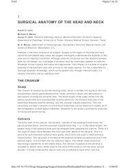
CHAPTER 5 GASTROINTESTINAL PROCEDURES
CHAPTER 5 GASTROINTESTINAL PROCEDURES
CHAPTER 5 GASTROINTESTINAL PROCEDURES

You also want an ePaper? Increase the reach of your titles
YUMPU automatically turns print PDFs into web optimized ePapers that Google loves.
Ovid:<br />
http://65.54.170.250/cgi-bin/getmsg/ManualofCommonBedsideSurgicalProcedures.html?cu...<br />
Página 1 de 28<br />
14/02/05<br />
<strong>CHAPTER</strong> 5<br />
<strong>GASTROINTESTINAL</strong> <strong>PROCEDURES</strong><br />
Robert C. Moesinger M.D.<br />
Disorders of the abdomen are, in many ways, the essence of general surgery. The surgeon<br />
should have expertise in the anatomy of the abdomen and confidence in examination of the<br />
abdomen. Similarly, gastrointestinal procedures should be an integral part of the<br />
armamentarium of the general surgeon.<br />
I. UPPER <strong>GASTROINTESTINAL</strong> <strong>PROCEDURES</strong><br />
Indications for intubation of the upper gastrointestinal (GI) tract include evacuation of the<br />
stomach (and occasionally more distal gastrointestinal tract) of gases and fluids for<br />
diagnostic and/or therapeutic purposes, or to deliver nutrients and medications. Modern GI<br />
tubes have a rich history; they are the product of many years of modifications in material<br />
and design.<br />
A. NASOGASTRIC TUBES<br />
1. Indications:<br />
a. Acute gastric dilatation<br />
b. Gastric outlet obstruction<br />
c. Upper gastrointestinal bleeding<br />
d. Ileus<br />
e. Small bowel obstruction<br />
f. Enteral feeding<br />
2. Contraindications:<br />
P.145<br />
a. Recent esophageal or gastric surgery<br />
b. Head trauma with possible basilar skull fracture<br />
3. Anesthesia:<br />
None or viscous lidocaine in the nose<br />
4. Equipment:<br />
a. Levin or Salem sump tube<br />
b. Water-soluble lubricant
Ovid:<br />
http://65.54.170.250/cgi-bin/getmsg/ManualofCommonBedsideSurgicalProcedures.html?cu...<br />
Página 2 de 28<br />
14/02/05<br />
c. Catheter-tip syringe (60 ml)<br />
d. Cup of ice<br />
e. Stethoscope<br />
f. Cup of water with a straw<br />
5. Positioning:<br />
Sitting or supine<br />
6. Technique:<br />
a. Measure tube from mouth to earlobe and down to anterior abdomen so that<br />
last hole on tube is below the xiphoid process. This marks the distance that<br />
the tube should be inserted.<br />
b. Some surgeons will place tip of tube in cup of ice to stiffen it or bend the tip<br />
downward to facilitate the tube's passage into the proximal esophagus.<br />
c. Apply lubricant liberally to tube.<br />
d. Ask patient to flex neck, and gently insert tube into a patent naris (see Figure<br />
5.1).<br />
Fig. 5.1.<br />
e. Advance tube into nasopharynx aiming posteriorly, asking the patient to<br />
swallow if possible.<br />
f. Once the tube has been swallowed, confirm that the patient can speak clearly<br />
and breathe without difficulty, and gently advance tube to estimated length. If<br />
the patient is able, instruct him or her to drink water through a straw; while the<br />
patient swallows, gently advance the tube.<br />
g. Confirm correct placement into the stomach by injecting approximately 20 ml<br />
of air with catheter-tip syringe while auscultating epigastric area. Return of a<br />
large volume of fluid through tube also confirms placement into stomach.<br />
h. Carefully tape tube to the patient's nose, ensuring that pressure is not applied<br />
by tube against naris. Tube should be kept well lubricated to prevent erosion<br />
at naris. With the use of tape and a safety pin, the tube can be secured to the<br />
patient's gown.<br />
P.146<br />
i. Irrigate tube with 30 ml of normal saline every 4 hours. Salem sump tubes will<br />
also require the injection of 30 ml of air through the sump (blue) port every 4<br />
hours to maintain proper functioning.<br />
j. Constant low suction may be applied to Salem sump tubes, whereas Levin
Ovid:<br />
http://65.54.170.250/cgi-bin/getmsg/ManualofCommonBedsideSurgicalProcedures.html?cu...<br />
Página 3 de 28<br />
14/02/05<br />
tubes should have only low intermittent suction.<br />
k. Monitor gastric pH every 4–6 hours and correct with antacids for pH < 4.5.<br />
l. Monitor gastric residuals if tube is used for enteral feeding. Obtain a chest<br />
radiograph to confirm correct placement before using any tube for enteral<br />
feeding.<br />
m. The tube ideally should not be clamped because it stents<br />
open the lower esophagus, increasing the risk of aspiration if the patient's<br />
stomach should distend.<br />
P.147<br />
7. Complications and Management:<br />
a. Pharyngeal discomfort<br />
• Common due to the large caliber of these tubes.<br />
• Throat lozenges or sips of water may provide relief.<br />
• Avoid using aerosolized anesthetic for the pharynx because this may<br />
inhibit the gag reflex, interfering with the protective mechanism of the<br />
airway.<br />
b. Erosion of the naris<br />
• Prevented by keeping tube well lubricated and ensuring that tube is<br />
taped so that pressure is not applied against naris. Tube should always<br />
be lower than the nose and never taped to the forehead of the patient.<br />
• Frequent checking of the tube position at the naris can help prevent this<br />
problem.<br />
c. Sinusitis<br />
• Occurs with long-term use of nasogastric tubes.<br />
• Remove the tube and place in other naris.<br />
• Antibiotic therapy if needed.<br />
d. Nasotracheal intubation<br />
• Results in airway obstruction that is fairly easy to diagnose in the awake<br />
patient (cough, inability to speak).<br />
• Obtain a chest radiograph to confirm placement prior to use for enteral<br />
feeding.<br />
e. Gastritis<br />
• Usually manifests itself as mild, self-limited upper gastrointestinal<br />
bleeding.<br />
• Prophylaxis consists of maintaining gastric pH > 4.5 with antacids via
Ovid:<br />
http://65.54.170.250/cgi-bin/getmsg/ManualofCommonBedsideSurgicalProcedures.html?cu...<br />
Página 4 de 28<br />
14/02/05<br />
the tube, intravenous (IV) histamine 2 receptor blockers, and removal of<br />
tube as soon as possible.<br />
f. Epistaxis<br />
• Usually self-limited.<br />
• If persists, remove the tube and assess location of bleed.<br />
• Refer to Chapter 1 for treatment of anterior and posterior epistaxis.<br />
B. OROGASTRIC TUBE<br />
1. Indications:<br />
The indications for orogastric (OG) tubes are generally the same as for NG tubes.<br />
However, because they are generally not<br />
tolerated well by the awake patient, they are used in intubated patients and<br />
newborns. The OG tube is the preferred tube for decompressing the stomach in the<br />
head trauma patient with a potential basilar skull fracture.<br />
P.148<br />
a. Acute gastric dilatation<br />
b. Gastric outlet obstruction<br />
c. Upper gastrointestinal bleeding<br />
d. Ileus<br />
e. Small bowel obstruction<br />
f. Enteral feeding<br />
2. Contraindications:<br />
Recent esophageal or gastric surgery<br />
3. Anesthesia:<br />
None<br />
4. Equipment:<br />
a. Levin or Salem sump tube<br />
b. Water-soluble lubricant<br />
c. Catheter-tip syringe (60 ml)<br />
d. Stethoscope<br />
5. Positioning:
Ovid:<br />
http://65.54.170.250/cgi-bin/getmsg/ManualofCommonBedsideSurgicalProcedures.html?cu...<br />
Página 5 de 28<br />
14/02/05<br />
Supine<br />
6. Technique:<br />
a. Measure tube from mouth to earlobe and down to anterior abdomen so that<br />
last hole on tube is below the xiphoid process. This marks the distance the<br />
tube should be inserted.<br />
b. Apply lubricant liberally to tube.<br />
c. Because the patients in whom OG tubes are used are generally unable to<br />
cooperate, the tube should be placed into the mouth, directed posteriorly, until<br />
the tip begins to pass downward into the esophagus.<br />
d. Advance the tube slowly and steadily. If any resistance is encountered, stop<br />
and withdraw the tube completely. Repeat step c.<br />
e. If the tube advances easily, with little resistance, continue until the<br />
premeasured distance is reached. Resistance, gagging,<br />
fogging of the tube, or hypoxia suggests errant placement of the tube into the<br />
trachea.<br />
P.149<br />
f. Confirm correct placement into stomach by injecting 20 ml of air with the<br />
catheter-tip syringe while auscultating over the epigastric area. Correct<br />
placement is also confirmed by aspiration of a large volume of fluid.<br />
g. Irrigate tube with 15–20 ml of saline every 4 hours. Salem sump tubes will<br />
require injection of 15–20 ml of air through the sump (blue) port every 4 hours<br />
to maintain proper functioning.<br />
h. Constant low suction may be applied to Salem sump tubes, whereas Levin<br />
tubes should have only low intermittent suction.<br />
i. Monitor gastric residuals if tube is used for enteral feeding. Obtain a chest<br />
radiograph to confirm placement before using for enteral feeding.<br />
j. Monitor gastric pH every 4–6 hours and correct with antacids for pH < 4.5.<br />
7. Complications and Management:<br />
a. Pharyngeal discomfort and gagging are a problem with OG tubes when they<br />
are placed in awake and alert patients, and essentially eliminates their use in<br />
such patients except in conjunction with an oral endotracheal tube.<br />
b. Tracheal intubation<br />
• Correct placement in the esophagus is usually evident by the ease of<br />
advancement of the tube. Any resistance suggests tracheal intubation or<br />
coiling within the posterior pharynx.<br />
• Obtain a chest radiograph to confirm placement prior to use for enteral<br />
feeding.
Ovid:<br />
http://65.54.170.250/cgi-bin/getmsg/ManualofCommonBedsideSurgicalProcedures.html?cu...<br />
Página 6 de 28<br />
14/02/05<br />
c. Gastritis<br />
• Usually manifests itself as mild, self-limited upper gastrointestinal<br />
bleeding.<br />
• Prophylaxis consists of maintaining gastric pH > 4.5 with antacids via<br />
the tube, IV histamine 2 receptor blockers, and removal of tube as soon<br />
as possible.<br />
C. NASODUODENAL TUBE<br />
1. Indications:<br />
Enteral feeding<br />
2. Contraindications:<br />
P.150<br />
Recent esophageal or gastric surgery<br />
3. Anesthesia:<br />
None or viscous lidocaine in the nose<br />
4. Equipment:<br />
a. Tip-weighted, small-caliber tube<br />
b. Guide wire<br />
c. Water-soluble lubricant<br />
d. Cup of water with a straw<br />
e. Stethoscope<br />
f. Catheter-tip syringe<br />
5. Positioning:<br />
Sitting or supine<br />
6. Technique:<br />
a. Measure tube length from mouth to earlobe and down to anterior abdomen so<br />
that tip is 6 cm below xiphoid process.<br />
b. Most duodenal tube tips are self-lubricating when moistened with water. If not,<br />
apply water-soluble lubricant to the tip of the tube.<br />
c. Ask patient to flex neck, and gently insert the tube containing the guide wire<br />
into a patent naris.<br />
d. Advance tube into pharynx aiming posteriorly, asking the patient to swallow if
Ovid:<br />
http://65.54.170.250/cgi-bin/getmsg/ManualofCommonBedsideSurgicalProcedures.html?cu...<br />
Página 7 de 28<br />
14/02/05<br />
possible.<br />
e. Once the tube has been swallowed, confirm that the patient can speak clearly<br />
and breathe without difficulty, and gently advance tube to estimated length. If<br />
the patient is able, instruct him or her to drink water through a straw, and<br />
while the patient swallows, gently advance the tube.<br />
f. Confirm correct placement into stomach by injecting approximately 20 ml of air<br />
with catheter-tip syringe while auscultating the epigastric area.<br />
g. Remove the guide wire and ask the patient to lie in a right decubitus position<br />
for 1–2 hours. An abdominal radiograph at this point will confirm transpyloric<br />
tube position or that the tube is coiled in the stomach; if coiled, withdraw tube<br />
for<br />
some distance and repeat this step. The tube should not be fixed to the nose.<br />
P.151<br />
h. The patient should first lie in a supine position for 1–2 hours and then in a left<br />
decubitus position for 1–2 hours to facilitate passage of the tube through the<br />
C-loop of the duodenum.<br />
i. At this point, position of the tube should be confirmed by radiograph. If the<br />
tube has not passed beyond the stomach by this time, then upper endoscopy<br />
or fluoroscopy may be necessary to advance the tube into the duodenum.<br />
7. Complications and Management:<br />
a. Epistaxis<br />
• Usually self-limited.<br />
• If persistent, remove the tube and assess location of bleed.<br />
• Refer to Chapter 1 for treatment of anterior and posterior epistaxis.<br />
b. Intestinal perforation<br />
• Presents usually as free air on chest radiograph.<br />
• Caused by inserting guide wire back through lumen of tube while it is in<br />
place. This should never be done.<br />
c. Obstruction of lumen (see section F below)<br />
D. LONG INTESTINAL TUBE<br />
1. Indications:<br />
Early partial small bowel obstruction<br />
2. Contraindications:
Ovid:<br />
http://65.54.170.250/cgi-bin/getmsg/ManualofCommonBedsideSurgicalProcedures.html?cu...<br />
Página 8 de 28<br />
14/02/05<br />
a. Uncooperative patient<br />
b. Indication for operative intervention (i.e., small bowel ischemia)<br />
3. Anesthesia:<br />
None or viscous lidocaine in the nose<br />
4. Equipment:<br />
a. Long intestinal tube<br />
b. Water-soluble lubricant<br />
c. Saline<br />
P.152<br />
d. 5-ml syringe, 22-gauge needle<br />
5. Positioning:<br />
Sitting up initially, then variable position as described below<br />
6. Technique:<br />
a. Using needle and syringe, inject 5 ml of saline into the balloon at the end of<br />
the tube (see Figure 5.2).<br />
Fig. 5.2.<br />
b. With the patient in an upright sitting position, roll up the balloon, apply a<br />
liberal amount of lubricant, and insert balloon into a patent naris.<br />
c. Carefully manipulate the tube such that the balloon falls into the nasopharynx<br />
without obstructing the airway.<br />
d. Instruct the patient to swallow the balloon as it is lowered slowly into the<br />
pharynx as though it were a bolus of food. Passage of the balloon in the<br />
patient who cannot swallow may be difficult. Often the balloon will advance<br />
along with the tube.<br />
e. After balloon has been swallowed, confirm that the patient can speak clearly<br />
and breathe easily, then advance it slowly<br />
into the stomach by instructing the patient to continue swallowing.<br />
P.153<br />
f. Insert the tube to the point at which the D mark is at the nose, and have the<br />
patient lie in a right decubitus position for 1–2 hours. The tube should not be<br />
fixed to the nose. Low intermittent suction may be applied.<br />
g. Obtain an abdominal radiograph to confirm the presence of the tip in the<br />
duodenum or that the tube is coiled in the stomach and may need to be
Ovid:<br />
http://65.54.170.250/cgi-bin/getmsg/ManualofCommonBedsideSurgicalProcedures.html?cu...<br />
Página 9 de 28<br />
14/02/05<br />
withdrawn for some distance.<br />
h. The patient should then be placed supine for 1–2 hours, then next in a left<br />
decubitus position for 1–2 additional hours to facilitate passage of the tube<br />
through the C-loop of the duodenum.<br />
i. At this point, position of the tube should be confirmed again by abdominal<br />
radiograph. If the tube has not passed beyond the stomach by this time,<br />
placement of the tip through the pylorus by flexible upper endoscopy or under<br />
fluoroscopy may be necessary.<br />
j. Once the tube is in the duodenum, it can be advanced 2–3 cm every 15<br />
minutes.<br />
k. Once the tube is no longer needed, removal should proceed slowly over<br />
several hours to prevent intussusception (withdraw tube 3–5 cm every 10–15<br />
minutes).<br />
7. Complications and Management:<br />
a. Airway obstruction<br />
• The balloon may occlude the upper airway during initial placement.<br />
• Withdraw the tube immediately.<br />
b. Epistaxis<br />
• Usually self-limited.<br />
• If it persists, remove the tube and assess location of bleed.<br />
• Refer to Chapter 1 for treatment of anterior and posterior epistaxis.<br />
c. Intussusception of small intestine during removal<br />
• Best avoided by withdrawing tube 3–5 cm every 10–15 minutes.<br />
E. SENGSTAKEN-BLAKEMORE TUBE<br />
The Sengstaken-Blakemore (SB) tube is an emergently placed tube that temporarily stops<br />
life-threatening hemorrhage from<br />
gastroesophageal varices. It is only a temporizing therapy before definitive operative,<br />
endoscopic, or transjugular intrahepatic portosystemic shunt procedure.<br />
P.154<br />
1. Indications:<br />
Exsanguinating hemorrhage from gastroesophageal varices<br />
2. Contraindications:
Ovid:<br />
http://65.54.170.250/cgi-bin/getmsg/ManualofCommonBedsideSurgicalProcedures.html?cu...<br />
Página 10 de 28<br />
14/02/05<br />
None<br />
3. Anesthesia:<br />
None or viscous lidocaine in the nose<br />
4. Equipment:<br />
a. SB tube<br />
b. Catheter-tip 60-ml syringe<br />
c. Hemostat clamps (two)<br />
d. Pressure manometer<br />
e. Levine or Salem sump NG tube<br />
f. Water-soluble lubricant<br />
g. Scissors<br />
5. Positioning:<br />
Supine or lateral decubitus<br />
6. Technique:<br />
a. Because potentially lethal complications can occur with the use of the SB tube,<br />
patients should be in a monitored setting, such as the intensive care unit,<br />
staffed by personnel experienced with the use of this device.<br />
b. Control of the airway by endotracheal intubation is strongly advised to<br />
minimize the risk of aspiration.<br />
c. Pass a large NG tube (see section I A) or OG tube (see section I B) to empty<br />
the stomach of blood, and then remove the tube.<br />
d. Inflate both esophageal and gastric balloons of the SB tube with air to test for<br />
leaks, then deflate.<br />
e. Apply lubricant liberally to the tube.<br />
f. Ask patient to flex neck, and gently insert tube into a patent naris.<br />
g. Advance tube into pharynx, aiming posteriorly and asking the patient to<br />
swallow if possible.<br />
P.155<br />
h. Once the tube has been swallowed, confirm that the patient can speak clearly<br />
and breathe without difficulty (if not intubated), and gently advance tube to<br />
approximately 45 cm.<br />
i. Apply low intermittent suction to the gastric aspiration port. Return of blood<br />
should confirm placement in the stomach. Otherwise inject 20 ml of air with the<br />
catheter-tip syringe while auscultating epigastric area (see Figure 5.3).
Ovid:<br />
http://65.54.170.250/cgi-bin/getmsg/ManualofCommonBedsideSurgicalProcedures.html?cu...<br />
Página 11 de 28<br />
14/02/05<br />
Fig. 5.3.<br />
j. Slowly inject 100 ml of air into the gastric balloon and then clamp the balloon<br />
port to prevent air leakage. Stop inflating the balloon immediately if the patient<br />
complains of pain because this could indicate that the balloon is in the<br />
esophagus. If this is the case, deflate the gastric balloon, advance the tube an<br />
additional 10 cm, and repeat the injection of air.<br />
P.156<br />
k. With the gastric balloon inflated, slowly withdraw the tube until resistance is<br />
met at the gastroesophageal junction. Anchor the tube to the patient's nose<br />
under minimal tension with padding.<br />
l. Obtain a chest radiograph to confirm correct gastric balloon positioning.<br />
m. Add an additional 150 ml of air to the gastric balloon and reapply the clamp.<br />
n. Irrigate the gastric port with saline. If no further gastric bleeding is found,<br />
leave the esophageal balloon deflated.<br />
o. If bleeding persists, connect the esophageal balloon port to the pressure<br />
manometer and inflate the esophageal balloon to 25–45 mm Hg.<br />
p. Transiently deflate the esophageal balloon every 4 hours to check for further<br />
bleeding (by aspirating through the gastric port) and to prevent ischemic<br />
necrosis of the esophageal mucosa.<br />
q. Apply low intermittent suction to both the gastric and esophageal aspiration<br />
tubes.<br />
r. After 24 hours without evidence of bleeding, deflate the esophageal and<br />
gastric balloons.<br />
s. The SB tube can be removed after an additional 24 hours without evidence of<br />
bleeding.<br />
7. Complications and Management:<br />
a. Esophageal perforation<br />
• Can result from intraesophageal inflation of the gastric balloon.<br />
• Deflate the gastric balloon and remove the SB tube.<br />
• Emergent surgical consult for operative therapy.<br />
b. Aspiration<br />
• Prevented by endotracheal intubation<br />
• Supportive therapy (oxygen, pulmonary toilet)
Ovid:<br />
http://65.54.170.250/cgi-bin/getmsg/ManualofCommonBedsideSurgicalProcedures.html?cu...<br />
Página 12 de 28<br />
14/02/05<br />
• Antibiotics as indicated<br />
c. Rebleeding<br />
P.157<br />
• Reinsert SB tube<br />
• Transjugular intrahepatic portosystemic shunt, endoscopy, or definitive<br />
surgery<br />
F. FEEDING TUBE TROUBLESHOOTING<br />
Feeding tubes in either the stomach or the jejunum are frequently used in patients who<br />
cannot eat. They can be placed through open techniques, laparoscopically and<br />
endoscopically, but when they malfunction, a surgeon is usually called. It is critical that<br />
after manipulation of a feeding tube, its position within the lumen of the gut be verified<br />
either by aspiration of intestinal contents or by a contrast study through the tube. Failure to<br />
do so can cause tube feeds to be injected directly into the peritoneal cavity, which is life<br />
threatening.<br />
1. Obstruction of Lumen<br />
a. Prevented by flushing of tube with water or saline at regular intervals.<br />
b. Avoid giving medications that are not easily liquefied through a feeding tube.<br />
c. Clearing of obstruction should be attempted with saline or carbonated liquids<br />
using a 1-ml (tuberculin-type) syringe. A difficult clog can sometimes be<br />
broken up by injecting a carbonated beverage and capping the tube, and<br />
repeating this multiple times over the course of a day.<br />
d. A guide wire can be used to break up inspissated tube feeds, but it must be<br />
used with extreme caution. It should be measured against the length of the<br />
feeding tube and not inserted more than 2–3 cm beyond the skin to prevent<br />
perforation of the bowel.<br />
e. Crushed pancrease has been used to break up obstructing tube feeds.<br />
2. Reinsertion of Feeding Tubes<br />
a. Accidental removal is prevented by frequent inspection of the feeding tube to<br />
ensure that it is well secured.<br />
b. Once a feeding tube has been in place for at least 2 weeks, if it falls out,<br />
reinsertion can usually be accomplished by passing a Foley catheter or MIC<br />
gastrostomy tube through the<br />
previous wound and into the stomach or jejunum. This should be done as soon<br />
as possible to prevent the tract from closing.<br />
P.158
Ovid:<br />
http://65.54.170.250/cgi-bin/getmsg/ManualofCommonBedsideSurgicalProcedures.html?cu...<br />
Página 13 de 28<br />
14/02/05<br />
c. In the stomach, the balloon can be fully inflated. In the jejunum, the balloon<br />
should be inflated with no more than 2–3 ml of saline to prevent intraluminal<br />
obstruction.<br />
d. A feeding tube that has been out for some time can often be replaced by<br />
interventional radiology. Insert a needle through the old site and place the<br />
feeding tube using the Seldinger technique under fluoroscopy.<br />
e. Placement must be confirmed radiographically.<br />
3. Changing Feeding Tubes<br />
a. After approximately 1 month, the feeding tube tract is so well developed that<br />
the tube can be changed without fear of losing the tract.<br />
b. Feeding tubes can be changed simply by deflating the balloon, removing the<br />
tube, and replacing with a new tube.<br />
c. PEG tubes have a disc-like button in the stomach that can be difficult to<br />
extract through the skin wound. In these cases, the percutaneous endoscopic<br />
gastrostomy PEG tube should be changed or removed endoscopically.<br />
4. Removing Feeding Tubes<br />
a. Feeding tubes should be left in place at least 2 weeks to ensure that the bowel<br />
has “healed” to the abdominal wall so that there is no intra-abdominal leak<br />
after removing a feeding tube.<br />
b. The enterocutaneous fistula resulting from the feeding tube tract usually<br />
closes over time with conservative therapy.<br />
II. LOWER <strong>GASTROINTESTINAL</strong> <strong>PROCEDURES</strong><br />
The anus and rectum are readily examined at the bedside using a number of<br />
straightforward techniques. Likewise, many lesions of the anorectal region are easily dealt<br />
with in the awake patient without the need for general anesthesia or operating room<br />
equipment. Although usually considered minor procedures, the direct benefit to the patient<br />
is often immense.<br />
P.159<br />
A. ANOSCOPY<br />
1. Indications:<br />
a. Anal lesions (fistulas, tumors, etc.)<br />
b. Rectal bleeding<br />
c. Rectal pain
Ovid:<br />
http://65.54.170.250/cgi-bin/getmsg/ManualofCommonBedsideSurgicalProcedures.html?cu...<br />
Página 14 de 28<br />
14/02/05<br />
d. Banding or injection of hemorrhoids<br />
2. Contraindications:<br />
a. Anal stricture<br />
b. Acute perirectal abscess<br />
c. Acutely thrombosed hemorrhoid<br />
3. Anesthesia:<br />
None<br />
4. Equipment:<br />
a. Clear polyethylene anoscope<br />
b. Water-soluble lubricant<br />
c. Directed light source or head-light<br />
5. Positioning:<br />
Lateral decubitus position or lithotomy position<br />
6. Technique:<br />
a. Examine anus by gently spreading anoderm and performing digital rectal<br />
examination.<br />
b. Insert the anoscope slowly, using a liberal amount of lubricant and with the<br />
obturator in place, until the flange at the base rests on perianal skin.<br />
c. Remove the obturator, and while withdrawing the anoscope, examine the anal<br />
mucosa in a systematic manner.<br />
d. Repeat the procedure as needed to ensure full inspection of the anal canal.<br />
7. Complications and Management:<br />
a. Fissure<br />
• Anal or perianal tears may occur and usually respond to conservative<br />
measures.<br />
b. Bleeding<br />
P.160<br />
• Unusual, but may occur especially in the setting of large internal<br />
hemorrhoids; usually self-limited.<br />
B. RIGID SIGMOIDOSCOPY
Ovid:<br />
http://65.54.170.250/cgi-bin/getmsg/ManualofCommonBedsideSurgicalProcedures.html?cu...<br />
Página 15 de 28<br />
14/02/05<br />
1. Indications:<br />
a. Rectal bleeding<br />
b. Lower abdominal and pelvic trauma<br />
c. Extraction of foreign bodies<br />
d. Stool cultures<br />
e. Evaluation and biopsy of ileoanal pouch<br />
2. Contraindications:<br />
a. Anal stricture<br />
b. Acute perirectal abscess<br />
c. Acutely thrombosed hemorrhoids<br />
3. Anesthesia:<br />
None<br />
4. Equipment:<br />
a. Rigid sigmoidoscope and obturator<br />
b. Light source<br />
c. Suction apparatus<br />
d. Insufflating bulb<br />
e. Water-soluble lubricant<br />
f. Long cotton-tipped swabs<br />
g. Biopsy forceps, if desired<br />
5. Positioning:<br />
Lateral decubitus, lithotomy, or prone jackknife<br />
6. Technique:<br />
a. Administer tap water or saline enema before procedure to empty distal colon<br />
of feces.<br />
b. Perform a digital rectal examination to assess for masses.<br />
c. Assemble sigmoidoscope by placing the obturator through<br />
the scope. Check light source and suction. Lubricate the scope thoroughly with<br />
water-soluble lubricant.<br />
P.161<br />
d. Gently insert the sigmoidoscope through the anus to 5 cm, remove the<br />
obturator, and attach the light source.
Ovid:<br />
http://65.54.170.250/cgi-bin/getmsg/ManualofCommonBedsideSurgicalProcedures.html?cu...<br />
Página 16 de 28<br />
14/02/05<br />
e. Judiciously insufflate air to visualize the lumen, using the minimum amount of<br />
air necessary to see.<br />
f. Slowly advance the sigmoidoscope as a unit to visualize the rectum. Air will<br />
leak during the procedure, and intermittent insufflation will be necessary.<br />
g. The lumen of the sigmoid will be posterior toward the sacrum and then gently<br />
curving to the patient's left. To minimize the risk of perforation, advance the<br />
sigmoidoscope only when the lumen is clearly visualized.<br />
h. If stool is obstructing the view, use the cotton-tipped swabs to clear the lumen.<br />
i. Advance the sigmoidoscope under direct vision as far as tolerated by the<br />
patient (most rigid scopes are 20 cm long) (see Figure 5.4).<br />
Fig. 5.4.<br />
j. To biopsy a mass or polyp, advance the scope until part of the mass is within<br />
the barrel of the scope. Insert the biopsy forceps<br />
into the barrel, and grasp a specimen of tissue. If needed, silver nitrate sticks<br />
may be used to achieve hemostasis.<br />
P.162<br />
k. Systematically inspect the mucosa while withdrawing the instrument slowly.<br />
7. Complications and Management:<br />
a. Bleeding<br />
• Usually self-limited, but may occur after biopsy.<br />
• Rarely will require treatment, but if bleeding is hemodynamically<br />
significant, then resuscitate and consider endoscopic treatment.<br />
b. Perforation<br />
• Manifested by abdominal pain, distention, and loss of hepatic dullness<br />
to percussion.<br />
• Obtain upright chest radiograph; free air under the diaphragm confirms<br />
the diagnosis.<br />
• IV fluids, IV antibiotics, urgent operative management.<br />
C. EXCISION OF THROMBOSED EXTERNAL HEMORRHOID<br />
1. Indications:
Ovid:<br />
http://65.54.170.250/cgi-bin/getmsg/ManualofCommonBedsideSurgicalProcedures.html?cu...<br />
Página 17 de 28<br />
14/02/05<br />
Painful thrombosed external hemorrhoid<br />
2. Contraindications:<br />
a. Coagulopathy (PT or PTT >1.3× control)<br />
b. Thrombocytopenia (platelet count < 50,000/mm 3 )<br />
c. Nonthrombosed prolapsed hemorrhoid<br />
3. Anesthesia:<br />
1% lidocaine (mixing lidocaine with 1/100,000 epinephrine may reduce bleeding)<br />
4. Equipment:<br />
a. Scalpel handle and #15 blade<br />
b. Sterile prep solution<br />
c. 25-gauge needle and syringe<br />
d. Forceps<br />
e. Small clamps<br />
f. Vaseline or Xeroform gauze<br />
5. Positioning:<br />
P.163<br />
Lateral decubitus or lithotomy<br />
6. Technique:<br />
a. Prep and drape the anal area with sterile prep solution.<br />
b. Identify the thrombosed external hemorrhoid. By definition, it lies exterior to<br />
the dentate line, and it is firm and tender (see Figure 5.5).<br />
Fig. 5.5.<br />
c. Perform a field block of the hemorrhoid by infiltrating the surrounding skin and<br />
soft tissues with lidocaine using a 25-gauge needle.<br />
d. Using a scalpel, make an elliptical incision over the thrombosed hemorrhoid<br />
(see Figure 5.6).<br />
Fig. 5.6.
Ovid:<br />
http://65.54.170.250/cgi-bin/getmsg/ManualofCommonBedsideSurgicalProcedures.html?cu...<br />
Página 18 de 28<br />
14/02/05<br />
e. Using the forceps to hold one side of the incision, enucleate the clot within the<br />
hemorrhoid with the aid of a clamp. Apply a Vaseline gauze or Xeroform<br />
dressing.<br />
f. The patient should be instructed to do sitz baths three times a day and after<br />
each bowel movement.<br />
7. Complications and Management:<br />
P.164<br />
a. Bleeding<br />
• A small amount of dark bloody ooze is to be expected. Bright red<br />
bleeding indicates that the hemorrhoid is not thrombosed, and the<br />
incision should be stopped.<br />
• Direct pressure or packing may be required to control bleeding.<br />
b. Fissure<br />
• Usually results from extending the incision beyond the hemorrhoid into<br />
anoderm.<br />
• Treat conservatively with sitz baths and Anusol suppositories.<br />
• Manage operatively if conservative treatment fails.<br />
D. REDUCTION OF RECTAL PROLAPSE<br />
1. Indications:<br />
a. Prolapse of rectum (full-thickness)<br />
b. Mucosal prolapse of rectum (mucosa only)<br />
2. Contraindications:<br />
a. Infarction or gangrene of prolapsed segment<br />
b. Severe tenderness of prolapsed segment<br />
c. Extreme edema of prolapsed segment<br />
3. Anesthesia:<br />
None<br />
4. Equipment:<br />
P.165<br />
a. Gloves<br />
b. Water-soluble lubricant
Ovid:<br />
http://65.54.170.250/cgi-bin/getmsg/ManualofCommonBedsideSurgicalProcedures.html?cu...<br />
Página 19 de 28<br />
14/02/05<br />
5. Positioning:<br />
Decubitus or dorsal lithotomy<br />
6. Technique:<br />
a. Don gloves and apply a liberal amount of water-soluble lubricant to the<br />
prolapsed segment.<br />
b. The concept is to apply steady, circumferential pressure on the prolapsed<br />
segment (to decrease edema) while simultaneously trying to reduce it. This is<br />
done by placing as many fingers of both hands as possible, oriented parallel to<br />
its longitudinal axis, around the segment and compressing it from all sides.<br />
c. Apply pressure firmly and steadily, with more pressure applied at the tip than<br />
at the base.<br />
d. Progress is typically slow and almost imperceptible. Be patient and squeeze<br />
for one to several minutes at a time, using plenty of lubricant.<br />
e. To prevent recurrence, the patient should be placed on stool softeners and<br />
should be instructed in the technique of manual self-reduction of prolapsed<br />
hemorrhoids, which may occur at each bowel movement.<br />
7. Complications and Management:<br />
Unsuccessful reduction<br />
• May result in infarction of prolapsed segment<br />
• Requires surgical management with excision of prolapsed portion<br />
III. ABDOMINAL <strong>PROCEDURES</strong><br />
These procedures are used to access the peritoneal cavity or to sample its contents. They<br />
are useful techniques that can provide diagnostic information or therapeutic benefit without<br />
the need for a major operative procedure.<br />
P.166<br />
A. PARACENTESIS<br />
1. Indications:<br />
a. Diagnostic studies<br />
b. Ascites<br />
c. Spontaneous bacterial peritonitis<br />
d. Therapeutic purposes<br />
e. Relief of respiratory compromise
Ovid:<br />
http://65.54.170.250/cgi-bin/getmsg/ManualofCommonBedsideSurgicalProcedures.html?cu...<br />
Página 20 de 28<br />
14/02/05<br />
f. Relief of abdominal pain and discomfort<br />
2. Contraindications:<br />
a. Coagulopathy (PT or PTT > 1.3)<br />
b. Thrombocytopenia (plt < 60,000)<br />
c. Bowel obstruction<br />
d. Pregnancy<br />
e. Infected skin or soft tissue at entry site<br />
3. Anesthesia:<br />
1% lidocaine<br />
4. Equipment:<br />
a. Sterile prep solution<br />
b. Sterile towels<br />
c. Sterile gloves<br />
d. 5-ml syringes, 20-ml syringes, 25-gauge and 22-gauge needles<br />
e. 3-way stopcock, IV tubing<br />
f. IV catheter (diagnostic: 20-gauge, therapeutic: 18-gauge) or long 16-gauge<br />
(CVP-type) catheter with 0.035-cm J wire<br />
g. 500- to 1000-ml vacuum bottles and IV drip set (for therapeutic paracentesis)<br />
5. Positioning:<br />
Supine<br />
a. Preferred sites of entry to prevent bleeding from epigastric vessels (see Figure<br />
5.7)<br />
Fig. 5.7.<br />
• Either lower quadrant (anterior iliac spine)<br />
• Lateral to the rectus muscle and at the level of or just below the<br />
umbilicus<br />
• Infraumbilically in the midline<br />
b. The entry site should not be the site of a prior incision and should be free of<br />
gross contamination and infection.<br />
P.167
Ovid:<br />
http://65.54.170.250/cgi-bin/getmsg/ManualofCommonBedsideSurgicalProcedures.html?cu...<br />
Página 21 de 28<br />
14/02/05<br />
c. The entry sites are percussed to confirm the presence of fluid and the absence<br />
of underlying bowel.<br />
d. The patient should empty his or her bladder prior to the procedure, and/or a<br />
Foley catheter should be placed to decrease the possibility of puncturing the<br />
bladder.<br />
6. Technique—Diagnostic Sampling:<br />
a. Prepare site with sterile prep solution and drape with sterile towels.<br />
b. Use 25-gauge needle to anesthetize skin and 22-gauge needle to anesthetize<br />
abdominal wall to peritoneum.<br />
P.168<br />
c. Introduce IV catheter into the abdominal cavity, aspirating as it is advanced.<br />
The needle should traverse the abdominal wall at an oblique angle to prevent<br />
persistent leak of ascites from the puncture site (see Figure 5.8).<br />
Fig. 5.8.<br />
d. When free flow of fluid occurs, the catheter should be advanced over the<br />
needle and the needle removed.<br />
e. Draw 20–30 ml of fluid into a sterile syringe for diagnostic studies and culture.<br />
P.169<br />
7. Technique—Therapeutic Drainage:<br />
a. Prepare site with sterile prep solution and drape with sterile towels.<br />
b. Use 25-gauge needle to anesthetize skin and 22-gauge needle to anesthetize<br />
abdominal wall to peritoneum.<br />
c. Introduce IV catheter into the abdominal cavity, aspirating as it is advanced.<br />
The needle should traverse the abdominal wall at an oblique angle to prevent<br />
persistent leak of ascites from the puncture site.<br />
d. When free flow of fluid occurs, the catheter should be advanced over the<br />
needle and the needle removed. Alternatively, a CVP-type catheter with extra<br />
side holes may be placed over a guide wire using the Seldinger technique.<br />
e. After insertion of the needle and aspiration of fluid, a J-tip guide wire is placed<br />
through the needle into the peritoneal space. The needle is removed, leaving<br />
the wire in place.<br />
f. A stiff plastic dilator is used to dilate the tract by placing it over the wire and<br />
into the abdomen. A #11-blade scalpel can be used to make a tiny nick at the<br />
entry site as well.
Ovid:<br />
http://65.54.170.250/cgi-bin/getmsg/ManualofCommonBedsideSurgicalProcedures.html?cu...<br />
Página 22 de 28<br />
14/02/05<br />
g. The dilator is removed, the catheter is placed over the wire and into the<br />
abdomen, and the wire is removed.<br />
h. Draw 20–30 ml of fluid into a sterile syringe for diagnostic studies and culture.<br />
i. IV tubing is hooked to the catheter and to a vacuum bottle to remove a large<br />
volume of fluid.<br />
j. Should the catheter become occluded, careful manipulation of the catheter to<br />
re-establish flow may be undertaken. Alternatively, asking the patient to turn<br />
on his or her side and again onto his or her back may also help re-establish<br />
flow. However, the needle or guide wire should not be reintroduced because of<br />
the risk of bowel injury. If less than an adequate volume is withdrawn, the<br />
catheter should be removed and replaced, possibly at another entry site.<br />
8. Complications and Management:<br />
a. Hypotension<br />
• Can occur during or after procedure due to rapid mobilization of fluid<br />
from intravascular space or due to vasovagal response.<br />
• IV hydration can prevent and correct the hypotension in most cases.<br />
P.170<br />
• 5% albumin solution or other colloid-based fluid is often used for this<br />
purpose.<br />
b. Bowel perforation<br />
• Rarely recognized at time of procedure<br />
• Can lead to infected ascites, peritonitis, and sepsis<br />
c. Hemorrhage<br />
• Rare, but can be caused by injury to mesentery or injury to inferior<br />
epigastric vessels.<br />
• Usually self-limited. Avoided by entering abdomen lateral to rectus and<br />
by correcting coagulopathy.<br />
• Hemodynamic instability requires laparotomy.<br />
d. Persistent ascites leak<br />
• Usually will seal in
Ovid:<br />
http://65.54.170.250/cgi-bin/getmsg/ManualofCommonBedsideSurgicalProcedures.html?cu...<br />
Página 23 de 28<br />
14/02/05<br />
• Obtain urology consult.<br />
B. DIAGNOSTIC PERITONEAL LAVAGE<br />
1. Indications:<br />
Blunt abdominal trauma, in the setting of an equivocal or unreliable abdominal<br />
examination (e.g., after head trauma or intoxication) in a patient with unexplained<br />
hypotension or blood loss. It is particularly useful in a patient who is too unstable to<br />
transport for computed tomography (CT) scan or when CT is not available.<br />
2. Absolute Contraindications:<br />
a. Indication for laparotomy is already present<br />
b. Pregnancy<br />
3. Relative Contraindications:<br />
a. Cirrhosis—Ascites can make the lavage fluid laboratory studies difficult to<br />
interpret.<br />
b. Morbid obesity—Makes diagnostic peritoneal lavage (DPL) technically more<br />
difficult.<br />
c. Prior abdominal surgery—Increases the risk of bowel injury during the<br />
procedure.<br />
P.171<br />
d. Suspected retroperitoneal injury—DPL results are often false-negative.<br />
4. Anesthesia:<br />
1% lidocaine with 1/100,000 epinephrine to decrease bleeding and false-positive<br />
results<br />
5. Equipment:<br />
a. Sterile prep solution<br />
b. Sterile towels, sterile gloves, gown, mask, cap<br />
c. Syringes: 5 ml, 10 ml, 20 ml<br />
d. 25-gauge needle<br />
e. Peritoneal dialysis catheter<br />
f. IV tubing<br />
g. 1000-ml bag of normal saline or Ringer's lactate<br />
h. Scalpel handle and #10 and #11 (or #15) blades
Ovid:<br />
http://65.54.170.250/cgi-bin/getmsg/ManualofCommonBedsideSurgicalProcedures.html?cu...<br />
Página 24 de 28<br />
14/02/05<br />
i. Surgical instruments: tissue forceps, hemostats, Allis clamps, retractors,<br />
suture<br />
6. Positioning:<br />
Supine. The stomach should be decompressed by an NG or an OG tube (OG if head<br />
trauma is present). The bladder should be drained by a Foley catheter.<br />
7. Technique:<br />
a. Prepare the entire abdomen with sterile prep solution and drape with sterile<br />
towels.<br />
b. With a 25-gauge needle and lidocaine with epinephrine, anesthetize a site in<br />
the lower midline approximately one-third the distance from the umbilicus to<br />
the symphysis pubis (see Figure 5.9).<br />
Fig. 5.9.<br />
c. Make a small incision down to the linea alba (the linea alba is midline in<br />
position and recognized by its decussating fibers and absence of muscle<br />
beneath it).<br />
d. Incise the fascia and peritoneum in the midline for a length of approximately 1<br />
cm, grasping the edges of the fascia with hemostats or Allis clamps (see<br />
Figure 5.10).<br />
Fig. 5.10.<br />
e. Introduce the dialysis catheter into the peritoneal cavity at<br />
an oblique angle aiming toward the cul-de-sac, and advance it carefully into<br />
the pelvis.<br />
P.172<br />
P.173<br />
f. Aspirate from the catheter with a syringe. Gross blood (5 ml or more) or gross<br />
enteric contents are indications for immediate laparotomy.<br />
g. If no gross blood or enteric contents are aspirated, instill 10 ml/kg of warmed<br />
saline or Ringer's lactate, up to 1000 ml, via the IV tubing. Drainage of<br />
dialysate into a chest tube or Foley catheter is also an indication for<br />
laparotomy.<br />
h. After waiting 5–10 minutes, allow the fluid to drain by gravity back into its<br />
original bag.
Ovid:<br />
http://65.54.170.250/cgi-bin/getmsg/ManualofCommonBedsideSurgicalProcedures.html?cu...<br />
Página 25 de 28<br />
14/02/05<br />
i. Send a sample of the fluid for cell count and amylase. Positive findings include<br />
a red blood cell count of >100,000/mm 3 , a white blood cell count >500/mm 3 , or<br />
amylase >175.<br />
j. Note: Criteria for positive lavage findings may vary among individual trauma<br />
surgeons.<br />
k. At the conclusion of the procedure, the catheter is removed and the fascia and<br />
skin are closed carefully using standard techniques (interrupted #1 Prolene,<br />
Vicryl, or PDS suture for fascia).<br />
8. Complications and Management:<br />
a. Bladder injury<br />
• Preventable by inserting Foley catheter prior to procedure.<br />
• Treated by Foley catheter drainage for a period of several days.<br />
b. Injury to bowel or other abdominal organ<br />
• Treated with nothing-by-mouth status, IV hydration, and IV antibiotics.<br />
• Bowel perforation with soilage requires laparotomy for repair.<br />
c. Hemorrhage<br />
• Rarely life-threatening, but may lead to false-positive results, especially<br />
if source is skin or subcutaneous tissue.<br />
• Treated with nothing-by-mouth status, IV hydration, transfusion, and<br />
laparotomy if it persists.<br />
d. Peritonitis<br />
• May be due to poor aseptic technique or bowel perforation.<br />
• Laparotomy may be necessary to rule out perforation.<br />
e. Wound infection<br />
• A potential late complication. Incidence may be<br />
diminished by a dose of broad-spectrum IV antibiotics prior to<br />
procedure.<br />
P.174<br />
• Treated with antibiotics and by opening the wound and packing it.<br />
C. TENCKHOFF CATHETER INSERTION<br />
1. Indications:<br />
Short-term or chronic ambulatory peritoneal dialysis
Ovid:<br />
http://65.54.170.250/cgi-bin/getmsg/ManualofCommonBedsideSurgicalProcedures.html?cu...<br />
Página 26 de 28<br />
14/02/05<br />
2. Contraindications:<br />
a. Obliterated peritoneal space (prior surgery, infection, carcinomatosis)<br />
b. Ruptured diaphragm<br />
c. Respiratory insufficiency<br />
d. Presence of a large ventral or umbilical hernia<br />
3. Anesthesia:<br />
1% lidocaine (1/100,000 epinephrine may reduce bleeding)<br />
4. Equipment:<br />
a. Surgical prep solution, sterile towels, sterile gloves<br />
b. Scalpel handle and #10 blade<br />
c. Tissue forceps<br />
d. Self-retaining retractor<br />
e. Double-cuff peritoneal dialysis catheter<br />
f. 3–0 absorbable suture on a taper-point curved needle<br />
g. 2–0 nylon suture on a curved cutting needle<br />
h. 25-gauge and 22-gauge needle<br />
i. 10-ml syringe<br />
5. Positioning:<br />
Supine. The stomach should be decompressed by an NG or an OG tube. The bladder<br />
should be drained by a Foley catheter.<br />
6. Technique:<br />
a. Prepare the entire abdomen with sterile prep solution and drape with sterile<br />
towels.<br />
b. With a 25-gauge needle and lidocaine, anesthetize a site lateral<br />
to the midline (over the rectus abdominus) approximately one-third the<br />
distance from the umbilicus to the symphysis pubis.<br />
P.175<br />
c. Make a longitudinal incision approximately 5 cm in length down to the level of<br />
fascia.<br />
d. Anesthetize a tract for the creation of a subcutaneous tunnel, to a point 8–12<br />
cm lateral to the incision, and make a small stab incision at this point (see<br />
Figure 5.11).
Ovid:<br />
http://65.54.170.250/cgi-bin/getmsg/ManualofCommonBedsideSurgicalProcedures.html?cu...<br />
Página 27 de 28<br />
14/02/05<br />
Fig. 5.11.<br />
e. Tunnel the dialysis catheter such that the proximal cuff lies in a subcutaneous<br />
location and the distal cuff lies in the first incision (see Figure 5.12).<br />
Fig. 5.12.<br />
f. Make an incision in the fascia and retract the rectus laterally, exposing the<br />
posterior fascia.<br />
g. Place a purse-string of 3–0 absorbable suture in the posterior fascia (see<br />
Figure 5.13).<br />
Fig. 5.13.<br />
h. Under direct vision, carefully incise the posterior fascia and peritoneum in the<br />
center of the purse-string suture. Locally<br />
explore the peritoneal cavity to be certain that adhesions or viscera are not in<br />
the way.<br />
P.176<br />
P.177<br />
i. Carefully insert the catheter into the peritoneal cavity, aiming inferiorly and<br />
posteriorly, such that the distal cuff lies just anterior to the peritoneum. The<br />
catheter should feed easily and without resistance into the pelvis. Flush the<br />
catheter with heparinized saline (100 units/ml) and be certain of the lack of<br />
significant resistance (see Figure 5.14).<br />
Fig. 5.14.<br />
j. Secure the catheter with the purse-string suture.<br />
k. Close the anterior fascia around the catheter such that the cuff lies within the<br />
muscle.<br />
l. The skin may be closed in the usual fashion.<br />
m. Secure the catheter where it exits the smaller incision with skin sutures.
Ovid:<br />
http://65.54.170.250/cgi-bin/getmsg/ManualofCommonBedsideSurgicalProcedures.html?cu...<br />
Página 28 de 28<br />
14/02/05<br />
n. The function of the catheter should be tested by infusing 1 l of saline or<br />
Ringer's lactate and then allowing it to drain by gravity.<br />
o. Peritoneal dialysis can begin the same day, using small volumes (1 L).<br />
7. Complications and Management:<br />
P.178<br />
a. Injury to intra-abdominal viscus<br />
• May occur in the setting of extensive adhesions or previous surgery<br />
b. Peritonitis<br />
• An ever-present risk that requires careful technique and surveillance<br />
• Treated with IV and/or intraperitoneal antibiotics<br />
• May occasionally require removal of catheter<br />
c. Catheter dysfunction<br />
• May be caused by ingrowth of tissue or adhesions to the catheter, and<br />
usually requires catheter removal.<br />
• If it is placed correctly deep in the pelvis, catheter is less likely to be<br />
occluded by omentum.<br />
Copyright (c) 2000-2004 Ovid Technologies, Inc.<br />
Version: rel9.2.0, SourceID 1.9998.1.313