Amoebiasis: A 10 Year Retrospective Study at the University ...
Amoebiasis: A 10 Year Retrospective Study at the University ...
Amoebiasis: A 10 Year Retrospective Study at the University ...

You also want an ePaper? Increase the reach of your titles
YUMPU automatically turns print PDFs into web optimized ePapers that Google loves.
I<br />
ORIGINAL ARTICLE<br />
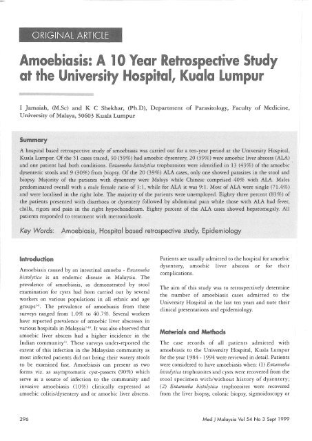
<strong>Amoebiasis</strong>: A <strong>10</strong> <strong>Year</strong> <strong>Retrospective</strong> <strong>Study</strong><br />
<strong>at</strong> <strong>the</strong> <strong>University</strong> Hospital, Kuala Lumpur<br />
I ]amaiah, (M. Se) and K C Shekhar, (Ph.D), Department of Parasitology, Faculty of Medicine,<br />
<strong>University</strong> of Malaya, 50603 Kuala Lumpur<br />
Introduction<br />
<strong>Amoebiasis</strong> caused by an intestinal amoeba - Entamoeba<br />
histolytica is an endemic disease in Malaysia. The<br />
prevalence of amoebiasis, as demonstr<strong>at</strong>ed by stool<br />
examin<strong>at</strong>ion for cysts had been carried out by several<br />
workers on various popul<strong>at</strong>ions in all ethnic and age<br />
groupsl-6. The prevalence of amoebiasis from <strong>the</strong>se<br />
surveys ranged from 1.0% to 40.7%. Several workers<br />
have reported prevalence of amoebic liver abscesses in<br />
various hospitals in Malaysia 7 - 1o • It was also observed th<strong>at</strong><br />
amoebic liver abscess had a higher incidence in <strong>the</strong><br />
Indian community". These surveys under-reported <strong>the</strong><br />
extent of this infection in <strong>the</strong> Malaysian community as<br />
most infected p<strong>at</strong>ients did not bring <strong>the</strong>ir w<strong>at</strong>ery stools<br />
to be examined fast. <strong>Amoebiasis</strong> can present as two<br />
forms viz. as asymptom<strong>at</strong>ic cyst-passers (90%) which<br />
serve as a source of infection to <strong>the</strong> community and<br />
invasive amoebiasis (<strong>10</strong>%) clinically expressed as<br />
amoebic colitis/dysentery and or amoebic liver abscess.<br />
P<strong>at</strong>ients are usually admitted to <strong>the</strong> hospital for amoebic<br />
dysentery, amoebic liver abscess or for <strong>the</strong>ir<br />
complic<strong>at</strong>ions.<br />
The aim of this study was to retrospectively determine<br />
<strong>the</strong> number of amoebiasis cases admitted to <strong>the</strong><br />
<strong>University</strong> Hospital in <strong>the</strong> last ten years and note <strong>the</strong>ir<br />
clinical present<strong>at</strong>ions and epidemiology.<br />
M<strong>at</strong>erials and Methods<br />
The case records of all p<strong>at</strong>ients admitted with<br />
amoebiasis to <strong>the</strong> <strong>University</strong> Hospital, Kuala Lumpur<br />
for <strong>the</strong> year 1984 - 1994 were reviewed in detail. P<strong>at</strong>ients<br />
were considered to have amoebiasis when: (1) Entamoeba<br />
histolytica trophozoites and cysts were recovered from <strong>the</strong><br />
stool specimen with/without history of dysentery;<br />
(2) Entamoeba histolytica trophozoites were recovered<br />
from <strong>the</strong> liver biopsy, colonic biopsy, sigmoidoscopy or<br />
296<br />
Med J Malaysia Vol 54 No 3 Sept 1999
AMOEBIASIS: A <strong>10</strong> YEAR RETROSPECTIVE STUDY<br />
from <strong>the</strong> anchovy pus; (3) Entamoeba histolytica antibody<br />
titres were gre<strong>at</strong>er than 1:64; (4) anchovy pus was<br />
drained from liver abscess and (5) when <strong>the</strong> p<strong>at</strong>ient<br />
responded to tre<strong>at</strong>ment with anti-amoebic drugs alone.<br />
The d<strong>at</strong>a collected were analysed on <strong>the</strong> basis of sex,<br />
ethnicity, age, socio-economic st<strong>at</strong>us, clinical fe<strong>at</strong>ures,<br />
complic<strong>at</strong>ions, management and type of amoebic<br />
infection. The blood chemistry of most of <strong>the</strong>se p<strong>at</strong>ients<br />
were not done as previous authors had indic<strong>at</strong>ed th<strong>at</strong> it<br />
was not diagnostic9•<br />
Results<br />
A total of 51 p<strong>at</strong>ients admitted with amoebiasis for <strong>the</strong><br />
periods 1984 - 1994 showed th<strong>at</strong> 30 (59%) had amoebic<br />
dysentery, 20 (39%) had amoebic liver abscess and one<br />
p<strong>at</strong>ient had both amoebic colitis and amoebic liver<br />
abscess. Table I shows th<strong>at</strong> of <strong>the</strong> 30 cases with amoebic<br />
dysentery, 13 (86.7%) were stool posltlve and nine<br />
(81.8%) specimens obtained from biopsy were positive<br />
for Entamoeba histolytica trophozoites. Of <strong>the</strong> 20 (39%)<br />
liver abscess cases, one (6.7%) showed parasites in <strong>the</strong><br />
stool specimens and one (6.7 %) was positive <strong>at</strong> biopsy.<br />
Parasites were identified in <strong>the</strong> stool and biopsy<br />
specimens in <strong>the</strong> p<strong>at</strong>ient with both amoebic dysentery<br />
and amoebic liver abscess. Of <strong>the</strong> 51 cases, 15 (30%)<br />
were stool positive thus proving an etiological link<br />
between <strong>the</strong> condition and <strong>the</strong> agent. In biopsy, 11<br />
(21.6%) were positive for <strong>the</strong> parasite.<br />
Table 11 shows th<strong>at</strong> of <strong>the</strong> 30 cases of amoebic dysentery,<br />
18 (60%) were males and 12 (40%) females. Twelve<br />
(40%) amoebic dysentery p<strong>at</strong>ients were Malays,<br />
followed by Chinese (33.3%) and 5 (16.7%) being<br />
Indians. Ninety percent (90%) of amoebic liver abscess<br />
cases were seen in males. Of <strong>the</strong> different types of<br />
clinical present<strong>at</strong>ions of amoebiasis, 37 (72.6%) were<br />
predominantly seen in males thus showing male<br />
Table I<br />
Classific<strong>at</strong>ion of <strong>the</strong> Various Clinical Forms of <strong>Amoebiasis</strong> Admitted to<br />
<strong>the</strong> <strong>University</strong> Hospital, Kuala Lumpur: 1984-1994<br />
Type of <strong>Amoebiasis</strong> Number of Cases (%) Stool Positive (%) Biopsy Specimen Positive (%)<br />
Amoebic dysentery<br />
Amoebic liver abscess<br />
Amoebic dysentery +<br />
Amoebic liver abscess<br />
Total<br />
30 (59) 13 (86.7) 9 (81.8)<br />
20 (39) 1 (6.7) 1 (9.1)<br />
1 (2) 1 (6.7) (9.1)<br />
51 15 11<br />
Table 11<br />
Types of <strong>Amoebiasis</strong> Classified According to Ethnicity for <strong>Year</strong>s 1984-1994<br />
Types of amoebiasis Malay Chinese Indians O<strong>the</strong>rs<br />
M F M F M F M F<br />
Amoebic dysentery 7 5 7 3 3 4 0<br />
Amoebic liver abscess 3 0 8 1 6 1 0<br />
Amoebic dysentery and 0 0 0 0 0 0 0<br />
amoebic liver abscess<br />
Total <strong>10</strong> 5 15 4 9 5 3 0<br />
Percentage (%) 19.6 9.8 29.4 7.8 17.7 9.8 5.9 0<br />
Total<br />
30<br />
20<br />
51<br />
<strong>10</strong>0<br />
Med J Malaysia Vol 54 No 3 Se pt 1999<br />
297
ORIGINAL ARTICLE<br />
preponderance. From Table Il, it can also be seen th<strong>at</strong><br />
male Chinese formed <strong>the</strong> core comprising 29.4%<br />
followed by male Malay (19.6%) and Indians (17.7%).<br />
Amoebic liver abscess was a condition seen largely<br />
among male Chinese who formed 40% of <strong>the</strong> cases<br />
followed by Indians (30%).<br />
Table III shows <strong>the</strong> type of abscesses encountered in this<br />
study. Of <strong>the</strong> total cases of amoebic liver abscess, 15<br />
(71.4%) had abscesses which were single and loc<strong>at</strong>ed in<br />
<strong>the</strong> right lobe while 2 (9.5%) were single abscesses<br />
loc<strong>at</strong>ed in <strong>the</strong> left lobe. Three (14.3%) were multiple<br />
abscesses.<br />
Table III<br />
Types of Abscesses in Amoebic Liver Abscess<br />
Types of Abscess Number of Cases (%)<br />
Single abscess in right lobe<br />
Single abscess of left lobe<br />
Multiple abscesses<br />
15 (75%)<br />
2 (<strong>10</strong>%)<br />
3 (15%)<br />
Table IV shows <strong>the</strong> distribution of amoebiasis according<br />
to age, sex and race. Thirty seven point three (37.3%)<br />
constituted <strong>the</strong> Chinese, 29.4% Malays and 27.5%<br />
Indians. The prevalence of amoebiasis was predominantly<br />
seen in two age groups: viz. 0 - 9 years and 50 - 59 years.<br />
Males formed 70.6% of <strong>the</strong> total number of amoebiasis<br />
cases of which 29.4% were Chinese in this study. Malays<br />
formed 19.6% of <strong>the</strong> cases while Indians constituted<br />
11.8%. When both sexes were combined, 37.3% of <strong>the</strong><br />
p<strong>at</strong>ients affected were Chinese. The Malays formed 29.4%<br />
and Indians 27.5%.<br />
Table V shows <strong>the</strong> socio-economic st<strong>at</strong>us or occup<strong>at</strong>ion<br />
of <strong>the</strong> affected p<strong>at</strong>ients. Most of <strong>the</strong> affected p<strong>at</strong>ients<br />
were people from <strong>the</strong> lower sodo-economic groups.<br />
Twenty percent were unemployed. Six of <strong>the</strong> p<strong>at</strong>ients<br />
were chronic smokers and chronic drinkers. All <strong>the</strong><br />
children came from poor families and four (20%) of<br />
amoebic liver abscesses reported were seen in children,<br />
<strong>the</strong> youngest, was a one year old male Indian child. An<br />
eleven-year old Indian child with amoebic liver abscess<br />
had just returned from India 6 days prior to admission.<br />
The child could have picked up <strong>the</strong> infection from India.<br />
Table IV<br />
Distribution of <strong>Amoebiasis</strong> by Age/ Sex and Race (1984 • 1994) UHKL<br />
Age Groups Race & Sex Total (%)<br />
Malay Chinese Indian O<strong>the</strong>rs<br />
M F M F M F M F<br />
0·9 3 0 3 0 0 9 17.7<br />
<strong>10</strong>·19 0 1 0 0 5 9.8<br />
20·29 2 3 0 0 0 0 7 13.7<br />
30d9 2 0 3 0 2 0 0 0 7 13.7<br />
40·49 0 4 0 0 8 15.7<br />
50 ·59 3 0 9 17.7<br />
60·69 1 1 0 1 1 2 0 0 6 11.8<br />
Total <strong>10</strong> 5 15 4 8 6 3 0 51<br />
(%) 19.6 9.8 29.4 7.8 15.7 11.8 5.9<br />
29.4% 37.3% 27.5% 5.9%<br />
298 Med J Malaysia Vol 54 No 3 Se pt 1 999
AMOEBIASIS: A <strong>10</strong> YEAR RETROSPECTIVE STUDY<br />
Table V<br />
S(l)ciOl"ec@n@mic Stah.iS> (l)ir OCCiJ!'JCilti(l)!il (l)f P<strong>at</strong>ients<br />
Typ~s @f O(:(up<strong>at</strong>ion Number (l)f CGses (%)<br />
Rubber tapper 2 (04)<br />
Labourer 5 (<strong>10</strong>)<br />
Security guard 2 (04)<br />
Pensioner 5 (<strong>10</strong>)<br />
Lorry driver 2 (04)<br />
House wife 4 (08)<br />
Clerk 3 (06)<br />
Unemployed <strong>10</strong> (20)<br />
Businessman 4 (08)<br />
Factory worker 2 (04)<br />
Ex-Army personnel (02)<br />
*Children, 11 (22%) were excluded from <strong>the</strong> above table<br />
Table VI shows <strong>the</strong> clinical present<strong>at</strong>ion of all <strong>the</strong><br />
amoebiasis p<strong>at</strong>ients admitted to <strong>the</strong> hospital. Majority<br />
(83%) of <strong>the</strong> p<strong>at</strong>ients with amoebic dysentery presented<br />
ei<strong>the</strong>r with diarrhoea or dysentery followed by<br />
abdominal pain. Majority (95%) of <strong>the</strong> p<strong>at</strong>ients with<br />
amoebic liver abscesses presented clinically with fever,<br />
chills, rigors and pain in <strong>the</strong> right hypochondrium.<br />
Eighty percent of <strong>the</strong> amoebic liver abscess cases showed<br />
hep<strong>at</strong>omegaly while (20%) presented with jaundice. It<br />
is interesting to note th<strong>at</strong> 20% of amoebic dysentery<br />
p<strong>at</strong>ients showed hep<strong>at</strong>omegaly.<br />
All p<strong>at</strong>ients responded to tre<strong>at</strong>ment with metronidazole<br />
except two cases: one who developed recurrent abscess<br />
and an ano<strong>the</strong>r elderly female p<strong>at</strong>ient who died of<br />
septicaemia secondary to peritonitis caused by amoebic<br />
colitis. Five of <strong>the</strong> amoebic liver abscess p<strong>at</strong>ients were<br />
tre<strong>at</strong>ed with both oral metronidazole and drainage of<br />
abscess.<br />
<strong>Amoebiasis</strong> is <strong>the</strong> second most important protozoan<br />
infection in Malaysia after malaria!. Surveys carried out<br />
by various authors show <strong>the</strong> prevalence of Entameoba<br />
histolytica infection to range from l.0% - 14.4% with a<br />
higher prevalence among aborigines of all age groups!.<br />
While survey prevalence of <strong>the</strong> infection is high within<br />
<strong>the</strong> community, <strong>the</strong> number of clinical cases of<br />
amoebiasis admitted to <strong>the</strong> hospitals appears to be low.<br />
The extent of <strong>the</strong> prevalence of intestinal and hep<strong>at</strong>ic<br />
amoebiasis is still unknown and could be a tip of iceberg<br />
problem in Malaysia, as <strong>the</strong>y are not reported. Even<br />
when <strong>the</strong>y are admitted ro <strong>the</strong> hospitals <strong>the</strong> cases are not<br />
reported except for" occasional public<strong>at</strong>ions as<br />
retrospective studies. Surgeons and gastro-enterologists<br />
report only amoebic liver abscess cases r<strong>at</strong>her than o<strong>the</strong>r<br />
clinical complic<strong>at</strong>ions. A number of hospital based<br />
studies have been carried out to determine <strong>the</strong> extent<br />
and prevalence of amoebiasis in Malaysia 7011 • Most of <strong>the</strong><br />
studies concentr<strong>at</strong>ed on amoebic liver abscess r<strong>at</strong>her<br />
than intestinal amoebiasis. The studies carried out<br />
indic<strong>at</strong>e th<strong>at</strong> amoebic liver abscess is a common<br />
Table VI<br />
Clinical PlI'e$ei'l~<strong>at</strong>icn@f All <strong>the</strong> Am@ebiasis P(!Itiel'llts<br />
Diarrhoea<br />
Abdominal pain<br />
Fever with chills and rigor<br />
Right hypochondrium pain<br />
Hep<strong>at</strong>omegaly<br />
Jaundice<br />
Am@ebic Dysentery (c/o)<br />
n:::30<br />
83<br />
63<br />
13<br />
o<br />
20<br />
o<br />
Amoebic Liver Abslte5$ (%)<br />
11:::::20<br />
50<br />
20<br />
95<br />
80<br />
80<br />
20<br />
*P<strong>at</strong>ient with both amoebic dysentery and liver abscess had all <strong>the</strong> symptoms listed above<br />
Med J Malaysia Vol 54 No 3 Sept 1999 299
ORIGINAL ARTICLE<br />
infection in Malaysia 7 - 13 • In this <strong>10</strong> year retrospective<br />
study, a total of 30 (59%) cases were diagnosed with<br />
amoebic dysentery, 20 (39%) were amoebic liver abscess<br />
and one p<strong>at</strong>ient had both amoebic dysentery and<br />
amoebic liver abscess. Thus, an average of 5.1<br />
amoebiasis cases was admitted to <strong>the</strong> hospital annually.<br />
The results of this study and o<strong>the</strong>rs show th<strong>at</strong> amoebic<br />
liver abscess and amoebic dysentery are important from<br />
<strong>the</strong> medical point of view but are not commonly<br />
reported.<br />
Clinical suspicion of amoebic liver abscess can best be<br />
confirmed by a positive serologic, sonographic or<br />
radiological procedure followed by a specific response to<br />
<strong>the</strong>rapy. Diagnostic aspir<strong>at</strong>ion may occasionally be<br />
needed to differenti<strong>at</strong>e between amoebic and pyogenic<br />
abscesses. The aspir<strong>at</strong>ion under <strong>the</strong>se circumstances<br />
needs to be carried out using ultrasound guidance. The<br />
percutaneous needle aspir<strong>at</strong>ion needs to be performed <strong>at</strong><br />
<strong>the</strong> site of maximal tenderness or over <strong>the</strong> area of<br />
maximum dullness to percussion. If <strong>the</strong>se signs are not<br />
present, <strong>the</strong> site of choice is along <strong>the</strong> anterior axillary<br />
line in <strong>the</strong> ninth inter-space.<br />
The most reliable and dependable diagnostic technique<br />
for amoebiasis is <strong>the</strong> demonstr<strong>at</strong>ion of <strong>the</strong> parasite<br />
(trophozoites) in specimens obtained from p<strong>at</strong>ient's i.e.<br />
<strong>the</strong> stools. Stool examin<strong>at</strong>ion is tedious and time<br />
-consuming. If <strong>the</strong> stools are not collected timely <strong>the</strong><br />
trophozoites are destroyed and diagnosis becomes<br />
inconclusive. A number of authors have testified to <strong>the</strong><br />
difficulties in confirming <strong>the</strong> caus<strong>at</strong>ive organism 7 ,IO-13.<br />
Trophozoites may not be found in biopsy m<strong>at</strong>erials<br />
because <strong>the</strong>y disintegr<strong>at</strong>e and disappear and hence<br />
confirm<strong>at</strong>ion of final diagnosis of amoebic liver abscess<br />
is seldom achieved. A number of serological tests have<br />
been found useful in confirming <strong>the</strong> diagnosis of<br />
invasive amoebiasis. This includes Fluorescent antibody<br />
test, gel diffusion, and immuno-electrophoresis, which<br />
are ideal but pose a limit<strong>at</strong>ion in routine use. The<br />
indirect immuno-fluorescent method has been found to<br />
be highly sensitive with 96% reactivity among<br />
confirmed liver abscess cases l4 • However, no serological<br />
diagnosis was carried out in <strong>the</strong>se cases. The reliance of<br />
<strong>the</strong> diagnosis was based purely on stool examin<strong>at</strong>ion and<br />
liver biopsy. If only 43% of <strong>the</strong> amoebic dysentery cases<br />
were stool positive and 30% were positive on biopsy, <strong>the</strong><br />
failure to detect <strong>the</strong> p<strong>at</strong>hogen in <strong>the</strong> remainder of <strong>the</strong> 17<br />
cases could be <strong>at</strong>tributed to misdiagnosis or failure to<br />
consider it in <strong>the</strong> differential diagnosis. It is obvious<br />
because any amoebic dysenteric stool would be positive<br />
for <strong>the</strong> trophozoites. On <strong>the</strong> o<strong>the</strong>r hand, in hep<strong>at</strong>ic liver<br />
abscess, trophozoites are never encountered in stools<br />
unless <strong>the</strong> p<strong>at</strong>ient has both intestinal and extraintestinal<br />
amoebiasis as was in <strong>the</strong> single case. Besides,<br />
amoebic liver abscess may mimic pyogenic liver abscess,<br />
tuberculous abscess or even indetermin<strong>at</strong>e abscess.<br />
Hence, an etiologic associ<strong>at</strong>ion of <strong>the</strong> abscess is<br />
mand<strong>at</strong>ory and must definitely be carried out by all<br />
personnel involved in <strong>the</strong> management.<br />
Previous authors have reported th<strong>at</strong> amoebic liver<br />
abscesses were predominantly seen in males and in<br />
Indians7- 8 ,lo-13. The male female r<strong>at</strong>io, in this study, for<br />
amoebic liver abscess was 9: 1 while for amoebic<br />
dysentery it was 3:2. Forty five percent of <strong>the</strong> amoebic<br />
liver abscess cases were seen in <strong>the</strong> Chinese followed by<br />
Indians (35%). Malays formed <strong>the</strong> predominant group<br />
to be affected with amoebic dysentery forming 40% of<br />
<strong>the</strong> cases followed by Chinese. Indians did not form <strong>the</strong><br />
predominant group affected for both conditions in this<br />
study. In Malaysia, previous reports showed high<br />
prevalence of amoebiasis among <strong>the</strong> Malays followed by<br />
<strong>the</strong> Aborigines l ,3.4, The higher prevalence among certain<br />
ethnic groups in Malaysia is not due to any ethnic<br />
predisposition but r,eflects <strong>the</strong> overall socio-economic<br />
st<strong>at</strong>us, level of environmental sanit<strong>at</strong>ion and hygiene<br />
practised as well as o<strong>the</strong>r socio-cultural practices<br />
pertaining to diet and e<strong>at</strong>ing habits l -4, The residence<br />
history of all <strong>the</strong> p<strong>at</strong>ients showed th<strong>at</strong> <strong>the</strong>y were from<br />
<strong>the</strong> rural and urban disadvantaged communities with<br />
poor toilet facilities,<br />
Goh et al,'s7 found <strong>the</strong> peak incidence of amoebic liver<br />
abscess in <strong>the</strong> 30 - 60 years age group while Teh et al,13<br />
study showed in <strong>the</strong> 40 - 70 years age group, Fock et <strong>at</strong>. 12<br />
noted incidence in 50 years and above, P<strong>at</strong>terson and<br />
Schoppe l5 however, reported <strong>the</strong> disease to be<br />
predominantly in young male contrary to <strong>the</strong> findings of<br />
o<strong>the</strong>rs, In this study, p<strong>at</strong>ient's age ranged from 0 - 59<br />
years with peak incidence noted in <strong>the</strong> 40 - 49 years age<br />
groups, However, 61.9% of <strong>the</strong> case were in <strong>the</strong> older<br />
age group i,e, 30 - 59 years concurring with <strong>the</strong> results<br />
of o<strong>the</strong>rs.<br />
300<br />
Med J Malaysia Vol 54 No 3 Sept 1999
AMOEBIASIS: A <strong>10</strong> YEAR RETROSPECTIVE STUDY<br />
The majority of <strong>the</strong> amoebic liver abscesses are solitary<br />
or single abscesses and usually loc<strong>at</strong>ed in <strong>the</strong> right lobe<br />
of <strong>the</strong> liver,,12.13, Amoebic abscesses were <strong>the</strong> only<br />
abscesses th<strong>at</strong> predomin<strong>at</strong>ed in <strong>the</strong> right lobe in <strong>the</strong><br />
Singapore studi' , Goh et al.' in <strong>the</strong>ir study found<br />
86,5% of <strong>the</strong> abscesses single with <strong>the</strong> majority (77 ,7%)<br />
in <strong>the</strong> right lobe. In this study, 75% of <strong>the</strong> liver<br />
abscesses were single and found in <strong>the</strong> right lobe. Only<br />
<strong>10</strong>% were found in <strong>the</strong> left lobe with multiple abscesses<br />
forming 15 % of <strong>the</strong> total. The results of this study<br />
concurs with Goh et <strong>at</strong>. 's' study which was carried out in<br />
<strong>the</strong> same hospital <strong>at</strong> a different period,<br />
The diagnosis of amoebic liver abscess depends on <strong>the</strong><br />
clinician suspicion and recognition of a well-described<br />
clinical syndrome, The common clinical present<strong>at</strong>ion is<br />
fever with chills and rigors, right hypochondrium pain,<br />
tender hep<strong>at</strong>omegaly, anorexia, malaise and nausea and<br />
<strong>the</strong>se have been observed in all <strong>the</strong> studies7-l0, It has been<br />
emphasised th<strong>at</strong> fever of unexplained origin should give<br />
a strong suspicion of amoebic liver abscess. This should<br />
be entertained in any case of prolonged fever particularly<br />
in <strong>the</strong> tropics 9 , Jaundice has been reported previously<br />
from rare to 55% of <strong>the</strong> p<strong>at</strong>ients7-l0, In this study, 20%<br />
showed jaundice, The clinical present<strong>at</strong>ion, however, in<br />
all <strong>the</strong> p<strong>at</strong>ients with amoebic liver abscess were similar<br />
to <strong>the</strong> findings of o<strong>the</strong>r researchers, In p<strong>at</strong>ients with<br />
amoebic dysentery diarrhoea, abdominal pain, fever<br />
with chills and rigor were p<strong>at</strong>hognomonic signs,<br />
<strong>Amoebiasis</strong> occurs in people from <strong>the</strong> lower S0C<strong>10</strong>-<br />
economic groups and is associ<strong>at</strong>ed with poverty, squ<strong>at</strong>ter<br />
settlements, improper sanit<strong>at</strong>ion, contamin<strong>at</strong>ed food<br />
and drinking w<strong>at</strong>er and poor nutrition l '. Most of <strong>the</strong><br />
p<strong>at</strong>ients in this survey were from poor socio-economic<br />
group, or unemployed (20%) and could have consumed<br />
food prepared in an unhealthy environment,<br />
Metronidazole (750-mg t.i,d for <strong>10</strong> days) is highly<br />
effective against invasive amoebiasis, as well as anaerobic<br />
bacteria, When scan or ultrasonography confirms a<br />
suspected liver abscess and while awaiting serologic<br />
confirm<strong>at</strong>ion of an amoebic aetiology, tre<strong>at</strong>ment with<br />
metronidazole should be initi<strong>at</strong>ed, This may obvi<strong>at</strong>e <strong>the</strong><br />
need for diagnostic needle aspir<strong>at</strong>ion, If clinical<br />
improvement is not obtained in about 3 days, aspir<strong>at</strong>ion<br />
should be performed, Some workers always recommend<br />
pre-tre<strong>at</strong>ment diagnostic aspir<strong>at</strong>ion when <strong>the</strong> aetiology<br />
is not clear. Tinidazole (Fasigyn) is a new drug th<strong>at</strong> has<br />
been used for <strong>the</strong> tre<strong>at</strong>ment of amoebiasis under many<br />
regimens: 600mg twice daily for 5 to <strong>10</strong> days; single<br />
daily doses of 2gm for 2 to 6 days; 800mg, 3 times daily<br />
for 5 days or single daily doses of 2gm repe<strong>at</strong>ed for 3<br />
days, This is an effective anti-amoebic drug with a<br />
longer half life and rel<strong>at</strong>ively fewer side effects, The<br />
efficacy of this drug awaits evalu<strong>at</strong>ion from our<br />
physicians and surgeons,<br />
In conclusion, this retrospective study shows th<strong>at</strong> <strong>the</strong> disease<br />
p<strong>at</strong>tern closely resembles th<strong>at</strong> of o<strong>the</strong>r countries in terms of<br />
its epidemiology and clinical present<strong>at</strong>ion, The morbidity<br />
and mortality d<strong>at</strong>a of this disease is unknown, While a<br />
number of serological tests are becoming available, it would<br />
be timely if <strong>the</strong>se tests are used to confirm <strong>the</strong> diagnosis in<br />
suspected individuals so th<strong>at</strong> a true incidence and prevalence<br />
of <strong>the</strong> disease can be determined in <strong>the</strong> Malaysian<br />
community. While .field surveys have indic<strong>at</strong>ed a high<br />
prevalence in <strong>the</strong> popul<strong>at</strong>ion this cannot be said for clinical<br />
cases of amoebiasis.<br />
The authors wish to thank Encik Awalluddin and Encik<br />
Sani from <strong>the</strong> records office <strong>University</strong> Hospital Kuala<br />
Lumpur for furnishing with <strong>the</strong> l<strong>at</strong>est d<strong>at</strong>a,<br />
Med J Malaysia Vol 54 No 3 Sept 1999<br />
301
ORIGINAL ARTICLE<br />
1. Kan, SP. Current st<strong>at</strong>us of protozoan infections in<br />
Malaysia. Yonsei Rep. Trop. Med. 1987; 18: 24-31.<br />
2. Lim KG. A review of diseases in Malaysia: Pelanduk<br />
Public<strong>at</strong>ions, 1992; 209-<strong>10</strong>.<br />
3. Lai, KPF. Intestinal protozoan infections in<br />
Malaysia. Sou<strong>the</strong>ast Asean ] Trop Med Public<br />
Health 1992; 23: 578-86.<br />
4. Karim, R, Rahmah, N, Khairul Anuar, A, Mehdi<br />
R,& Abdullah B. Parasitic infections in <strong>the</strong><br />
Aboriginal co=unity <strong>at</strong> Temengor, Hulu Perak,<br />
Malaysia. Malayan N<strong>at</strong>ure ]ournal1995; 48:425-32.<br />
5. Hamimah I, Zahedi M and Ainiyah AJ. The<br />
prevalence of intestinal parasites among children <strong>at</strong><br />
<strong>the</strong> General Hospital, Kuala Lumpur, Malaysia.<br />
Med.] Malaysia 1982; 37 (4): 373-77.<br />
6. Che Ghani M, Mohamed AM and Oothman P.<br />
Infection with Entamoeba histolytica and Giardia<br />
lamblia in an urban slum, rural villages and medical<br />
students in Peninsular Malaysia. Trop. Biomed<br />
1987; 4: 150-4.<br />
7. Goh KL,Wong Nw, Paramsothy M, Nojeg M and<br />
Somasundram K. Liver abscess in <strong>the</strong> tropics:<br />
experience in <strong>the</strong> <strong>University</strong> Hospital, Kuala Lumpur.<br />
Postgradu<strong>at</strong>e Medical]ournal1987; 63: 551-4.<br />
8. Balasegaram M. Management of hep<strong>at</strong>ic abscess.<br />
Curr Probl Surg 1981; 18: 282-340.<br />
9. Chellappa M, Rangabashyam N. Amoebic liver<br />
abscess a review and study in 167 cases. Med. J.<br />
Malaysia 1977; 31 (3): 192-6.<br />
<strong>10</strong>. Vijendran M. The diagnosis and current tre<strong>at</strong>ment<br />
of liver abscess. Med] Malaysia 1977; 32: 133-8.<br />
11. Manukaran MN, Ahmad H & Abdullah I.<br />
<strong>Amoebiasis</strong> with multiple colonic perfor<strong>at</strong>ions and<br />
ruptured liver abscess - a case report. Med ]<br />
Malaysia 1983; 38 (1): 71-3.<br />
12. Fock KM, Ong NT & Chua KL. Liver abscess: A<br />
two-year study. Singapore Med] 1985; 26: 350-3.<br />
13. Teh LB, Ng HS, Kwok KC et al. Liver abscess - a<br />
clinical study. Ann Acad Med Singapore 1986; 15<br />
(2): 176-81.<br />
14. Thomas V, Sinniah B & Yap PL. Assessment of <strong>the</strong><br />
sensitivity, specificity, and reproducibility of <strong>the</strong><br />
indirect immunofluorescent technique for <strong>the</strong><br />
diagnosis of amoebiasis. Am] Trop Med Hyg 1980;<br />
30 (1): 57-62.<br />
15. P<strong>at</strong>terson M & Schoppe LE. The present<strong>at</strong>ion of<br />
amoebiasis. Med- Clin- North- Am .1982 May; 66<br />
(3): 689-705.<br />
302<br />
Med J Malaysia Vol 54 No 3 Sep! 1999