Morphology of Mosses (Phylum Bryophyta)
Morphology of Mosses (Phylum Bryophyta)
Morphology of Mosses (Phylum Bryophyta)

You also want an ePaper? Increase the reach of your titles
YUMPU automatically turns print PDFs into web optimized ePapers that Google loves.
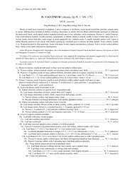
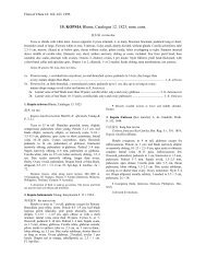
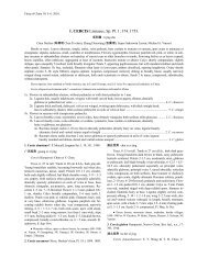
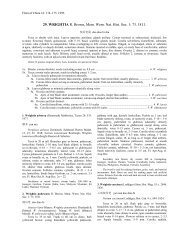
MORPHOLOGY 5<br />
FIGURE 1. General morpohological features <strong>of</strong> mosses. The peristome illustrated for the acrocarpic moss<br />
Funaria is <strong>of</strong> the diplolepidous, opposite type, with endostome segments directly located to the inside <strong>of</strong> the<br />
exostome teeth. In this genus the exostome teeth remain attached to a small part <strong>of</strong> the columella, and the<br />
divisural or median line is not easily seen. The terms acrocarpic, cladocarpic and pleurocarpic refer respectively<br />
to perichaetia and the subsequent sporophytes borne at the apex <strong>of</strong> the main shoot, or on a normal branch, or on<br />
a modified lateral branch.<br />
or arise all along the stem where it is in contact with the<br />
substrate. In some taxa, e.g., Tomentypnum, densely<br />
packed rhizoids form a feltlike tomentum on the stem.<br />
Most rhizoids are slender and only sparingly branched<br />
(micronematal type) but others are larger in diameter and<br />
extensively branched (macronematal type). The former<br />
arise from any <strong>of</strong> the epidermal cells <strong>of</strong> the stem, but the<br />
latter type is associated only with branch primordia as<br />
discussed earlier. Rhizoids are not major sites <strong>of</strong> water<br />
and nutrient uptake, but can enhance capillary movement<br />
<strong>of</strong> water along the outer surface <strong>of</strong> the stem (M. C. F.<br />
Proctor 1984). They function primarily as anchoring<br />
structures and in some taxa, e.g., Leptobryum and<br />
Pyramidula, form asexual propagules that aid in the<br />
spread <strong>of</strong> the colony over unoccupied substrate.<br />
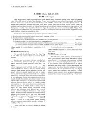
Stem Anatomy<br />
In many mosses, the stem is anatomically complex,<br />
consisting <strong>of</strong> a differentiated epidermal layer, a cortex,<br />
and a central strand <strong>of</strong> thin-walled, hydrolyzed waterconducting<br />
cells, called hydroids (Fig. 2). The epidermal<br />
cells are typically elongate in surface view and have thick,<br />
pigmented walls and small lumina. Various types <strong>of</strong> waxy<br />
deposits, analogous to cuticle or epicuticular waxes, may<br />
cover the surface <strong>of</strong> the epidermal cell wall, in both stems<br />
and leaves, especially in endohydric mosses (M. C. F.<br />
Proctor 1979). When these waxes are abundantly<br />
produced, the plants appear glaucous or iridescent to the<br />
naked eye. Although a thick-walled epidermis is<br />
common, in a variety <strong>of</strong> taxa, e.g., elements <strong>of</strong> the<br />
Pottiaceae as well as taxa <strong>of</strong> very wet habitats like<br />
Hygrohypnum, epidermal cells are thin-walled and<br />
swollen, forming a unistratose hyalodermis (Fig. 1). The<br />
cortex is generally divided into two zones. The outer<br />
zone, or sclerodermis, just to the inside <strong>of</strong> the epidermis,<br />
is comprised <strong>of</strong> stereids. These are elongated, thick-walled<br />
cells, generally with living protoplasts, that function in<br />
mechanical support, analogous to collenchyma in<br />
vascular plants. The inner zone <strong>of</strong> the cortex, or central<br />
cylinder, contains photosynthetic parenchyma cells that