SAM 314 - aphis
SAM 314 - aphis
SAM 314 - aphis
You also want an ePaper? Increase the reach of your titles
YUMPU automatically turns print PDFs into web optimized ePapers that Google loves.
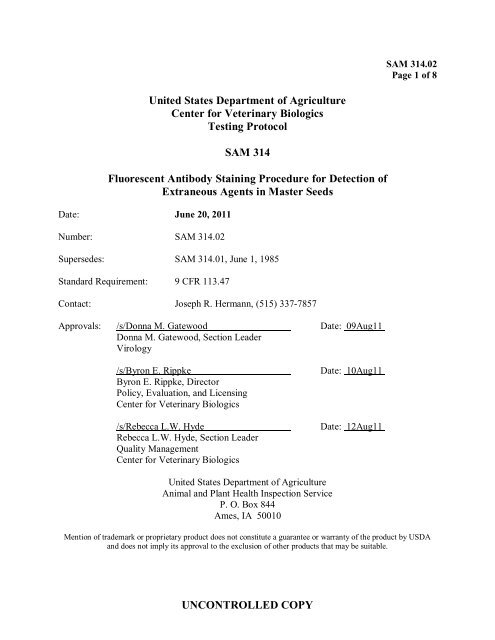
<strong>SAM</strong> <strong>314</strong>.02<br />
Page 1 of 8<br />
United States Department of Agriculture<br />
Center for Veterinary Biologics<br />
Testing Protocol<br />
<strong>SAM</strong> <strong>314</strong><br />
Fluorescent Antibody Staining Procedure for Detection of<br />
Extraneous Agents in Master Seeds<br />
Date: June 20, 2011<br />
Number: <strong>SAM</strong> <strong>314</strong>.02<br />
Supersedes: <strong>SAM</strong> <strong>314</strong>.01, June 1, 1985<br />
Standard Requirement: 9 CFR 113.47<br />
Contact: Joseph R. Hermann, (515) 337-7857<br />
Approvals: /s/Donna M. Gatewood Date: 09Aug11<br />
Donna M. Gatewood, Section Leader<br />
Virology<br />
/s/Byron E. Rippke<br />
Byron E. Rippke, Director<br />
Policy, Evaluation, and Licensing<br />
Center for Veterinary Biologics<br />
/s/Rebecca L.W. Hyde<br />
Rebecca L.W. Hyde, Section Leader<br />
Quality Management<br />
Center for Veterinary Biologics<br />
Date: 10Aug11<br />
Date: 12Aug11<br />
United States Department of Agriculture<br />
Animal and Plant Health Inspection Service<br />
P. O. Box 844<br />
Ames, IA 50010<br />
Mention of trademark or proprietary product does not constitute a guarantee or warranty of the product by USDA<br />
and does not imply its approval to the exclusion of other products that may be suitable.<br />
UNCONTROLLED COPY
Center for Veterinary Biologics <strong>SAM</strong> <strong>314</strong>.02<br />
Testing Protocol Page 2 of 8<br />
Fluorescent Antibody Staining Procedure for Detection of Extraneous Agents in Master Seeds<br />
Table of Contents<br />
1. Introduction<br />
2. Materials<br />
2.1 Equipment/instrumentation<br />
2.2 Reagents/supplies<br />
3. Preparation for the Test<br />
3.1 Personnel qualifications/training<br />
3.2 Preparation of equipment/instrumentation<br />
3.3 Preparation of reagents/control procedures<br />
3.4 Preparation of the sample<br />
3.5 Fixation of cell cultures<br />
4. Performance of the Test<br />
4.1 Direct FA/Indirect FA<br />
4.2 Reading<br />
5. Interpretation of the Test Results<br />
6. Report of Test Results<br />
7. References<br />
8. Summary of Revisions<br />
UNCONTROLLED COPY
Center for Veterinary Biologics <strong>SAM</strong> <strong>314</strong>.02<br />
Testing Protocol Page 3 of 8<br />
Fluorescent Antibody Staining Procedure for Detection of Extraneous Agents in Master Seeds<br />
1. Introduction<br />
This Supplemental Assay Method (<strong>SAM</strong>) describes fluorescent antibody staining techniques for<br />
detection of extraneous agents in Master Seeds (MS). The procedure utilizes either the direct or<br />
indirect method, and uses fluorescein isothiocyanate (FITC) conjugated specific antibody which<br />
enables visualization of viral antigen-antibody complexes by ultraviolet (UV) light microscopy.<br />
For the direct fluorescent antibody (FA) method, a specific fluorescein-labeled specific antibody<br />
binds to the viral antigen. The indirect fluorescent antibody (IFA) staining employs two specific<br />
antibodies: 1) The unlabeled primary antibody binds to a specific viral antigen if present in the<br />
infected cell; and 2) The fluorescein-labeled anti-species secondary antibody binds to the<br />
primary antibody-antigen complex. The antigen-antibody complexes can be visualized with a<br />
UV light microscope.<br />
FA or IFA testing is conducted to detect a variety of extraneous agents, and is also used to<br />
determine if the MS was adequately neutralized.<br />
The procedures described here are performed on third pass materials generated from MSinoculated<br />
cell cultures and controls as described in the current version of VIRPRO1013,<br />
Neutralization and Passage of Master Seed in Cell Cultures.<br />
2. Materials<br />
2.1 Equipment/instrumentation<br />
Equivalent equipment or instrumentation may be substituted for any brand names listed<br />
below.<br />
2.1.1 Aerobic incubator, 36° ± 2°C, humidified<br />
2.1.2 Laminar flow biological safety cabinet<br />
2.1.3 Ultraviolet (UV) light microscope<br />
2.1.4 Chemical fume hood<br />
2.2 Reagents/supplies<br />
Equivalent reagents or supplies may be substituted for any brand name listed below.<br />
2.2.1 Cultures prepared in chamber slides or other vessels in accordance with<br />
the current version of VIRPRO1013.<br />
2.2.2 Deionized water (DI)<br />
UNCONTROLLED COPY
Center for Veterinary Biologics <strong>SAM</strong> <strong>314</strong>.02<br />
Testing Protocol Page 4 of 8<br />
Fluorescent Antibody Staining Procedure for Detection of Extraneous Agents in Master Seeds<br />
2.2.3 Acetone (100% or 80% in DI) will be referenced throughout this<br />
document; however other fixatives may be used as appropriate.<br />
2.2.4 Phosphate buffered saline (PBS), 0.01M, pH 7.2, National Centers for<br />
Animal Health (NCAH) Media #30054.<br />
2.2.5 Evan’s blue biological stain (EBBS), 1.0% stock solution in DI.<br />
2.2.6 Antigen-specific conjugated polyclonal antiserum or conjugated<br />
monoclonal antibody (direct FA)<br />
2.2.7 Primary antibody, polyclonal antiserum or monoclonal antibody, specific<br />
to the virus (IFA)<br />
2.2.8 Conjugated secondary antibody or anti-species conjugate specific to the<br />
species in which the primary antibody was produced (IFA)<br />
2.2.9 Microscope slide glass staining dish with rack or Coplin staining jar<br />
2.2.10 Miscellaneous lab supplies: Beakers, pipettes, wash bottle<br />
2.2.11 Mounting medium (e.g., 10% glycerol, 50% glycerol, etc.)<br />
3. Preparation for the Test<br />
3.1 Personnel qualifications/training<br />
Personnel performing this procedure must have experience or training in: aseptic<br />
biological laboratory techniques, cell culture, preparation, handling, and disposal of<br />
biological agents and chemicals. Personnel must also have knowledge of safe operating<br />
procedures and training in the operation of the necessary laboratory equipment listed in<br />
Section 2.1.<br />
3.2 Preparation of equipment/instrumentation<br />
Turn on biological safety cabinet at least 15 minutes before use.<br />
3.3 Preparation of reagents/control procedures<br />
On the day of the FA staining, dilute reagents in Sections 2.2.6 through 2.2.8 with PBS<br />
to a previously determined optimal concentration for the detection of antigens.<br />
UNCONTROLLED COPY
Center for Veterinary Biologics <strong>SAM</strong> <strong>314</strong>.02<br />
Testing Protocol Page 5 of 8<br />
Fluorescent Antibody Staining Procedure for Detection of Extraneous Agents in Master Seeds<br />
3.4 Preparation of the sample<br />
The following procedures are followed for cell cultures grown in chambered slides.<br />
plates, or dishes.<br />
Cell cultures are prepared for FA staining according to the current version of<br />
VIRSOP2007, Master Seed Testing in the Virology Section. Seven days after the last<br />
subculturing, MS inoculated and control monolayers are fixed and FA stained. Positive<br />
and negative control slides, prepared concurrently with the MS slides, are used for<br />
verification of the assay. To ensure or enhance fluorescence detection, additional MS<br />
and positive control monolayers may be fixed before day 7. Regardless of when<br />
monolayers are fixed, they shall be stained concurrently.<br />
Previously prepared rabies virus positive control slides are available through the Center<br />
for Veterinary Biologics (CVB).<br />
3.5 Fixation of cell cultures<br />
Caution: Use fixative in a fume hood or biological safety cabinet. Gloves will be<br />
worn to avoid skin contact. Fixatives may be flammable; keep away from sources of<br />
heat or flame.<br />
3.5.1 Chamber Slides<br />
1. Decant the media from the chamber slides into an autoclavable<br />
container and remove the plastic walls, leaving the slides with the gasket<br />
attached. Handle and dispose of all viral fluids according safe laboratory<br />
practices.<br />
2. Place the slides in a slide rack and immerse the loaded slide rack in a<br />
staining dish filled with PBS, and wash for 10 ± 5 minutes<br />
3. Remove the slides from the PBS and place in a staining dish filled with<br />
acetone for 10 Use 100% acetone for glass slides and 80%<br />
acetone for plastic slides. Remove the slides and allow to air dry Used<br />
acetone should be collected for proper disposal.<br />
4. If cell preparations are not immediately FA stained, store the slides at<br />
-20° C or colder until use. Length of storage will vary for each antigen.<br />
Adequacy for use is monitored by FA staining.<br />
UNCONTROLLED COPY
Center for Veterinary Biologics <strong>SAM</strong> <strong>314</strong>.02<br />
Testing Protocol Page 6 of 8<br />
Fluorescent Antibody Staining Procedure for Detection of Extraneous Agents in Master Seeds<br />
3.5.2 Cell culture plates or dishes<br />
1. Decant the media from the plate/dish into an autoclavable container.<br />
Handle and dispose of all viral fluids according to safe laboratory<br />
practices.<br />
2. Gently wash cell monolayer with PBS and decant.<br />
3. Fix cell monolayer with 80% acetone for 10 ± 5 minutes.<br />
4. Decant the acetone into a suitable container; gently blot the plate/dish<br />
onto an absorbent surface and allow the plate/dish to air dry. Used<br />
acetone should be collected for proper disposal.<br />
5. Stain plate immediately or store at -20° C or colder until use. Length<br />
of storage will vary for each viral antigen. Adequacy for use is monitored<br />
by FA or IFA fluorescence of positive control.<br />
4. Performance of the Test<br />
Note: Volumes of reagents used in part 4.1 are for 2-chambered slides. When different<br />
vessels are stained, volumes may be adjusted to compensate for differences in cell<br />
monolayer area. Regardless of vessel used, a minimum surface area of 6 cm 2 is required<br />
for the final reading for both material under test and each of the controls.<br />
4.1 Direct FA/Indirect FA<br />
4.1.1 Apply 300 ± 25 µL of fluorescein-labeled antibody (FA) or primary<br />
antibody (IFA) to cover the fixed cell monolayer. Slides should not be allowed to<br />
dry during staining process.<br />
4.1.2 Incubate at 36° ± 2° ± 10 minutes in a humidified chamber.<br />
4.1.3 Decant the antibody from the slides. Rinse slides three times in PBS for<br />
approximately 5 ± 2 minutes per rinse. After final rinse, gently tap slide onto<br />
absorbent material to remove excess PBS.<br />
4.1.4 For IFA, repeat sections 4.1.1 through 4.1.3 using a secondary<br />
fluorescein-labeled antibody.<br />
4.1.5 Following PBS rinses, stained slides should be quickly rinsed in DI.<br />
UNCONTROLLED COPY
Center for Veterinary Biologics <strong>SAM</strong> <strong>314</strong>.02<br />
Testing Protocol Page 7 of 8<br />
Fluorescent Antibody Staining Procedure for Detection of Extraneous Agents in Master Seeds<br />
4.1.6 Gently tap the DI from the slides.<br />
Note: Counterstaining fluorescein-stained cell culture is optional and<br />
sometimes desirable to quench nonspecific background fluorescence or to<br />
heighten contrast between specific staining and background refer to Section<br />
4.1.7.<br />
4.1.7 For counterstaining (optional), make a predetermined dilution of the<br />
EBBS stock solution in DI which is based on the requirements for individual<br />
viruses. Apply the diluted EBBS to slides and incubate at 36°± 2°C for 20 ± 10<br />
minutes in a humidified chamber. Decant the EBBS and quickly rinse with<br />
distilled water. Gently tap the slides onto an absorbent surface.<br />
4.1.8 Every effort should be made to read the slides immediately after staining.<br />
Alternatively, slides may be stored in the dark at 4°± 2°C for up to 7 days. Prior to<br />
storage, slides must be air dried or overlaid with mounting medium. Air dried<br />
slides must be moistened using DI or mounting medium prior to reading.<br />
4.2 Reading<br />
4.2.1 The stained cell cultures are observed at 100X to 200X magnification in a<br />
darkened room with the use of a UV light microscope.<br />
4.2.2 Results of observations to be recorded.<br />
5. Interpretation of the Test Results<br />
Cells exhibiting specific apple-green fluorescence are considered positive. For a valid test, the<br />
positive cell culture controls must contain a sufficient number of infected cells to easily<br />
determine positive status; however, non-infected cells should also be present in order to<br />
differentiate positive cells from negative cells. Negative control wells must be free of specific<br />
fluorescence. Additionally, the MS inoculated cell cultures will be stained to confirm<br />
neutralization and must remain free of specific fluorescence.<br />
If agent specific fluorescence is observed in the MS inoculated cell monolayers, the MS will be<br />
determined to be unsatisfactory. The MS may be retested to confirm original results if warranted<br />
by the agent contact or reviewer.<br />
UNCONTROLLED COPY
Center for Veterinary Biologics <strong>SAM</strong> <strong>314</strong>.02<br />
Testing Protocol Page 8 of 8<br />
Fluorescent Antibody Staining Procedure for Detection of Extraneous Agents in Master Seeds<br />
6. Report of Test Results<br />
All records are kept in accordance with the current recordkeeping practices. Test results will be<br />
reviewed and signed by the agent contact. Test results are then entered into the current reporting<br />
system and released to the reviewer for distribution to the firm.<br />
7. References<br />
Code of Federal Regulations, Title 9, Part 113.309, U.S. Government Printing Office,<br />
Washington, DC.<br />
8. Summary of Revisions<br />
This is a complete revision of an existing document to reflect current practices in place at the<br />
Center for Veterinary Biologics.<br />
The Contact information has been updated.<br />
UNCONTROLLED COPY


