Inhibition of lipid metabolic enzymes using Mangifera indica extracts
Inhibition of lipid metabolic enzymes using Mangifera indica extracts
Inhibition of lipid metabolic enzymes using Mangifera indica extracts

You also want an ePaper? Increase the reach of your titles
YUMPU automatically turns print PDFs into web optimized ePapers that Google loves.
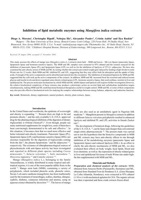
<strong>Inhibition</strong> <strong>of</strong> <strong>lipid</strong> <strong>metabolic</strong> <strong>enzymes</strong> <strong>using</strong> <strong>Mangifera</strong> <strong>indica</strong> <strong>extracts</strong><br />
Diego A. Moreno 1 , Christophe Ripoll 1 , Nebojsa Ilic 2 , Alexander Poulev 1 , Cristin Aubin 3 and Ilya Raskin 1*<br />
1<br />
Rutgers – The State University <strong>of</strong> New Jersey, Biotech Center, Cook College, 228 Foran Hall, 59 Dudley Road, New<br />
Brunswick, New Jersey 08901-8520, U.S.A. *e-mail: raskin@aesop.rutgers.edu. 2 Phytomedics Inc., 65 Stults Road, Dayton, NJ<br />
08810-1523, USA. 3 Children’s Hospital Boston, Division <strong>of</strong> Endocrinology, 300 Longwood Ave., Boston, MA 02115, U.S.A<br />
Received 28 August 2004, accepted 27 November 2005.<br />
Abstract<br />
This study assesses the effects <strong>of</strong> mango tree (<strong>Mangifera</strong> <strong>indica</strong> L.) <strong>extracts</strong> (stem bark – MSB and leaves – ML) on lipases (pancreatic lipase,<br />
lipoprotein lipase and hormone-sensitive lipase). The MSB and ML samples were extracted in 95% ethanol and the <strong>extracts</strong> assayed for the<br />
inhibition <strong>of</strong> pancreatic lipase (PL) and lipoprotein lipase (LPL) as well as for the inhibition <strong>of</strong> lipolysis <strong>of</strong> 3T3-L1 adipocytes. We have also<br />
examined the anti-obesity action <strong>of</strong> MSB and ML by testing whether the <strong>extracts</strong> prevented weight gain induced by feeding a high-fat diet to male<br />
Wistar rats for 12 weeks. Both MSB and ML inhibited PL and LPL, suggesting that they may affect both fat absorption and the uptake <strong>of</strong> fatty<br />
acids, if enough <strong>of</strong> the active components can be absorbed and entered into the circulation. The inhibition <strong>of</strong> stimulated lipolysis by MSB and ML<br />
suggested that the cells took up the active components <strong>of</strong> the <strong>extracts</strong>. In addition, MSB and ML increased fecal fat excretion and reduced serum<br />
glucose and insulin levels and down-regulated some obesity-related genes (LPL, hormone-sensitive lipase, fatty acid synthase, resistin) in liver and<br />
epididymal fat. The precise molecular mechanisms by which MSB and ML inhibit lipases and lipolysis still requires further investigation.However,<br />
the relative biochemical complexity <strong>of</strong> these <strong>extracts</strong> may produce a pleiotropic action on several <strong>lipid</strong> and carbohydrate metabolism targets<br />
simultaneously, making MSB and ML multifunctional botanical therapeutics useful in weight control. MSB and ML or some <strong>of</strong> their components<br />
may also provide effective biochemical tools for studying the complex relationships between energy balance, adiposity and endocrine function.<br />
Key words: Botanicals, dietary supplements, natural products, obesity, plant <strong>extracts</strong>, lipase.<br />
Introduction<br />
In the United States and worldwide, the epidemic <strong>of</strong> overweight<br />
and obesity is expanding 1, 2 . Western diets are high in fat and<br />
promote obesity 3 , and the only available U.S.-F.D.A. approved<br />
drug for the pharmacological inhibition <strong>of</strong> the digestion <strong>of</strong> dietary<br />
triglycerides is Orlistat (Xenical ® ) 4 . Even though, people use<br />
many nutritional supplements for weight loss, none <strong>of</strong> them have<br />
been convincingly demonstrated to be safe and effective 5 . In<br />
this situation, it becomes clear that we need more effective and<br />
better tolerated anti-obesity treatments. Pancreatic lipase (PL),<br />
lipoprotein lipase (LPL) and hormone-sensitive lipase (HSL) are<br />
<strong>enzymes</strong> responsible for the digestion <strong>of</strong> triglycerides coming<br />
from the diet 6 , the plasma lipoproteins 7 and the adipocytes 8, 9 ,<br />
respectively. The existence <strong>of</strong> ethnopharmacological sources <strong>of</strong><br />
phytochemicals with anti-lipase activity has been investigated<br />
and reported in different plant species including Cassia<br />
mimosoides 10 , Salacia reticulata 11 , Salix matsudara 12 ,<br />
Dioscorea nipponica 13 and Camelia sinensis 14 .<br />
Mango (<strong>Mangifera</strong> <strong>indica</strong> L.), belonging to the family<br />
Anacardiaceae, is widely distributed in many tropical and subtropical<br />
regions; it is one <strong>of</strong> the most popular edible fruits in the<br />
world 15 . Mango stem bark (MSB), containing a variety <strong>of</strong><br />
polyphenols, which included phenolic acids, phenolic esters,<br />
flavan-3-ols and a xanthone (mangiferin), has been traditionally<br />
used for the treatment <strong>of</strong> menorrhagia, scabies, diarrhea, allergies,<br />
syphilis, diabetes, cutaneous infections and anemia, <strong>using</strong> an<br />
aqueous extract obtained by decoction 16, 17 . The leaves <strong>of</strong> mango<br />
(ML) are also used as an antidiabetic agent in Nigerian folk<br />
medicine 18 . Mangiferin present in <strong>Mangifera</strong> <strong>extracts</strong> in addition<br />
to different Salacia reticulata polyphenols resulted in enhanced<br />
lipolysis and inhibited PL and LPL activities in female Zucker<br />
rats 11 .<br />
The development <strong>of</strong> botanical drugs, following the guidelines<br />
<strong>of</strong> the U.S.-F.D.A. 19 , can be faster and cheaper than conventional<br />
single-entity pharmaceuticals 20 . The present study was carried<br />
out to test the hypothesis that the bioactive compounds in MSB<br />
and ML <strong>extracts</strong> may have anti-obesity effects in rats through<br />
inhibition <strong>of</strong> fat metabolizing <strong>enzymes</strong> (pancreatic lipase and<br />
lipoprotein lipase) and reduced lipolysis (HSL). In an effort to<br />
clarify the anti-obesity mechanisms <strong>of</strong> MSB and ML, we also<br />
evaluated their effects on body weight, fecal <strong>lipid</strong>s, blood and<br />
liver chemistry as well as the expression <strong>of</strong> obesity-related genes<br />
in liver and epididymal fat tissue <strong>of</strong> male Wistar rats under highfat<br />
diet supplemented with MSB and ML.<br />
Materials and Methods<br />
Preparation and chemical analysis <strong>of</strong> the <strong>extracts</strong>: MSB,<br />
obtained from Amazon Herbs (Paramaribo, Suriname) and ML,<br />
obtained from Standard Fruit de Honduras S.A. (Zona Mazapan<br />
La Ceiba Atlantida, Honduras), were extracted in 95% ethanol<br />
(1:10 w:v) with mechanical agitation for 24 h. The organic solvent<br />
was then evaporated and these crude <strong>extracts</strong> freeze-dried.
Pancreatic lipase [PL; E.C. 3.1.1.3]: Lipase-PS TM reagents were<br />
obtained from Sigma Diagnostics (Procedure No. 805, Sigma-<br />
Aldrich, St. Louis, MO). Human pancreatic lipase (Lipase-PS<br />
standard, 230 U l -1 ) was obtained from Sigma Diagnostics (Sigma-<br />
Aldrich, St. Louis, MO). Aliquots (30 µl) <strong>of</strong> lipase standard, blank<br />
(water as reference) and MSB and ML samples were added to 400<br />
µl <strong>of</strong> substrate solution, mixed gently and incubated for 5 min at<br />
37°C. Activator reagent (300 µl) was added to the samples, mixed<br />
by gentle inversion and incubated for an additional 3 min at 37°C.<br />
The increase in absorbance at 550 nm, due to the formation <strong>of</strong><br />
quinone diimine dye, was measured to determine the activity in<br />
the samples 4, 6 .<br />
Lipoprotein lipase [LPL; E.C. 3.1.1.34]: LPL was measured<br />
according to the method <strong>of</strong> Nilsson-Ehle and Schotz 22 . A pool <strong>of</strong><br />
LPL was made by incubating human adipose tissue fragments<br />
with 10 U ml -1 heparin (500 mg 5 ml -1 ) for 45 min at 24°C. Aliquots<br />
<strong>of</strong> this heparin eluate were preincubated with varying<br />
concentrations <strong>of</strong> MSB and ML (Table 2) for 30 min at 4°C. After<br />
addition <strong>of</strong> 3 H-triolein substrate, samples were incubated for 60<br />
min at 37°C and the released 3 H-oleic acid was measured 23 .<br />
Hormone-sensitive lipase [HSL, E.C. 3.1.1.]: Lipolytic activity<br />
in cultured mouse 3T3-L1 adipocytes was used as a measure <strong>of</strong><br />
HSL activity 21, 24 . 3T3-L1 cells were cultured with DMEM (4500<br />
mg glucose l -1 ) supplemented with 100 g l -1 fetal bovine serum, 2<br />
mM glutamine, 100 units ml -1 penicillin, 100 µg ml -1 streptomycin,<br />
110 µg ml -1 sodium pyruvate and 8 µg ml -1 biotin, in a 50 g kg -1 CO 2<br />
atmosphere at 37°C. The differentiation <strong>of</strong> 3T3-L1 cells was<br />
initiated by the addition <strong>of</strong> 10 µM dexamethasone, 0.5 mM<br />
isobutyl-methylxanthine and 10 µg ml -1 insulin to the culture<br />
medium <strong>of</strong> confluent cells for 3 days, followed by the cultivation<br />
<strong>of</strong> cells without supplements for an additional 3 or more days. We<br />
added MSB and ML <strong>extracts</strong> as <strong>indica</strong>ted in Fig. 1; after 18 hours<br />
<strong>of</strong> incubation, the stimulation <strong>of</strong> lipolysis was accomplished by<br />
incubating differentiated adipocytes with 10 µM isoproterenol<br />
-<br />
and 20 g l 1 fatty acid-free bovine serum albumin for 1 h. Lipolysis<br />
was determined by measuring glycerol release <strong>using</strong> a fluorometric<br />
23, 24<br />
enzymatic assay .<br />
In vivo study: This study was approved by the Animal Care and<br />
Facilities Committee in the Office <strong>of</strong> Research and Sponsored<br />
Programs at Rutgers University. 7-9-week-old healthy male Wistar<br />
rats (Charles River Laboratories Inc., MA, USA) were kept, one<br />
per collection cage, in a temperature-controlled room at 22°C with<br />
a 12h/12h light/darkness cycle with lights on at 7:00 AM. There<br />
was an average <strong>of</strong> 10-15 air changes per room per h. Rats were<br />
allowed free access to water and food and adapted to the facility<br />
for 1 week before treatment.<br />
The rats were divided into three groups. The control group was<br />
fed a high-fat AIN-76A purified rodent diet (45% kcal fat, Dyets<br />
Inc. Bethlehem, PA). The other two groups were fed a modified<br />
diet to match the energy supply <strong>of</strong> the diets and including 10 g<br />
-<br />
kg 1 <strong>of</strong> either MSB or ML <strong>extracts</strong> for 12 weeks. Body weight was<br />
measured between 9:00-11:00 AM every Thursday morning. Daily<br />
(24 h) food intake was measured on a per-animal basis once per<br />
__________________________________________________________________________________<br />
Abbreviations: BWG: body weight gain; FAS: Fatty acid synthase; FFA: Free fatty acids; HSL: Hormonesensitive<br />
lipase; ISO: Isoproterenol; LPL: Lipoprotein lipase; PL: Pancreatic lipase; MSB: Mango stem<br />
bark; ML: Mango leaves<br />
week. Feces were collected once per 2 weeks. Feces from each rat<br />
were pooled and dried to constant weight. Fecal <strong>lipid</strong>s were<br />
extracted by the method <strong>of</strong> Folch et al. 25 .<br />
After 12 weeks <strong>of</strong> treatment, we kept rats fasting overnight<br />
and euthanized by decapitation for necrospy. Liver and fat<br />
deposits (right-half epididymal fat depots) were excised, weighed<br />
and immediately frozen in liquid nitrogen and stored at –80°C for<br />
future analyses. Blood was collected for biochemical analyses<br />
on a Hitachi 747 chemistry analyzer (Ani Lytics Inc., Gaithersburg,<br />
MD): glucose (Hexokinase, Roche Molecular Biochemicals,<br />
Germany) and insulin (Radioinmunoassay specific for rat, Linco<br />
Research Inc., Missouri); total cholesterol (Cholesterol/HP assay<br />
kit Roche Molecular biochemicals, Indianapolis) and triglycerides<br />
(Glycerol Phosphate Peroxidase, Roche reagents). These<br />
experiments were carried out at the Cook Animal Facility (Cook<br />
College, Rutgers University), an A.A.A.L.A.C. Intl.-accredited<br />
facility. Animals were cared for in accordance with national<br />
guidelines <strong>of</strong> Public Health for the Care and Use <strong>of</strong> Laboratory<br />
Animals.<br />
Obesity-related gene expression analyses: Total RNA from liver<br />
and epididymal fat tissues was extracted following the TRI-<br />
Reagent® protocol (Sigma, St Louis, MO) and pooled from the 6<br />
rats per treatment before RT-PCR analysis. RNA was treated with<br />
RNase-free RQ1 DNase (Promega, Madison, WI) and then<br />
submitted to a reverse transcription with superscript II H-<br />
(Invitrogen, Carslad, CA) according to the manufacturer’s<br />
instructions.<br />
Obesity-related gene expression levels were quantified <strong>using</strong><br />
a Stratagene Mx 3000P TM Real-Time PCR System (Stratagene, La<br />
Jolla, CA). Primers for each gene were designed <strong>using</strong> Primer<br />
Express® ver. 2.0 (Applied Biosystems, Foster City, CA) as<br />
presented in Table 1.<br />
Real-time PCR analyses were carried out in a Brilliant® SYBR®<br />
Green PCR master mix kit (Stratagene) according to kit instructions.<br />
Samples were amplified <strong>using</strong> the following program: 2 minutes<br />
incubation at 50°C; initial denaturation and polymerase activation<br />
at 95ºC for 10 min; 40 PCR cycles consisting <strong>of</strong> 15 s at 95°C and 60<br />
s at 60°C each. The RNA expression was analyzed by ‘∆∆Ct’<br />
methods 26 , <strong>using</strong> the β-actin gene as a normalizer. Amplification<br />
<strong>of</strong> specific transcripts was further confirmed by obtaining melting<br />
curve pr<strong>of</strong>iles. All samples were assayed in duplicate and 5<br />
independent analyses were performed.<br />
Statistical analysis: All data were subjected to analysis <strong>of</strong><br />
variance (ANOVA). The data (means ± SEM) shown are mean<br />
values and the significance <strong>of</strong> the differences was compared <strong>using</strong><br />
the Duncan’s Multiple Range Test at Least Significant Difference<br />
(P
Table 1. Primers sequence for RT-PCR (5’-3’) <strong>of</strong> selected obesity-related genes.<br />
Gene (accession number) Forward Reverse<br />
Medium Chain Acyl-CoA<br />
GCTAGTAAAGCCTTCACCGGATT TTAGTTCCTTTTTTCCGATGTGTATTC<br />
(NM_016986)<br />
Acyl-CoA oxidase (NM_01734) TGGCCAACTATGGTGGACATC TACCAATCTGGCTGCACGAA<br />
Fatty Acid Synthase (NM_017332) GGCATCATTGGGCACTCCTT GCTGCAAGCACAGCCTCTCT<br />
Lipoprotein Lipase (NM_012598) CTGAAAGTGAGAACATTCCCTTCA CCGTGTAAATCAAGAAGGAGTAGGTT<br />
Hormone sensitive lipase<br />
CCAAGTGTGTGAGCGCCTATT CACGCCCAATGCCTTCTG<br />
(NM_012859)<br />
UCP-2 (NM_019354) TCCGGACACAATAGTATCTTTAAG GCCTGATCCCCTTGATTTCC<br />
Adiponectin (NM_144740) CCCAGGGTCCAGATTCAACTC GGTGTAATGGTGGGCTTGCT<br />
Resistin (NM_144741) ACTGCCAGTGCGGAAGCATAG ATCAACCGTCCTCAGGAACCA<br />
Actin (NM_031144) GGGAAATCGTGCGTGACATT GCGGCAGTGGCCATCTC<br />
Table 2. Inhibitory effects <strong>of</strong> <strong>Mangifera</strong> <strong>indica</strong> <strong>extracts</strong> on the activity <strong>of</strong> human<br />
pancreatic lipase (PL) and lipoprotein lipase (LPL).<br />
Extract (mg ml -1 ) PL (U l -1 )<br />
Inhibitory<br />
effect (%)<br />
LPL (U ml -1 )<br />
(µmol glycerol released<br />
h -1 ml -1 )<br />
0 230.00.2 a † 0.628 0.016 a †<br />
MSB 0.01 204.52.1 b 11 0.620 0.015 a<br />
0.1 193.32.8 c 16 0.560 0.021 b 11<br />
1 115.52.4 d 50 0.158 0.015 c 75<br />
ML 0.01 169.52.1 b 26 0.618 0.020 a<br />
0.1 159.32.1 c 31 0.508 0.017 b 19<br />
1 129.81.3 d 44 0.478 0.011 c 24<br />
Inhibitory<br />
effect (%)<br />
Inhibitory effect shown as the lowering <strong>of</strong> relative activity (%) compared to control (0 mg ml -1 = 100% activity).†Values followed<br />
by different lowercase letters are significantly different (ANOVA) from the control at p
200<br />
a<br />
b<br />
b<br />
a<br />
35<br />
***<br />
a<br />
Control HFD MSB +HFD ML +HFD<br />
a<br />
µM Released Glycerol (well) -1<br />
150<br />
100<br />
50<br />
d<br />
c<br />
b<br />
c<br />
Fecal Lipids (mg g -1 feces d.w.) d -1 rat -1<br />
30<br />
25<br />
20<br />
15<br />
10<br />
5<br />
b<br />
**<br />
a<br />
b<br />
b<br />
a<br />
a<br />
a<br />
a<br />
a<br />
a<br />
0<br />
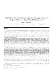
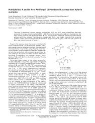
Figure 1. Inhibitory effect <strong>of</strong> <strong>Mangifera</strong> <strong>indica</strong> <strong>extracts</strong> on HSL activity<br />
in cultured murine 3T3-L1 adipocytes. Each column represents the<br />
mean ±SEM (n= 4). Means followed by the same letter are not significantly<br />
different at p
Table 4. Effect <strong>of</strong> treatment with <strong>Mangifera</strong> <strong>indica</strong> <strong>extracts</strong><br />
<strong>extracts</strong> on gene expression in the liver and<br />
epididymal fat tissues <strong>of</strong> male Wistar rats.<br />
Gene HFD Control MSB +HFD<br />
ML +HFD<br />
Mango leaf<br />
Liver<br />
MCAD 100 90.5 10.0 86.3 14.0<br />
-Amylase 100 95.5 8.1 86.1 8.0<br />
ACO 100 74.3 11.4 108.0 11.4<br />
FAS 100 109.5 11.3 88.9 8.3<br />
Epididymal fat<br />
LPL 100 a† 77.5 3.6 b 69.4 3.7 b<br />
HSL 100 a 87.3 4.7 b 73.0 1.2 c<br />
UCP-2 100 107.1 3.6 101.2 5.1<br />
FAS 100 a 64.8 3.5 b 38.7 8.6 c<br />
Adiponectin 100 101.0 5.0 111.6 8.1<br />
Resistin 100 a 90.3 4.7 a 67.5 7.7 b<br />
Results represents means ± SEM (n = 6).†Values followed by different lowercase letters are<br />
significantly different (ANOVA) from the control at p
Chemistry 37:361-368.<br />
7<br />
Goldberg, I.J. and Merkel, M. 2002. Lipoprotein lipase: physiology,<br />
biochemistry, and molecular biology. Front Biosciences 6: D388-4051.<br />
8<br />
Holm, C., Osterlund, T., Laurell, H. and Contreras, J. 2000. Molecular<br />
mechanisms regulating hormone-sensitive lipase and lipolysis. Annual<br />
Review Nutrition 20:365-93.<br />
9<br />
Slee, D.H., Bhat, A.S., Nguyen, T.N., Kish, M., Lundeen, K., Newman,<br />
M.J. and McConnell, S.J. 2003. Pyrrolopyrazinedione-based inhibitors<br />
<strong>of</strong> human hormone-sensitive lipase. Journal Medicinal Chemistry 46:<br />
1120-1122.<br />
10<br />
Yamamoto, M., Shimura, S., Itoh, Y., Ohsaka, T., Egawa, M. and Inoue,<br />
S. 2000. Anti-obesity effects <strong>of</strong> lipase inhibitor CT-II, an extract from<br />
edible herbs, Nomame Herba, on rats fed a high-fat diet. International<br />
Journal Obesity 24:758-764.<br />
11<br />
Yoshikawa, M., Shimoda, H., Nishida, N., Takada, M. and Matsuda,<br />
H. 2002. Salacia reticulata and its polyphenolic constituents with<br />
lipase inhibitory and lipolytic activities have mild antiobesity effects<br />
in rats. Journal <strong>of</strong> Nutrition 132:1819-1824.<br />
12<br />
Han, L.-K., Sumiyoshi, M., Zhang, J., Liu, M.-X., Zhang, X.-F., Zheng,<br />
Y.-N., Okuda, H. and Kimura, Y. 2003. Anti-obesity action <strong>of</strong> Salix<br />
matsudana leaves (Part 1). Anti-obesity action by polyphenols <strong>of</strong><br />
Salix matsudana in high fat-diet treated rodent animals. Phytotherapy<br />
Research 17:1188-1194.<br />
13<br />
Kwon, C.-S., Sohn, H.-Y., Kim, S.-H., Kim, J.-H., Son, K.-H., Lee, J.-<br />
S., Lim, J.-K. and Kim, J.-S. 2003. Anti-obesity effect <strong>of</strong> Dioscorea<br />
nipponica Makino with lipase-inhibitory activity in rodents.<br />
Bioscience, Biotechnology and Biochemistry 67:1451-1456.<br />
14<br />
Wu, L.-Y., Juan, C.-C., Ho, L.-T., Hsu, Y.-P. and Hwang, L.-S. 2004.<br />
Effect <strong>of</strong> green tea supplementation on insulin sensitivity in Sprague-<br />
Dawley Rats. Journal Agricultural Food Chemistry 52:643-648.<br />
15<br />
Ross, I.A. 2003. <strong>Mangifera</strong> <strong>indica</strong> L. In Medicinal Plants <strong>of</strong> the World,<br />
Volume 1. Chemical Constituents, Traditional and Modern Uses. 2 nd<br />
Edition. Humana Press, Totowa NJ, pp. 315-328.<br />
16<br />
Nunez-Selles, A.J., Velez-Castro, H.T., Agüero-Agüero, J., Gonzalez-<br />
Gonzalez, J., Naddeo, F., De Simone, F. and Rastrelli, L. 2002. Isolation<br />
and quantitative analysis <strong>of</strong> phenolic antioxidants, free sugars, and<br />
polyols from mango (<strong>Mangifera</strong> <strong>indica</strong> L.) stem bark aqueous decoction<br />
used in Cuba as nutritional supplement. Journal Agricultural Food<br />
Chemistry 50:762-766.<br />
17<br />
Garcia, D., Escalante, M., Delgado, R., Ubeira, F.M. and Leiro, J. 2003.<br />
Anthelminthic and antiallergic activities <strong>of</strong> <strong>Mangifera</strong> <strong>indica</strong> L. stem<br />
bark components vimang and mangiferin. Phytotherapy Research 17:<br />
1203-1208.<br />
18<br />
Aderibigbe, A.O., Emudianughe, T.S. and Lawal, B.A.S. 1999.<br />
Antihyperglycaemic effect <strong>of</strong> <strong>Mangifera</strong> <strong>indica</strong> in Rat. Phytotherapy<br />
Research 13:504-507.<br />
19<br />
U.S. -F.D.A. 2004. Guidance for Industry Botanical Drug Products.<br />
Center for Drug Evaluation and Research, US Food and Drug<br />
Administration. URL: http://www.fda.gov/cder/guidance/4592fnl.htm<br />
(Access 25 May, 2005).<br />
20<br />
Ribnicky, D., Poulev, A., O’Neal, J., Wnorowski, G., Malek, D.E.,<br />
Jäger, R. and Raskin, I. 2004. Toxicological evaluation <strong>of</strong> the ethanolic<br />
extract <strong>of</strong> Artemisia dracunculus L. for use as a dietary supplement<br />
and in functional foods. Food Chemistry Toxicology 42:585-598.<br />
21<br />
Moreno, D.A., Ilic, N., Poulev, A., Brasaemle, D.L., Fried, S.K. and<br />
Raskin, I. 2003. Inhibitory effects <strong>of</strong> grape seed extract on lipases.<br />
Nutrition 19:876-879.<br />
22<br />
Nilsson-Ehle, P. and Schotz, M.C. 1976. A stable, radioactive substrate<br />
emulsion for assay <strong>of</strong> lipoprotein lipase. Journal Lipid Research 17:<br />
546-541.<br />
23<br />
Brasaemle, D.L., Barber, T., Wolins, N.E., Serrero, G., Blanchette-<br />
Mackie, E.J. and Londos, C. 1997. Adipose differentiation-related<br />
protein is an ubiquitously expressed <strong>lipid</strong> storage droplet-associated<br />
protein. Journal Lipid Resesearch 38:2249-2263.<br />
24<br />
Laurell, S. and Tibbling, G. 1966. An enzymatic fluorometric<br />
micromethod for the determination <strong>of</strong> glycerol. Clinical Chimica Acta<br />
13:317-322.<br />
25<br />
Folch, J., Lees, M. and Sloane-Stanley, G.M. 1957. Simple method for<br />
isolation and purification <strong>of</strong> total <strong>lipid</strong>s from animal tissues. Journal<br />
Biological Chemistry 226:497-509.<br />
26<br />
Winer, J., Jung, C.K.S., Shackel, I. and Williams, P.M. 1999.<br />
Development and validation <strong>of</strong> real-time quantitative reverse<br />
transcriptase-polymerase chain reaction for monitoring gene expression<br />
in cardia myocytes in vitro. Analytical Biochemistry 270:41-49.<br />
27<br />
Harkness, J.E. and Wagner, J.E. 1995. The Biology and Medicine <strong>of</strong><br />
Rabbits and Rodents. 4 th Edition. Lea & Febiger Editors, Philadelphia,<br />
PA, 372 pp.<br />
28<br />
Akiyama, T., Tachibana, I., Shirohara, H., Watanabe, N. and Otsuki,<br />
M. 1996. High-fat hypercaloric diet induces obesity, glucose intolerance<br />
and hyper<strong>lipid</strong>emia in normal adult male Wistar rat. Diabetes Research<br />
Clinical Practice 31:27-35.<br />
29<br />
Muruganandan, S., Srinivasan, K., Gupta, S., Gupta, P.K. and Lal, J.<br />
2005. Effect <strong>of</strong> mangiferin on hyperglycemia and atherogenicity in<br />
streptozotocin diabetic rats. Journal Ethnopharmacology 97:497-501.<br />
30<br />
Choo, J.J. 2003. Green tea reduces body fat accretion caused by highfat<br />
diet in rats through β-adrenoceptor activation <strong>of</strong> thermogenesis in<br />
brown adipose tissue. Journal Nutritional Biochemistry 11: 671-676.<br />
31<br />
Ouchi, N., Kihara, S., Funahashi, T., Matsuzawa, Y. and Walsh, K.<br />
2003. Obesity, adiponectin and vascular inflammatory disease. Current<br />
Opinion Lipidology 14:561-566.<br />
32<br />
Sul, H.S., Latasa, M.J., Moon, Y. and Kim, K.H. 2000. Regulation <strong>of</strong><br />
the fatty acid synthase promoter by insulin. Journal <strong>of</strong> Nutrition 130:<br />
315S-320S.<br />
33<br />
Vendrell, J., Broch, M., Vilarrasa, N., Molina, A., Gomez, J.M.,<br />
Gutierrez, C., Simon, I., Soler, J. and Richart, C. 2004. Resistin,<br />
adiponectin, ghrelin, leptin, and proinflammatory cytokines:<br />
relationships in obesity. Obesity Research 12:962-971.