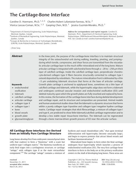
The Cartilage-Bone Interface
The Cartilage-Bone Interface
The Cartilage-Bone Interface
Create successful ePaper yourself
Turn your PDF publications into a flip-book with our unique Google optimized e-Paper software.
Special Focus Section<br />
<strong>The</strong> <strong>Cartilage</strong>-<strong>Bone</strong> <strong>Interface</strong><br />
Caroline D. Hoemann, Ph.D. 1, 2, 3 Charles-Hubert Lafantaisie-Favreau, M.Sc. 2<br />
Viorica Lascau-Coman, M.Sc. 1, 3 Gaoping Chen, M.D. 1 Jessica Guzmán-Morales, Ph.D. 1<br />
1 Department of Chemical Engineering, Ecole Polytechnique,<br />
Montreal, Quebec, Canada<br />
2 Institute of Biomedical Engineering, Ecole Polytechnique, Montreal,<br />
Quebec, Canada<br />
3 Groupe de Recherche en Sciences et Technologies Biomédicales<br />
(GRSTB), Ecole Polytechnique, Montreal, Quebec, Canada<br />
Address for correspondence and reprint requests Caroline D.<br />
Hoemann, Ph.D., Department of Chemical Engineering, Ecole<br />
Polytechnique, 2900 Boulevard Edouard-Montpetit, Montreal, Quebec<br />
H3G 1J4, Canada (e-mail: caroline.hoemann@polymtl.ca).<br />
J Knee Surg<br />
Abstract<br />
Keywords<br />
► endochondral<br />
ossification<br />
► tidemark<br />
► calcified cartilage<br />
► articular cartilage<br />
► collagen type II<br />
► collagen type I<br />
► bone<br />
► blood vessels<br />
► growth plate<br />
► glycosaminoglycan<br />
In the knee joint, the purpose of the cartilage-bone interface is to maintain structural<br />
integrity of the osteochondral unit during walking, kneeling, pivoting, and jumping–<br />
during which tensile, compressive, and shear forces are transmitted from the viscoelastic<br />
articular cartilage layer to the much stiffer mineralized end of the long bone. Mature<br />
articular cartilage is integrated with subchondral bone through a 20 to 250 µm thick<br />
layer of calcified cartilage. Inside the calcified cartilage layer, perpendicular chondrocyte-derived<br />
collagen type II fibers become structurally cemented to collagen type I<br />
osteoid deposited by osteoblasts. <strong>The</strong> mature mineralization front is delineated by a thin<br />
5 µm undulating tidemark structure that forms at the base of articular cartilage.<br />
Growth plate cartilage is anchored to epiphyseal bone, sometimes via a thin layer of<br />
calcified cartilage and tidemark, while the hypertrophic edge does not form a tidemark<br />
and undergoes continual vascular invasion and endochondral ossification (EO) until<br />
skeletal maturity upon which the growth plates are fully resorbed and replaced by bone.<br />
In this review, the formation of the cartilage-bone interface during skeletal development<br />
and cartilage repair, and its structure and composition are presented. Animal models<br />
and human anatomical studies show that the tidemark is a dynamic structure that forms<br />
within a purely collagen type II-positive and collagen type I-negative hyaline cartilage<br />
matrix. <strong>Cartilage</strong> repair strategies that elicit fibrocartilage, a mixture of collagen type I<br />
and type II, are predicted to show little tidemark/calcified cartilage regeneration and to<br />
develop a less stable repair tissue-bone interface. <strong>The</strong> tidemark can be regenerated<br />
through a bone marrow-driven growth process of EO near the articular surface.<br />
All <strong>Cartilage</strong>-<strong>Bone</strong> <strong>Interface</strong>s Are Derived<br />
from an Initially Pure <strong>Cartilage</strong> Structure<br />
Long bones develop first from embryonic mesenchymal stem<br />
cells that coalesce to form a “blastema,” with a scant but<br />
uniform type I collagen matrix. 1 <strong>The</strong> blastema transforms at<br />
early fetal stages into a cartilaginous structure, or cartilage<br />
“anlage,” with collagen type II as the main extracellular<br />
component. 2–6 <strong>The</strong> cartilage “anlage” contains a mixture of<br />
fusiform and round chondroblast cells, 7 that upon terminal<br />
differentiation will hypertrophy (become unusually large),<br />
and begin to express proteins that attract blood vessels and<br />
facilitate biomineralization. 8<br />
Mineralized bone begins to form when the fetal cartilage<br />
undergoes focal hypertrophy which launches a process of<br />
endochondral ossification (EO). <strong>The</strong> very first cartilage-bone<br />
interfaces to form in the body are at the primary ossification<br />
centers in the shaft of developing long bones. <strong>The</strong>se areas are<br />
received<br />
November 21, 2011<br />
accepted after revision<br />
March 26, 2012<br />
Copyright © 2012 by Thieme Medical<br />
Publishers, Inc., 333 Seventh Avenue,<br />
New York, NY 10001, USA.<br />
Tel: +1(212) 584-4662.<br />
DOI http://dx.doi.org/<br />
10.1055/s-0032-1319782.<br />
ISSN 1538-8506.
<strong>The</strong> <strong>Cartilage</strong>-<strong>Bone</strong> <strong>Interface</strong><br />
Hoemann et al.<br />
Figure 1 Three dynamic cartilage-bone interfaces are established in the postnatal knee femoral end: (1) articular cartilage-epiphyseal bone,<br />
(2) growth plate-epiphyseal bone, and (3) growth plate-metaphyseal bone. A macroscopic transverse view of a skeletally immature 4-month<br />
rabbit knee trochlea shows the articular cartilage (AC, white arrowheads, A & B) and growth plate (GP, white arrows, A & C), and distribution<br />
of collagen type II (D, E, F) and collagen type I (G, H, I) in serial sections from the area of the dashed squares in panel A. <strong>The</strong> distal femur was fixed<br />
in 4% paraformaldehyde/100 mM cacodylate, decalcified in EDTA with a Milestone microwave, cryoembedded and cryosectioned using the<br />
CryoJane tape system; the sections were predigested in hyaluronidase and protease to remove glycosaminoglycans and immunostained for<br />
collagen type II (monoclonal II6B3, DSHB, USA) or collagen type I (monoclonal antibody I-H85, VWR, Canada), using secondary biotinylated goat<br />
antimouse and avidin-alkaline phosphatase red substrate detection with iron hematoxylin counterstain. 91,92 In panels A and B, the rough articular<br />
surface is a cutting artifact from the isomet diamond saw. <strong>The</strong> tear/crack in the growth plate indicated by a white asterisk in panel D is a<br />
cryosectioning artifact. 92 EO, endochondral ossification; BP, subchondral bone plate; AC, articular cartilage; GP, growth plate.<br />
marked by remodeling and vascular invasion in parallel with<br />
the deposition of a mineralizing collagen type I matrix that<br />
ensheathes and mechanically protects the blood vessels. 4,9<br />
After the secondary ossification centers appear in the distal<br />
tibia and femur, three types of dynamic cartilage-bone interface<br />
are established, as illustrated in the immature rabbit<br />
knee (►Fig. 1A–C). Articular cartilage sits on top of the<br />
epiphyseal subchondral bone (►Fig.1B,E,H). <strong>The</strong> growth<br />
plate is a cartilage structure firmly sandwiched between two<br />
layers of bone: the epiphyseal and the metaphyseal bone<br />
(►Fig.1C,F,I).<br />
Growth Plate <strong>Cartilage</strong>-<strong>Bone</strong> <strong>Interface</strong>s<br />
during Postnatal Development<br />
In the developing knee, epiphyseal bone will continue to<br />
expand into the cartilage anlage until the cartilage interface<br />
forms a thin calcified layer that arrests vascular invasion.<br />
Calcified cartilage forms at the base of the articular cartilage,<br />
and in certain growth plate reserve zones (►Fig. 2), through<br />
mechanisms that are still not fully understood. Haines 10<br />
previously noticed that growth plate reserve zones fused to<br />
“permanent” epiphyseal lines develop a thin layer of calcified<br />
cartilage/tidemark (►Fig. 2A, proximal trochlea) while other<br />
reserve zones do not form tidemarks (►Fig. 2B, distal trochlea)<br />
and eventually close without leaving a scar.<br />
Growth plate hypertrophic cartilage (HTC) does not form a<br />
tidemark. This interface is actually a mixture of cartilage and<br />
bone, by definition of the primary spongiosa, where new bone is<br />
deposited on the cartilage trabeculae carved out by invading<br />
blood vessels and marrow (►Figs. 1F, I, and 3). In trabecular<br />
bone maturing below the growth plate, an initially pure collagen<br />
type II-glycosaminoglycan (GAG) extracellular matrix is slowly<br />
incorporating collagen type I (EO, ►Fig. 3). Vascular invasion of<br />
<strong>The</strong> Journal of Knee Surgery
<strong>The</strong> <strong>Cartilage</strong>-<strong>Bone</strong> <strong>Interface</strong><br />
Hoemann et al.<br />
secretion of angiogenic factors, 19 MMP-13, 11,13 and gelatinases<br />
capable of untethering matrix-bound vascular endothelial<br />
growth factor. 15 Osteoclasts that remodel the base of<br />
endochondral bone are also known to release angiogenic<br />
factors and can also promote vascular invasion and osteogenesis.<br />
20,21 Apoptosis of hypertrophic chondrocytes is also<br />
implicated as an important driver of the endochondral<br />
growth process. 15,22<br />
Articular <strong>Cartilage</strong>-<strong>Bone</strong> <strong>Interface</strong> during<br />
Postnatal Development<br />
Figure 2 <strong>The</strong> growth plate-epiphyseal bone interface sometimes<br />
includes a layer of calcified cartilage and a tidemark in the reserve zone<br />
(A, proximal trochlea), and in other areas is devoid of calcified cartilage<br />
or tidemark and fused to a more vascular bone (B, distal trochlea).<br />
Representative decalcified transverse sections from 4-month-old<br />
rabbit trochlear growth plates stained with hematoxylin and eosin are<br />
shown, from N ¼ 7 distinct New Zealand white rabbit femurs,<br />
4 months old. TM, tidemark (white arrows); BV, blood vessels;<br />
CC, calcified cartilage; EO, endochondral ossification (cartilage remnant).<br />
the hypertrophic zone spurs a continual endochondral expansion<br />
of the distal femur (arrows, ►Fig. 4A), in tandem with<br />
appositional cartilage and bone growth (►Fig. 4B, C).<br />
<strong>The</strong> growth cartilage-metaphyseal bone interface is a<br />
dynamic and ever-expanding front of HTC undergoing vascular<br />
invasion and ossification. Interestingly, in newly formed<br />
endochondral bone, hypertrophic chondrocytes express zymogen<br />
forms of enzymes capable of remodeling collagen<br />
matrix, including matrix metalloproteinases (MMP-13,<br />
MMP-9) and complement C1s. 11,12 In knockout mice for<br />
MMP-13 or MMP-9, conversion of collagen type II HTC to<br />
collagen type I trabecular bone is inhibited. 13–15 Remodeling<br />
of the HTC front by osteoclasts, chondroclasts, and bonemarrow-derived<br />
metalloproteinases drives the replacement<br />
of HTC with vascularized bone. 13–15 Growth plate cell proliferation<br />
and vascular invasion can be diminished by nutritional<br />
deprivation, ischemia, or supraphysiologic<br />
loading. 16–18 Blood vessel invasion of the HTC layer is believed<br />
to be naturally driven by hypertrophic chondrocyte<br />
A distinct and more advanced EO process is going on during<br />
postnatal articular cartilage growth (►Fig. 1E and H). In 3- to<br />
6-month-old rabbit articular cartilage, most chondrocytes are<br />
no longer proliferating, and a tidemark has formed at the base<br />
of the hypertrophic zone. 23 <strong>Bone</strong> is not being deposited along<br />
cartilage trabeculae, it has developed layer-by-layer to form a<br />
thick osteoid around blood vessels subjacent to the calcified<br />
cartilage layer. Only small patches of cartilage persist in the<br />
subchondral bone (EO, ►Fig. 1E). <strong>The</strong> remnants of GAG and<br />
collagen type II in trabecular bone are the hallmarks of EO.<br />
Neonatal articular cartilage is relatively thick; it is filled<br />
with a system of endothelial-lined canals distinct from the<br />
normal vasculature. 7 <strong>Cartilage</strong> canals have been described in<br />
immature articular cartilage in a variety of large animals and<br />
in human (fetal ovine, 2-week-old calf, 2-year-old human). 7,24<br />
Postnatal weight-bearing activity is associated with regression<br />
of the canals, and a thinning and anisotropic organization<br />
of the articular cartilage layer. <strong>The</strong> articular layer<br />
continues to grow postnatally through an appositional or<br />
asymmetric layer-by-layer expansion, through cell division<br />
near the superficial zone 6,25,26 (►Fig. 4). In the deep zone<br />
near the articular cartilage-bone interface, chondrocytes<br />
terminally differentiate into hypertrophic chondrocytes,<br />
cease to proliferate, and express collagen type II, collagen<br />
type X, alkaline phosphatase, and osteopontin, a highly<br />
phosphorylated hydroxylapatite-binding protein. 27–31 Like<br />
articular cartilage, the growth plate hypertrophic zone also<br />
contains collagen type X and alkaline phosphatase, but a<br />
tidemark is notably absent. 32,33 <strong>The</strong> tidemark that forms at<br />
the base of mature articular cartilage develops slightly below<br />
the region of chondrocytes expressing collagen type X. 28<br />
Mineral deposits form in the neonatal calcified layer of the<br />
articular cartilage in line with the collagen fibers. 34 Using<br />
fluorescent pulse labeling of the mineral phase, Oegema et al<br />
observed that the tidemark advances above the pulse-labeled<br />
mineralization front in 4-month-old rabbit patella at a rate of<br />
8 µm/week, compared with 1 µm/week in the 7-month-old<br />
rabbit patella. 26 Creeping advancement of the tidemark is<br />
associated with thinning of the articular cartilage layer. 23<br />
Vascular channels are branched structures that supply the<br />
calcified cartilage of the articular layer, 7,9,35 and have similarities<br />
with vascular channels in the vertebral endplate where<br />
bone abuts the cartilaginous nucleus pulposus. 36 <strong>The</strong> calcified<br />
cartilage zone is thus normally vascularized, while the nonmineralized<br />
cartilage above the tidemark is normally avascular.<br />
In a study by Bonde et al, 37 blood vessels were observed to<br />
<strong>The</strong> Journal of Knee Surgery
<strong>The</strong> <strong>Cartilage</strong>-<strong>Bone</strong> <strong>Interface</strong><br />
Hoemann et al.<br />
Figure 3 Newly synthesized endochondral metaphyseal bone below the growth plate of a 4-month-old rabbit contains abundant sulfated<br />
glycosaminoglycans (GAG, red Safranin O stain, panels A, B) and collagen type II (pink immunostain, panels C, D) in addition to collagen type I<br />
(in this figure, the collagen type I matrix has been counterstained green by fast green in A, B, or blue by hematoxylin in C, D). In panel B, fast green<br />
counterstain also shows bone marrow and blood vessels. A 4-month-old rabbit knee femur end was fixed in formalin, decalcified in 0.5N HCl/0.1%<br />
glutaraldehyde, cut transversely in the trochlea, embedded in paraffin, and stained with Safranin O/Fast green/Iron hematoxylin or<br />
immunostained for collagen type II as previously described. 60,72,91 Examples of cartilage remnant present in the primary spongiosa formed by EO<br />
are indicated in B and D. EO, endochondral ossification; HTC, hypertrophic cartilage.<br />
Figure 4 Illustration of femur growth and appositional growth. (A) <strong>The</strong> distal femur grows (as illustrated in faithful tracings of a transverse section<br />
through the immature rabbit trochlea and proximal condyles) through vascular invasion which drives endochondral ossification of the<br />
hypertrophic zone of the growth plates (arrows, thick dashed lines) while blood vessels from the epiphyseal bone supply the articular cartilage<br />
hypertrophic zone (arrowheads). During appositional growth of articular cartilage (B), proliferating chondroblasts deposit fibrillar collagen type II<br />
above a previously existing layer of collagen type II; the proliferating chondrocytes are situated above invading blood vessels frequently<br />
capped with collagen type I-positive mineralized bone. In intramembranous bone growth (C), osteoblasts and blood vessels are closely associated<br />
during inframembranous generation of lamellar bone. <strong>The</strong> mechanisms of endochondral bone growth are not illustrated in this diagram.<br />
<strong>The</strong> Journal of Knee Surgery
<strong>The</strong> <strong>Cartilage</strong>-<strong>Bone</strong> <strong>Interface</strong><br />
Hoemann et al.<br />
Table 1 Published Histomorphometric and Stereological Measures of Articular <strong>Cartilage</strong>, Tidemark, Calcified <strong>Cartilage</strong>, <strong>Bone</strong> Plate<br />
Thickness, and Tidemark Number or Area Normal and OA Human Subjects, and Different Animal Species<br />
N/OA Age Site Articular<br />
<strong>Cartilage</strong><br />
(µm)<br />
Tidemark<br />
(Number or Area)<br />
Calcified<br />
<strong>Cartilage</strong><br />
(µm)<br />
Lane and Bullough 1980 44 NH 20–39 FH — 1.2 0.1 193 28 —<br />
Lane and Bullough 1980 44 NH 40–59 FH — 1.2 0.1 141 18 —<br />
Lane and Bullough 1980 44 NH 60–93 FH — 1.8 0.4 119 24 —<br />
Frisbie et al 2006 48 NH — MFC 2200 — 125 490<br />
Frisbie et al 2006 48 NE — MFC 2000 — 210 375<br />
Frisbie et al 2006 48 NO — MFC 450 — 125 250<br />
Frisbie et al 2006 48 NR — MFC 200 — 100 250<br />
Hunziker et al 2002 46 NH 23–49 MFC 2410 — 134 190<br />
Wang et al 2009 43 NH 20–45 MFC a — — 104 21 —<br />
Bonde et al 2005 37 NH 65–85 PAT — 2.5 cm 2 (1.8–3.9) b — —<br />
Bonde et al 2005 37 OA 47–86 MFC — 7.7 cm 2 (2.4–13.3) c — —<br />
a Weight-bearing area.<br />
b Mean 0.5 (0 to 10) penetrating blood vessels in non-OA patellar tidemark.<br />
c Mean 9 penetrating blood vessels (2 to 47) in OA tidemark area, 3 out of 21 vessels had thrombosis.<br />
—, not done; FH, femoral head; MFC, medial femoral condyle; NH, normal human; OA, OA human; NE, normal equine; NO, normal ovine; NR, normal<br />
rabbit, PAT, patella.<br />
<strong>Bone</strong><br />
Plate<br />
only sporadically penetrate the tidemark into cartilage in<br />
normal patellar cadaveric subjects (less than 1 average blood<br />
vessel per normal patella from subjects 75 to 89 years old)<br />
compared with an average of nine tidemark-penetrating vessels<br />
per subject in osteoarthritis (OA) femoral condyle samples.<br />
Pathologic blood vessel passage beyond the tidemark is associated<br />
with occasional thrombosis, a thicker calcified cartilage<br />
layer, and tidemark duplication 37 (►Table 1).<br />
<strong>The</strong> calcified cartilage layer is semipermeable and permits<br />
passage of small molecules (
<strong>The</strong> <strong>Cartilage</strong>-<strong>Bone</strong> <strong>Interface</strong><br />
Hoemann et al.<br />
Figure 5 <strong>The</strong> mineralization front at the cartilage-bone interface, in a skeletally aged 30-month-old rabbit trochlea articular cartilagesubchondral<br />
bone plate (A, B), and skeletally immature 4-month-old rabbit articular cartilage-epiphyseal bone interface (C, D) and hypertrophic<br />
growth plate-metaphyseal bone interface (E, F). Panels A, C, and E show 3D reconstructions from a microcomputed tomography scan (SkyScan<br />
1172 instrument, 9.8 µm/pixel resolution, NRecon and CTAn software) from the 30-month-old rabbit (A), and the 4-month-old rabbit<br />
trochlea shown in ►Fig. 1C, E. Panels B, D, and F show the histological appearance of the cartilage-bone interface from the 30-month-old (B), and<br />
4-month-old rabbit trochlea (D, F), Hematoxylin & eosin stain. <strong>The</strong> small black arrows show the tidemark. Open arrowheads show vestigial or newly<br />
duplicating tidemarks (B) and the solid black arrowheads show areas of bone osteoid.<br />
Structure and Mineral Content of the Mature<br />
Articular <strong>Cartilage</strong>-<strong>Bone</strong> <strong>Interface</strong><br />
Articular calcified cartilage is a mineralized layer where<br />
extracellular matrix is chiefly composed of collagen type II,<br />
collagen type X, and GAG 19 ; the layer also contains extracellular<br />
alkaline phosphatase. 31 Alkaline phosphatase can generate<br />
free phosphate from organophosphates such as<br />
β-glycerol phosphate, for incorporation into hydroxylapatite<br />
mineral (Ca-P). 40–42 In the calcified cartilage layer of normal<br />
human femoral condyles, chondrocytes are quiescent and<br />
present at a much lower density compared with hyaline<br />
cartilage (average of 51 cells/mm 2 versus 152 cells/mm 2 ). 43<br />
<strong>The</strong> calcified cartilage layer is flanked by an undulating<br />
tidemark, and an even more irregular cement line adjacent<br />
to the bone. Wang et al 43 analyzed normal adult human bone<br />
(20- to 45-year-old cadaveric) by histomorphometry and<br />
stereology to show that hyaline cartilage is interlocked tightly<br />
in a “ravine-engomphosis” structure with the calcified cartilage<br />
zone, which is then attached in a “comb-anchor” to<br />
bone. 43 <strong>The</strong> surface roughness was determined to be 1.14<br />
(tidemark) and 1.99 (cement line). 43 <strong>The</strong> more irregular<br />
cement line-bone interface is the end result of an inhomogeneous<br />
vascular invasion during development of the calcified<br />
cartilage layer (white arrowheads, ►Fig. 1H).<br />
In normal human subjects, the mean calcified cartilage<br />
thickness is variable, from 20 µm to 250 µm. 44–46 <strong>The</strong><br />
calcified cartilage is tightly fused to the articular cartilage<br />
and subchondral bone plate composed of lamellar bone, along<br />
with punctate regions where the calcified cartilage is in direct<br />
contact with vascular channels. 7,47 In any group of individuals,<br />
the mean calcified cartilage thickness and mineral density<br />
will vary according to age, site in the joint, and mechanical<br />
loading (►Table 1). 44,48,49 In a study of normal femoral head<br />
cadaveric specimens with no signs of OA by Lane and Bullough,<br />
44 the calcified cartilage thickness in the femoral head<br />
varied from 79 µm to 243 µm, with a thicker calcified cartilage<br />
in less stressed areas of the hip joint. Muller-Gerbl et al 45<br />
performed a similar study in normal cadaveric femur heads,<br />
and found the calcified cartilage thickness varied from 20 µm<br />
to 230 µm, and that the ratio of calcified cartilage to total<br />
cartilage thickness was relatively constant. <strong>The</strong> calcified<br />
cartilage layer shows gradual thinning with age, along with<br />
tidemark duplication in subjects over 70 years old<br />
(see ►Table 1). 44 Lane and Bullough concluded that the<br />
calcified layer is undergoing continual resorption and endochondral<br />
advancement over time. 44 <strong>The</strong>se observations are<br />
consistent with the measured dwindling rate of advancement<br />
of the tidemark with age in rabbit patella. 26 In 31 normal<br />
human femoral condyles, an age-dependent loss in bone mass<br />
was measured in the subchondral bone plate. 50 <strong>The</strong> bone<br />
volume fraction (<strong>Bone</strong> Volume/Total Volume%) of the bone<br />
plate region was observed to decline from 36% for subjects<br />
in their 20’sto27% for those >80 years old. 50 <strong>Bone</strong> loss was<br />
<strong>The</strong> Journal of Knee Surgery
<strong>The</strong> <strong>Cartilage</strong>-<strong>Bone</strong> <strong>Interface</strong><br />
Hoemann et al.<br />
attributed to thinning of the subchondral trabeculae with age<br />
(as opposed to diminished trabecular number), and this<br />
occurred at a relatively steady rate (Trabecular Thickness ¼<br />
141 µm-0.63 age). 50 By contrast, in OA, a pathological<br />
increase in the calcified cartilage layer thickness arises, along<br />
with abnormal tidemark duplication 37 (see Bonde, ►Table 1),<br />
and this is frequently accompanied by subchondral bone<br />
plate thickening and sclerosis. 51 <strong>The</strong> consequence or implication<br />
of tidemark duplication is not known, although Burr<br />
has proposed that microcracks at the bone-cartilage interface<br />
may be implicated in the etiology. 51<br />
<strong>The</strong> mineral component in the calcified cartilage layer is<br />
similar but distinct from that found in bone, and notably<br />
influenced by the uniform presence of GAG. In a study by Rey<br />
et al, pulverized calcifying cartilage from 2-month-old calves<br />
(collagen type II-positive and type I-negative) had a very low<br />
mineral content (2.8% by weight), with an immature, very<br />
poorly crystalline and low carbonate apatite mineral [calcium/<br />
(phosphate + carbonate)], compared with bone (normally<br />
0.20 carbonate/P). 41 <strong>The</strong> calcified cartilage mineral phase<br />
was also characterized by a large proportion of nonapatite<br />
“brushite-like” phosphate. 41 Unlike bone, this high nonmineral,<br />
labile phosphate content actually increases over time. 41<br />
<strong>The</strong> low mineral content measured in immature calcified<br />
cartilage by Rey et al 41 is consistent with the quite irregular<br />
mineral surface of the epiphyseal growth plate shown<br />
in ►Fig. 5C. In this sample, the mineral surface most probably<br />
corresponds to mineralized collagen type I, as the threshold<br />
level used in this 3-dimensional reconstruction model would<br />
remove hypomineralized calcified cartilage from the image<br />
(i.e., areas between the black arrows and black<br />
arrowheads, ►Fig. 5D).<br />
In situ elemental analyses of the skeletally mature normal<br />
human or OA human cartilage-bone interface has revealed<br />
the presence of calcium, phosphorus, potassium, sulfur, zinc,<br />
and strontium. 52,53 In mature osteochondral samples, the<br />
mineralized surface has a smoother texture and corresponds<br />
to the tidemark, with no visible difference by micro-CT<br />
between the calcified cartilage and osteoid immediately<br />
below (►Fig. 5A, B). 54 In a normal human patella, mineralized<br />
cartilage showed a slightly but significantly higher calcium<br />
content than adjacent bone (25% vs. 23% w/v), and the<br />
mineral particles in bone and articular calcified cartilage<br />
were found to align with the direction of collagen organization.<br />
55 Using 2-dimensional nuclear magnetic resonance (2D-<br />
NMR) spectroscopy and X-ray diffraction, Duer et al 56 analyzed<br />
the mineral component of pulverized calcified cartilage<br />
samples from skeletally mature horse phalanx and distal<br />
radius. <strong>The</strong>y concluded that the calcified cartilage mineral<br />
signal is similar to hydroxyapatite of bone, but with smaller<br />
peaks indicating small crystals or disorder in the mineral<br />
component. <strong>The</strong>y also found evidence that unlike bone, GAG<br />
present in calcified cartilage provides a more hydrated matrix,<br />
and provides anionic side-chains (carboxylate and sulfate)<br />
for binding calcium in the mineral crystal surfaces, and<br />
the hydroxyl groups to H-bond with surface water, mineral<br />
hydroxyl, and phosphate ions. 56 <strong>The</strong> spectra were also consistent<br />
with the presence of Gla residues (γ-carboxy glutamic<br />
acid) in the calcified cartilage layer; Gla-domain proteins are<br />
found in a variety of mineral-binding proteins such as<br />
osteocalcin. 27<br />
It is not well understood how the tidemark is formed, and<br />
knowledge of its precise composition is also limited. <strong>The</strong><br />
tidemark is a 5 µm thick structure that appears at the<br />
cartilage-calcified cartilage junction and can be visualized<br />
with hematoxylin, a blue dye that is intensified by metal ions.<br />
<strong>The</strong> tidemark could potentially arise simply by the accumulation<br />
and precipitation of chondrocyte-derived extracellular<br />
matrix species and ions at the calcified cartilage front, due to<br />
the sharp decrease in tissue permeability. <strong>The</strong> tidemark could<br />
serve to inhibit calcifying matrix vesicles released from the<br />
bone from penetrating into hyaline cartilage. Lectin staining<br />
has suggested that the tidemark contains variously branched<br />
alkali-resistant glycans with β-galactosyl or N-acetyl-lactosamine<br />
termini. 57 Zoeger et al 53 detected a specific accumulation<br />
of lead at the tidemark in normal cadaveric femoral head<br />
and patella. Oegema et al suggested that the superficial<br />
articular layer could be involved in a paracrine loop that<br />
controls deep zone chondrocyte hypertrophy and calcification,<br />
which could potentially explain thickening of the calcified<br />
cartilage in OA following loss of the superficial zone. 26<br />
Alternatively, microcracks in the calcified layer could permit<br />
diffusion of bone-derived matrix vesicles farther into the<br />
deep zone, resulting in tidemark advancement.<br />
Duplication of the tidemark in aging and OA is well<br />
documented. 26,44,51,58 In aging subjects, up to 5 duplicated<br />
tidemarks were observed in normal human subjects over<br />
70 years old, 44 and as many as 10 tidemarks in primates over<br />
20 years old. 58 Asignificant correlation was observed between<br />
increasing tidemark duplication, mineral density, and<br />
carbonate content in primates. 58 Repetitive knee microtrauma<br />
in a rabbit model during 9 weeks of loading was<br />
shown to lead to a mean 25% increase in the proximal tibial<br />
calcified cartilage layer thickness, and tidemark duplication,<br />
with no change in mean articular cartilage thickness. 59<br />
Multiple tidemarks were observed to form at the cartilagebone<br />
interface in tissues surrounding an osteochondral defect<br />
in rabbit trochlea 6 months postoperative, 60 and in sheep<br />
above a metal implant placed in the subchondral bone. 59<br />
Tidemark duplication could be related to uneven load-sharing<br />
following softening of a focal area of damaged cartilage. 60<br />
<strong>Cartilage</strong>-<strong>Bone</strong> <strong>Interface</strong> in <strong>Cartilage</strong> Repair<br />
In articular cartilage lesions, the tidemark is either fully<br />
retained (Outerbridge grade I to III partial-thickness lesions),<br />
or missing to a variable extent (grade IV full-thickness<br />
lesions). 61 Most cartilage repair procedures start with debridement<br />
of the surface of the lesion to remove degenerated<br />
articular cartilage. 62 Depending on the repair approach, the<br />
debridement step may aim to retain the calcified cartilage<br />
layer for cell delivery 63–65 or to completely remove it, as<br />
during microfracture or marrow stimulation. 66–68 However if<br />
present, the tidemark and calcified cartilage are technically<br />
very challenging to debride with precision. Light curettage<br />
usually leaves a thin layer of noncalcified deep zone articular<br />
<strong>The</strong> Journal of Knee Surgery
<strong>The</strong> <strong>Cartilage</strong>-<strong>Bone</strong> <strong>Interface</strong><br />
Hoemann et al.<br />
cartilage, while shaving or vigorous curettage often removes a<br />
considerable amount of subchondral bone plate with the<br />
calcified cartilage. 54,64,65 Vascular channels containing erythrocytes<br />
terminate normally inside the calcified cartilage<br />
layer. 7 <strong>The</strong>refore, in a joint with only one tidemark, debridement<br />
of the tidemark along with as little as 50 µm of the<br />
superficial mineralized layer is expected to generate some<br />
bleeding at the debrided surface, although bleeding from<br />
these tiny capillaries may not be macroscopically visible.<br />
Skeletally immature animals have a greater ease of debridement<br />
and different cell populations present in the epiphysis<br />
compared with the adult knee (►Figs. 1 and 5). In addition,<br />
the epiphyseal blood vasculature in skeletally immature<br />
knees has active endothelial cell proliferation while adult<br />
vasculature has postmitotic endothelia and the subchondral<br />
bone no longer contains osteoclasts. 21 It is for these reasons<br />
that skeletally immature animals are improper cartilage<br />
repair models for adult knees. 69 <strong>The</strong>se same cautionary notes<br />
hold for rats and mice, whose growth plates never close.<br />
Scarce information is available on tidemark regeneration.<br />
One may reasonably wonder whether tidemark regeneration<br />
should be one of the goals of cartilage repair strategies. Frisbie<br />
et al showed that tidemark could regenerate in equine microfractured<br />
defects at 12 months postoperative only if the<br />
calcified cartilage layer were completely debrided. 70 In lesions<br />
that retained calcified cartilage and original tidemark,<br />
the new repair tissue had poor tissue integration with the<br />
base of the defect. 70 Sheep femoral condyle microfracture<br />
defects treated or not with a chitosan-based implant showed<br />
partial tidemark regeneration at 6 months postoperative, at<br />
the base of hyaline-like cartilage (HyC) repair with a collagen<br />
type II-positive and collagen type I-negative deep zone fully<br />
integrated with bone. 71,72 In an equine case study, a cartilage<br />
defect treated by deep debridement and a composite implant<br />
regenerated a tidemark at 12 months postoperative in repair<br />
cartilage with a deep zone containing appropriate collagen<br />
fiber organization. 73 In a rabbit microdrill model of cartilage<br />
repair, a tidemark was observed at 6 months postoperative<br />
where the bone and cartilage tissue formed an integrated<br />
unit. 60<br />
Several groups have shown that chondrogenic foci will<br />
spontaneously form in drill or microfracture holes generated<br />
in skeletally mature knee cartilage defects. 74–77 Subchondral<br />
cartilage repair tissue contains cells with chondrocyte morphology<br />
that normally progress to hypertrophy, vascular<br />
invasion, and replacement by bone. 21,60,74–78 Chevrier et<br />
al 75 concluded that chondrogenic foci that appear near the<br />
top of the drill holes can mature to acquire a stratified<br />
structure with vascular invasion and endochondral resorption<br />
at the base after 2 months in a rabbit model, leaving an<br />
articular layer of hyaline repair cartilage.<br />
In various models of marrow stimulation in the rabbit,<br />
microdrill holes that are created and left to bleed (as in<br />
clinical practice 79 ) will spontaneously regenerate a fibrocartilage<br />
repair tissue that contains both collagen type I and<br />
collagen type II. 21,60,78 This type of spontaneous repair in a<br />
rabbit is illustrated in ►Fig. 6, at 2.5 months postoperative. In<br />
this rabbit, using a small arthrotomy, a 1.4 mm diameter,<br />
2 mm deep microdrill hole was created in the distal femoral<br />
knee trochlea and allowed to bleed without further treatment.<br />
After 2.5 months postoperative, the drilled defect is still<br />
undergoing EO and repair. <strong>The</strong> drill hole has spontaneously<br />
regenerated fibrocartilage repair at the top of the drill hole,<br />
which is anchored to HTC that sits below the tidemark within<br />
the flanking articular cartilage (►Fig. 6A–C). Endochondral<br />
vascular invasion and mineralization are occurring at base of<br />
the HTC (black arrowheads, ►Fig. 6A–E). Patches of GAG and<br />
collagen type II in the new repair bone trabeculae reveal the<br />
“tell-tale signs” of EO (►Fig. 6A–D). From the histology<br />
drawing shown in ►Fig. 6D, we can appreciate that EO has<br />
been initiated at a previous time point in this defect, because<br />
the patches of GAG which reveal remnants of hyaline cartilage<br />
in the newly formed bone occur in areas up to 400 µm below<br />
the blunted mineralization front (►Fig. 6D–F). <strong>The</strong> morphology<br />
of the endochondral repair tissue at this point, including<br />
cryosectioning tears, resembles that of the growth plate and<br />
no tidemark is visible.<br />
In the same animal described above, a 1.4 mm diameter,<br />
2 mm deep microdrill hole was created in the left knee<br />
trochlea and further treated by press-fitting a presolidified<br />
chitosan-blood implant into the hole. 80 Relative to the contralateral<br />
untreated drill hole, after 2.5 months of repair, a<br />
delayed and altered EO process is seen in the treated defect<br />
(►Fig. 7). HyC repair tissue containing low levels of collagen<br />
type II and no collagen type I is observed above the mineralization<br />
front (►Fig. 7B, C). <strong>The</strong> hyaline-like repair is overlaid<br />
with undifferentiated mesenchyme surrounded by collagen<br />
type I (►Fig. 7C). In this implant-treated defect, the mineralization<br />
front has a more irregular appearance and consists in<br />
bony vascular invasion of hyaline tissue (►Fig. 7F). One can<br />
appreciate that in this osteochondral defect, the implant has<br />
delayed osteochondral ossification because the mineralized<br />
GAG is only beginning to form at the repair cartilage-bone<br />
interface (EO, ►Fig.7A,D,E). Unlike the endochondral bone<br />
formed during spontaneous repair, hypertrophic chondrocytes<br />
are scarcely present at the advancing interface of new<br />
bone and blood vessels (►Fig. 8). Collagen type I of newly<br />
formed bone is being deposited from inside the invading bone<br />
marrow channels. Given that the chondrocytes present in the<br />
collagen type II repair matrix are not yet terminally differentiated<br />
to hypertrophic cells, the proximity of repair cells and<br />
invading blood vasculature can still drive cell proliferation<br />
and appositional growth of more collagen type II hyaline-like<br />
matrix. At one edge of the drill hole, the bone has regenerated<br />
to the native tidemark level, and a new tidemark can be<br />
observed (►Fig. 9).<br />
In some rabbit cartilage repair models involving complete<br />
debridement of the calcified cartilage layer, subchondral<br />
bone plate advancement beyond the native tidemark in<br />
flanking cartilage has been observed after 3 to 9 months of<br />
repair. 81,82 <strong>Bone</strong> plate advancement could be a consequence<br />
of delayed or failed tidemark regeneration during bone<br />
marrow-driven EO below hyaline-like repair tissue.<br />
In human cartilage repair, the extent of tidemark formation<br />
in repair osteochondral biopsies has been added to a new<br />
histological scoring system generated by the International<br />
<strong>The</strong> Journal of Knee Surgery
<strong>The</strong> <strong>Cartilage</strong>-<strong>Bone</strong> <strong>Interface</strong><br />
Hoemann et al.<br />
Figure 6 Spontaneous repair of a 1.4 mm diameter, 2 mm deep osteochondral drill hole at 2.5 months postoperative in the knee trochlea of a<br />
skeletally aged (32 months) rabbit. Decalcified serial cryosections through the drill hole were stained for SafraninO/Fast green (A, E), or<br />
immunostained for collagen type II (B) or type I (C). <strong>The</strong> articular cartilage repair tissue is characterized as fibrocartilage or fibrous because it<br />
contains mainly collagen type I with little collagen type II and is depleted of GAG. Panel D shows faithful tracings of structures from Panel A,<br />
including bone-associated GAG as a marker of endochondral ossification (EO, black), angiogenic bone marrow cavities (red), hypertrophic<br />
cartilage (HTC), and fibrocartilage (FC). Mineral formation below hypertrophic cartilage during endochondral ossification is shown in Panel F, by a<br />
reconstructed 3-D image from a micro-CTscan (SkyScan 1172, 9.8 µm/pixel resolution, area corresponding to panel E) that was performed prior to<br />
decalcification. All protocols involving animals were approved by Institutional Ethics Committees. Symbols: AC, articular cartilage; FC,<br />
fibrocartilage; TM, tidemark; CC, calcified cartilage; EO, endochondral ossification; HTC, hypertrophic cartilage area; VB, vascular bone;<br />
arrowheads, bone mineralization front.<br />
Figure 7 Repair of a 1.4 mm diameter, 2 mm deep osteochondral drill hole at 2.5 months postsurgery in the knee trochlea of a skeletally mature<br />
(32 months) rabbit, where the drill hole was treated at surgery by press-fitting a presolidified chitosan-NaCl/autologous whole blood clot implant into the<br />
hole. 80 <strong>The</strong> defect was generated in the contralateral knee of the rabbit defect shown in ►Fig. 6, under institutionally approved animal protocols. At<br />
2.5 months postoperative, femur ends were fixed, micro-CT scanned (Skyscan 1172, 9.8 µm/pixel resolution), decalcified in EDTA, and cryosections stained<br />
for Safranin O/Fast green (panels A, E), immunostained for collagen type II (B), or collagen type I (C). Panel D shows tracings of structures in Panel A,<br />
including bone-associated GAG as a marker of EO (black), angiogenic marrow cavities (red), HyC, area of HTC, and UM. Panel F shows a reconstructed 3D<br />
image from a micro-CT scan corresponding to the area shown in (E). Black arrowheads: mineralization front. <strong>The</strong> three white arrows in panel F show a boneencased<br />
blood vessel very similar to a branched vascular invasion histology image previously published by Oegema. 26 HyC, hyaline-like cartilage; HTC,<br />
hypertrophic cartilage area; EO, endochondral ossification; UM, undifferentiated mesenchyme; arrowheads, mineralization front.<br />
<strong>The</strong> Journal of Knee Surgery
<strong>The</strong> <strong>Cartilage</strong>-<strong>Bone</strong> <strong>Interface</strong><br />
Hoemann et al.<br />
Figure 8 Developing cartilage-bone interface in the subchondral area<br />
of an osteochondral defect treated with a presolidified chitosan-based<br />
implant at 2.5 months postoperative (from the same histology<br />
image shown in ►Fig. 7C). <strong>The</strong> section was immunostained for<br />
collagen type I (red stain) with iron hematoxylin counterstain. Arrows<br />
show round and crescent-shaped chondrocyte cells in hyaline-like<br />
repair cartilage and arrowheads show collagen type I-expressing cells<br />
that cap the invading blood vessel bone marrow cavities with new<br />
osteoid, similar to that described by Gilmore and Palfrey in neonatal<br />
human lateral femoral articular cartilage. 7 Both round and cuboidal<br />
cellsexpresscollagentypeI,asisseeningrowthplateendochondral<br />
bone. Scale bar: 50 µm.<br />
<strong>Cartilage</strong> Repair Society (ICRS II). 83 <strong>The</strong> score uses a visual<br />
analog scale (VAS) where the reader marks a line on a 10 mm<br />
scale that is then converted to a percentage between 0% (no<br />
tidemark) to 100%. In one randomized controlled clinical trial<br />
comparing characterized chondrocyte implantation (CCI) and<br />
microfracture (MFX), osteochondral repair biopsies were<br />
Figure 9 Evidence of tidemark formation at 2.5 months postoperative<br />
at the edge of the repairing rabbit trochlear osteochondral hole at the<br />
level of the tidemark, in an osteochondral drill hole treated with<br />
presolidified chitosan-blood implant. Hematoxylin-eosin stained EDTAdecalcified<br />
cryosection. <strong>The</strong> arrows show the new tidemark (TM) and<br />
the arrowhead shows a terminally differentiated hypertrophic<br />
chondrocyte inside the newly forming calcified cartilage layer. <strong>The</strong><br />
image was taken from a field near the upper right corner of ►Fig. 7A.<br />
Scale bar: 50 µm.<br />
analyzed in a blinded fashion using the ICRS II scoring system.<br />
At 12 months postoperative, both CCI and MFX groups<br />
showed the same 16-point mean clinical improvement<br />
from baseline in overall KOOS score 84 that progressed to<br />
20 CCI versus 15 MFX mean change from baseline KOOS<br />
at 5 years postoperative (p ¼ 0.116). 85 Biopsies collected at<br />
12 months postoperative from 48 out of 61 MFX-treated<br />
patients showed a mean 18% tidemark formation along<br />
the biopsy width compared with a mean 32% tidemark<br />
formation in biopsies from 38 out of 51 CCI-allocated patients<br />
(p ¼ 0.036). 84 Given that cell therapy aims to retain the<br />
calcified cartilage layer at surgery, 86 significantly greater<br />
tidemark present in CCI biopsies could be partly due to a<br />
lighter debridement of the initial tidemark in the CCI lesions.<br />
Another randomized controlled clinical trial compared<br />
MFX to MFX and chitosan-glycerol phosphate/blood implant<br />
(BST-CarGel), at 12 months postoperative. 87,88 Blinded ICRS II<br />
scoring of osteochondral biopsies revealed more tidemark<br />
present in nine biopsies from MFX-treated defects compared<br />
with 12 implant-treated defects. 88 Some biopsies from this<br />
study showed zonal collagen organization resembling native<br />
articular cartilage, 89 and a hyaline-like deep zone containing<br />
collagen type II and no collagen type I. 87,88 One MFX biopsy<br />
collected at 12 months postoperative consisted in a collagen<br />
type I + /collagen type II+ fibrocartilage repair with an irregular<br />
bone interface, and 0% tidemark formation. 72<br />
To summarize, tidemark has been observed in some<br />
human cartilage repair osteochondral biopsies 12 months<br />
following bone marrow stimulation or cell therapy. <strong>The</strong><br />
presence of a tidemark could arise through hyaline cartilage<br />
regeneration via EO, or by incomplete debridement and<br />
persistence of the native tidemark in the treated lesion.<br />
Finally, despite great care, it is also possible that some<br />
biopsies with a complete tidemark may have been taken<br />
from outside the area of the initial lesion. 72<br />
<strong>Cartilage</strong> repair is a complex process that takes place over<br />
a long period of time. <strong>The</strong> notion of cartilage repair as an<br />
isolated event should be discarded for the more comprehensive<br />
view of osteochondral repair, given the extensive<br />
cross-talk between cartilage repair tissues, bone, and blood<br />
vessels in the developing interface. New calcified cartilage<br />
layer/tidemark can be regenerated in a pure type II collagen<br />
matrix containing GAG integrated to endochondral bone<br />
near the articular surface. Residual cartilage and calcified<br />
cartilage can block cell migration and vascular invasion<br />
during marrow-derived cartilage regeneration, therefore,<br />
new tools or methods that permit the surgeon to verify<br />
the presence of residual cartilage and calcified cartilage at<br />
the debridement step would help control this important<br />
variable. To better evaluate the progression and success of<br />
different cartilage repair therapies, patient-reported outcomes<br />
90 need to be correlated with repair tissue architecture.<br />
72 A better understanding of cartilage repair tissue<br />
maturity will be reached with new histological methods<br />
that can distinguish between native and regenerated tidemark,<br />
standardized measures of tidemark and calcified<br />
cartilage formation, and further research on the mechanisms<br />
of cartilage calcification.<br />
<strong>The</strong> Journal of Knee Surgery
<strong>The</strong> <strong>Cartilage</strong>-<strong>Bone</strong> <strong>Interface</strong><br />
Hoemann et al.<br />
Summary Points and Clinical Relevance<br />
1. All cartilage-bone interfaces develop from an initially<br />
cartilaginous structure that undergoes coordinated invasion<br />
by blood vessels and osteoblasts. Formation of a<br />
tidemark anatomically stabilizes the cartilage-bone interface<br />
and arrests cartilage calcification and blood vessel<br />
invasion. Vascularization of the calcified cartilage layer<br />
and subchondral bone plate is an important feature of a<br />
healthy cartilage-bone interface.<br />
2. <strong>The</strong> cartilage-bone interface is a mineralized blood vessel<br />
boundary where collagen type II is integrated with collagen<br />
type I.<br />
3. Animals that have permanently open growth plates (mice<br />
and rats), and skeletally immature animals with open<br />
growth plates (rabbits less than 7-months old, and large<br />
animals less than 2 years old), are improper cartilage<br />
repair models for establishing the efficacy of therapies<br />
intended for use in adult human knees.<br />
4. Chronic medications (i.e., steroids), drugs (i.e., smoking), or<br />
surgical procedures that produce chronic ischemia in the<br />
epiphyseal bone may contribute to articular cartilage<br />
degeneration and/or suppress cartilage regeneration. Conversely,<br />
treatments that stimulate revascularization of<br />
subchondral bone damaged by drilling or microfracture<br />
have the potential to drive epiphyseal endochondral<br />
repair.<br />
5. With increasing age, microtrauma, and advanced OA, the<br />
calcified cartilage layer either thins out, or thickens and<br />
becomes more mineralized (►Fig. 5A vs. 5C, ►Table 1).<br />
<strong>The</strong>refore, when carrying out clinical surgical procedures<br />
that aim to debride the calcified cartilage layer, the potential<br />
thickness and extent of mineralization of the calcified<br />
cartilage layer should be taken into account.<br />
6. Calcified cartilage and osteoid in the adult subchondral<br />
bone have a similar mineral level, which means that during<br />
debridement procedures, the tidemark can only be fully<br />
removed by cleanly and carefully scraping off a specific<br />
mineralized depth from the entire lesional surface.<br />
7. <strong>The</strong> tidemark and calcified cartilage layer are technically<br />
very challenging to debride with precision. Light<br />
curettage usually leaves a thin layer of noncalcified deep<br />
zone articular cartilage, while shaving or vigorous curettage<br />
often removes a considerable amount of subchondral<br />
bone plate with the calcified cartilage. In a joint with only<br />
one tidemark, debridement of the tidemark along with as<br />
little as 50 μm of the superficial mineralized layer is<br />
expected to generate bleeding, that may or may not be<br />
immediately visible.<br />
8. <strong>Bone</strong> plate advancement could be a consequence of delayed<br />
or failed tidemark regeneration during bone marrow-driven<br />
EO below hyaline-like repair tissue.<br />
9. When evaluating outcomes of cartilage repair procedures,<br />
it is important to realize that the presence of a tidemark<br />
could arise through true hyaline cartilage regeneration or<br />
by incomplete debridement and persistence of the native<br />
tidemark.<br />
Acknowledgments<br />
Funding for this work was from the Canadian Institutes of<br />
Health Research (CIHR, 185810-BME), Natural Sciences<br />
and Engineering Research Council of Canada (NSERC,<br />
STGP 365025), the Canada Foundation for Innovation<br />
(CFI), and Fonds de Recherche Santé du Québec (FRSQ,<br />
Groupe de Recherche en Sciences et Technologies Biomédicales,<br />
GRSTB). Salary support for C.D.H. was from an<br />
FRSQ bourse Chercheur National/National Researcher Career<br />
Award. We thank Geneviève Picard and Jun Sun for<br />
excellent technical contributions, and Julie Tremblay for<br />
Quality Assurance.<br />
References<br />
1 Sasano Y, Mizoguchi I, Kagayama M, Shum L, Bringas P Jr, Slavkin<br />
HC. Distribution of type I collagen, type II collagen and PNA<br />
binding glycoconjugates during chondrogenesis of three distinct<br />
embryonic cartilages. Anat Embryol (Berl) 1992;186(3):205–213<br />
2 Craig FM, Bentley G, Archer CW. <strong>The</strong> spatial and temporal pattern<br />
of collagens I and II and keratan sulphate in the developing chick<br />
metatarsophalangeal joint. Development 1987;99(3):383–391<br />
3 Olsen BR, Reginato AM, Wang W. <strong>Bone</strong> development. Annu Rev<br />
Cell Dev Biol 2000;16(10810706):191–220<br />
4 Sandberg M, Vuorio E. Localization of types I, II, and III collagen<br />
mRNAs in developing human skeletal tissues by in situ hybridization.<br />
J Cell Biol 1987;104(4):1077–1084<br />
5 Shapiro F. <strong>Bone</strong> development and its relation to fracture repair. <strong>The</strong><br />
role of mesenchymal osteoblasts and surface osteoblasts. Eur Cell<br />
Mater 2008;15:53–76<br />
6 Hunziker EB. <strong>The</strong> elusive path to cartilage regeneration. Adv Mater<br />
(Deerfield Beach Fla) 2009;21(32-33):3419–3424<br />
7 Gilmore RS, Palfrey AJ. A histological study of human femoral<br />
condylar articular cartilage. J Anat 1987;155:77–85<br />
8 Leboy PS, Vaias L, Uschmann B, Golub E, Adams SL, Pacifici M.<br />
Ascorbic acid induces alkaline phosphatase, type X collagen, and<br />
calcium deposition in cultured chick chondrocytes. J Biol Chem<br />
1989;264(29):17281–17286<br />
9 Clark JM. <strong>The</strong> structure of vascular channels in the subchondral<br />
plate. J Anat 1990;171:105–115<br />
10 Haines RW. <strong>The</strong> histology of epiphyseal union in mammals. J Anat<br />
1975;120(Pt 1):1–25<br />
11 D’Angelo M, Yan Z, Nooreyazdan M, et al. MMP-13 is induced<br />
during chondrocyte hypertrophy. J Cell Biochem 2000;77(4):<br />
678–693<br />
12 Sakiyama H, Inaba N, Toyoguchi T, et al. Immunolocalization of<br />
complement C1s and matrix metalloproteinase 9 (92kDa gelatinase/type<br />
IV collagenase) in the primary ossification center of the<br />
human femur. Cell Tissue Res 1994;277(2):239–245<br />
13 Stickens D, Behonick DJ, Ortega N, et al. Altered endochondral bone<br />
development in matrix metalloproteinase 13-deficient mice. Development<br />
2004;131(23):5883–5895<br />
14 Little CB, Meeker CT, Hembry RM, et al. Matrix metalloproteinases<br />
are not essential for aggrecan turnover during normal skeletal<br />
growth and development. Mol Cell Biol 2005;25(8):3388–3399<br />
15 Vu TH, Shipley JM, Bergers G, et al. MMP-9/gelatinase B is a key<br />
regulator of growth plate angiogenesis and apoptosis of hypertrophic<br />
chondrocytes. Cell 1998;93(3):411–422<br />
16 Reich A, Jaffe N, Tong A, et al. Weight loading young chicks inhibits<br />
bone elongation and promotes growth plate ossification and<br />
vascularization. J Appl Physiol 2005;98(6):2381–2389<br />
17 Farnum CE, Lee AO, O’Hara K, Wilsman NJ. Effect of short-term<br />
fasting on bone elongation rates: an analysis of catch-up growth in<br />
young male rats. Pediatr Res 2003;53(1):33–41<br />
<strong>The</strong> Journal of Knee Surgery
<strong>The</strong> <strong>Cartilage</strong>-<strong>Bone</strong> <strong>Interface</strong><br />
Hoemann et al.<br />
18 Kim HK, Su P-H, Qiu Y-S. Histopathologic changes in growth-plate<br />
cartilage following ischemic necrosis of the capital femoral epiphysis.<br />
An experimental investigation in immature pigs. J <strong>Bone</strong> Joint<br />
Surg Am 2001;83-A(5):688–697<br />
19 Alini M, Matsui Y, Dodge GR, Poole AR. <strong>The</strong> extracellular matrix of<br />
cartilage in the growth plate before and during calcification:<br />
changes in composition and degradation of type II collagen. Calcif<br />
Tissue Int 1992;50(4):327–335<br />
20 Cackowski FC, Anderson JL, Patrene KD, et al. Osteoclasts are<br />
important for bone angiogenesis. Blood 2010;115(1):140–149<br />
21 Chen G, Sun J, Lascau-Coman V, Chevrier A, Marchand C, Hoemann<br />
CD. Acute osteoclast activity following subchondral drilling is<br />
promoted by chitosan and associated with improved cartilage<br />
tissue integration. <strong>Cartilage</strong> 2011;2:173–185<br />
22 Hashimoto S, Ochs RL, Rosen F, et al. Chondrocyte-derived apoptotic<br />
bodies and calcification of articular cartilage. Proc Natl Acad<br />
Sci U S A 1998;95(6):3094–3099<br />
23 Hunziker EB, Kapfinger E, Geiss J. <strong>The</strong> structural architecture of<br />
adult mammalian articular cartilage evolves by a synchronized<br />
process of tissue resorption and neoformation during postnatal<br />
development. Osteoarthritis <strong>Cartilage</strong> 2007;15(4):403–413<br />
24 Namba RS, Meuli M, Sullivan KM, Le AX, Adzick NS. Spontaneous<br />
repair of superficial defects in articular cartilage in a fetal lamb<br />
model. J <strong>Bone</strong> Joint Surg Am 1998;80(1):4–10<br />
25 Hayes AJ, MacPherson S, Morrison H, Dowthwaite G, Archer CW.<br />
<strong>The</strong> development of articular cartilage: evidence for an appositional<br />
growth mechanism. Anat Embryol (Berl) 2001;203(6):<br />
469–479<br />
26 Oegema TR Jr, Carpenter RJ, Hofmeister F, Thompson RC Jr. <strong>The</strong><br />
interaction of the zone of calcified cartilage and subchondral bone<br />
in osteoarthritis. Microsc Res Tech 1997;37(4):324–332<br />
27 Heinegård D, Oldberg A. Structure and biology of cartilage and<br />
bone matrix noncollagenous macromolecules. FASEB J 1989;<br />
3(9):2042–2051<br />
28 Gannon JM, Walker G, Fischer M, Carpenter R, Thompson RC Jr,<br />
Oegema TR Jr. Localization of type X collagen in canine growth<br />
plate and adult canine articular cartilage. J Orthop Res 1991;9(4):<br />
485–494<br />
29 Jiang J, Leong NL, Mung JC, Hidaka C, Lu HH. Interaction between<br />
zonal populations of articular chondrocytes suppresses chondrocyte<br />
mineralization and this process is mediated by PTHrP. Osteoarthritis<br />
<strong>Cartilage</strong> 2008;16(1):70–82<br />
30 Sundaramurthy S, Mao JJ. Modulation of endochondral development<br />
of the distal femoral condyle by mechanical loading. J Orthop<br />
Res 2006;24(2):229–241<br />
31 Stephens M, Kwan APL, Bayliss MT, Archer CW. Human articular<br />
surface chondrocytes initiate alkaline phosphatase and type X<br />
collagen synthesis in suspension culture. J Cell Sci 1992;103(Pt 4):<br />
1111–1116<br />
32 Ali SY, Sajdera SW, Anderson HC. Isolation and characterization of<br />
calcifying matrix vesicles from epiphyseal cartilage. Proc Natl Acad<br />
Sci U S A 1970;67(3):1513–1520<br />
33 Shum L, Nuckolls G. <strong>The</strong> life cycle of chondrocytes in the developing<br />
skeleton. Arthritis Res 2002;4(2):94–106<br />
34 Robinson RA, Cameron DA. Electron microscopy of the<br />
primary spongiosa of the metaphysis at the distal end of the femur<br />
in the newborn infant. J <strong>Bone</strong> Joint Surg Am 1958;40-A(3):<br />
687–697<br />
35 Boyde A, Firth EC. Articular calcified cartilage canals in the third<br />
metacarpal bone of 2-year-old thoroughbred racehorses. J Anat<br />
2004;205(6):491–500<br />
36 Kobayashi S, Baba H, Takeno K, et al. Fine structure of<br />
cartilage canal and vascular buds in the rabbit vertebral endplate.<br />
Laboratory investigation. J Neurosurg Spine 2008;9(1):<br />
96–103<br />
37 Bonde HV, Talman MLM, Kofoed H. <strong>The</strong> area of the tidemark in<br />
osteoarthritis—a three-dimensional stereological study in 21 patients.<br />
APMIS 2005;113(5):349–352<br />
38 Arkill KP, Winlove CP. Solute transport in the deep and calcified<br />
zones of articular cartilage. Osteoarthritis <strong>Cartilage</strong> 2008;16(6):<br />
708–714<br />
39 Pan J, Zhou XZ, Li W, Novotny JE, Doty SB, Wang LY. In situ<br />
measurement of transport between subchondral bone and articular<br />
cartilage. J Orthop Res 2009;27(10):1347–1352<br />
40 Bellows CG, Aubin JE, Heersche JNM. Initiation and progression of<br />
mineralization of bone nodules formed in vitro: the role of alkaline<br />
phosphatase and organic phosphate. <strong>Bone</strong> Miner 1991;14(1):<br />
27–40<br />
41 Rey C, Beshah K, Griffin R, Glimcher MJ. Structural studies of the<br />
mineral phase of calcifying cartilage. J <strong>Bone</strong> Miner Res 1991;6<br />
(5):515–525<br />
42 Hoemann CD, El-Gabalawy H, McKee MD. In vitro osteogenesis<br />
assays: influence of the primary cell source on alkaline phosphatase<br />
activity and mineralization. Pathol Biol (Paris) 2009;57(4):<br />
318–323<br />
43 Wang FY, Ying Z, Duan XJ, et al. Histomorphometric analysis of<br />
adult articular calcified cartilage zone. J Struct Biol 2009;168(3):<br />
359–365<br />
44 Lane LB, Bullough PG. Age-related changes in the thickness of the<br />
calcified zone and the number of tidemarks in adult human<br />
articular cartilage. J <strong>Bone</strong> Joint Surg Br 1980;62(3):372–375<br />
45 Müller-Gerbl M, Schulte E, Putz R. <strong>The</strong> thickness of the calcified<br />
layer of articular cartilage: a function of the load supported? J Anat<br />
1987;154:103–111<br />
46 Hunziker EB, Quinn TM, Häuselmann HJ. Quantitative structural<br />
organization of normal adult human articular cartilage. Osteoarthritis<br />
<strong>Cartilage</strong> 2002;10(7):564–572<br />
47 Lyons TJ, McClure SF, Stoddart RW, McClure J. <strong>The</strong> normal human<br />
chondro-osseous junctional region: evidence for contact of uncalcified<br />
cartilage with subchondral bone and marrow spaces. BMC<br />
Musculoskelet Disord 2006;7:52<br />
48 Frisbie DD, Cross MW, McIlwraith CW. A comparative study of<br />
articular cartilage thickness in the stifle of animal species used in<br />
human pre-clinical studies compared to articular cartilage thickness<br />
in the human knee. Vet Comp Orthop Traumatol 2006;19(3):<br />
142–146<br />
49 Madry H, van Dijk CN, Mueller-Gerbl M. <strong>The</strong> basic science of the<br />
subchondral bone. Knee Surg Sports Traumatol Arthrosc 2010;<br />
18(4):419–433<br />
50 Koszyca B, Fazzalari NL, Vernon-Roberts B. Calcified cartilage,<br />
subchondral and cancellous bone morphometry within the knee<br />
of normal subjects. Knee 1996;3:15–22<br />
51 Burr DB, Schaffler MB. <strong>The</strong> involvement of subchondral mineralized<br />
tissues in osteoarthrosis: quantitative microscopic evidence.<br />
Microsc Res Tech 1997;37(4):343–357<br />
52 Kaabar W, Iaklouk A, Bunk O, Baily M, Farquharson MJ, Bradley D.<br />
Compositional and structural studies of the bone-cartilage interface<br />
using PIXE and SAXS techniques. Nuclear Instruments &<br />
Methods in Physics Research Section Accelerators Spectrometers<br />
Detectors and Associated Equipment 2010;619(1–3):78–82<br />
53 Zoeger N, Roschger P, Hofstaetter JG, et al. Lead accumulation in<br />
tidemark of articular cartilage. Osteoarthritis <strong>Cartilage</strong> 2006;<br />
14(9):906–913<br />
54 Marchand C, Chen HM, Buschmann MD, Hoemann CD. Standardized<br />
three-dimensional volumes of interest with adapted surfaces<br />
for more precise subchondral bone analyses by micro-computed<br />
tomography. Tissue Eng Part C Methods 2011;17(4):475–484<br />
55 Zizak I, Roschger P, Paris O, et al. Characteristics of mineral<br />
particles in the human bone/cartilage interface. J Struct Biol<br />
2003;141(3):208–217<br />
56 Duer MJ, Friscić T, Murray RC, Reid DG, Wise ER. <strong>The</strong> mineral phase<br />
of calcified cartilage: its molecular structure and interface with the<br />
organic matrix. Biophys J 2009;96(8):3372–3378<br />
57 Lyons TJ, Stoddart RW, McClure SF, McClure J. <strong>The</strong> tidemark of the<br />
chondro-osseous junction of the normal human knee joint. J Mol<br />
Histol 2005;36(3):207–215<br />
<strong>The</strong> Journal of Knee Surgery
<strong>The</strong> <strong>Cartilage</strong>-<strong>Bone</strong> <strong>Interface</strong><br />
Hoemann et al.<br />
58 Miller LM, Novatt JT, Hamerman D, Carlson CS. Alterations in<br />
mineral composition observed in osteoarthritic joints of cynomolgus<br />
monkeys. <strong>Bone</strong> 2004;35(2):498–506<br />
59 Burr DB. Anatomy and physiology of the mineralized tissues: role<br />
in the pathogenesis of osteoarthrosis. Osteoarthritis <strong>Cartilage</strong><br />
2004;12(Suppl A):S20–S30<br />
60 Marchand C, Chen G, Tran-Khanh N, et al. Microdrilled cartilage<br />
defects treated with thrombin-solidified chitosan/blood implant<br />
regenerate a more hyaline, stable, and structurally integrated<br />
osteochondral unit compared to drilled controls. Tissue Eng Part<br />
A 2012;18(5–6):508–519<br />
61 Cameron ML, Briggs KK, Steadman JR. Reproducibility and reliability<br />
of the outerbridge classification for grading chondral lesions of the<br />
knee arthroscopically. Am J Sports Med 2003;31(1):83–86<br />
62 Mithoefer K, McAdams TR, Scopp JM, Mandelbaum BR. Emerging<br />
options for treatment of articular cartilage injury in the athlete.<br />
Clin Sports Med 2009;28(1):25–40<br />
63 Peterson L, Minas T, Brittberg M, Nilsson A, Sjögren-Jansson E,<br />
Lindahl A, Sjögren-Jansson E. Two- to 9-year outcome after<br />
autologous chondrocyte transplantation of the knee. Clin Orthop<br />
Relat Res 2000;374(374):212–234<br />
64 Drobnic M, Radosavljevic D, Cör A, Brittberg M, Strazar K. Debridement<br />
of cartilage lesions before autologous chondrocyte implantation<br />
by open or transarthroscopic techniques: a comparative study using<br />
post-mortem materials. J <strong>Bone</strong> Joint Surg Br 2010;92(4):602–608<br />
65 Mika J, Clanton TO, Pretzel D, Schneider G, Ambrose CG, Kinne RW.<br />
Surgical preparation for articular cartilage regeneration without<br />
penetration of the subchondral bone plate: in vitro and in vivo<br />
studies in humans and sheep. Am J Sports Med 2011;39(3):624–631<br />
66 Steadman JR, Rodkey WG, Singleton SB, Briggs KK. Microfracture<br />
technique for full-thickness chondral defects: technique and clinical<br />
results. Oper Tech Orthop 1997;7(4):300–304<br />
67 Mithoefer K, Williams RJ III, Warren RF, et al. <strong>The</strong> microfracture<br />
technique for the treatment of articular cartilage lesions in the<br />
knee. A prospective cohort study. J <strong>Bone</strong> Joint Surg Am 2005;87<br />
(9):1911–1920<br />
68 Knutsen G, Engebretsen L, Ludvigsen TC, et al. Autologous chondrocyte<br />
implantation compared with microfracture in the knee. A<br />
randomized trial. J <strong>Bone</strong> Joint Surg Am 2004;86-A(3):455–464<br />
69 Hurtig MB, Buschmann MD, Fortier LA, et al. Preclinical Studies for<br />
<strong>Cartilage</strong> Repair: Recommendations from the International <strong>Cartilage</strong><br />
Repair Society. <strong>Cartilage</strong>. 2011;2(2):137–152<br />
70 Frisbie DD, Morisset S, Ho CP, Rodkey WG, Steadman JR, McIlwraith<br />
CW. Effects of calcified cartilage on healing of chondral<br />
defects treated with microfracture in horses. Am J Sports Med<br />
2006;34(11):1824–1831<br />
71 Hoemann CD, Hurtig M, Rossomacha E, et al. Chitosan-glycerol<br />
phosphate/blood implants improve hyaline cartilage repair in<br />
ovine microfracture defects. J <strong>Bone</strong> Joint Surg Am 2005;87(12):<br />
2671–2686<br />
72 Hoemann CD, Kandel R, Roberts S, et al. International <strong>Cartilage</strong><br />
Repair Society (ICRS) Recommended Guidelines for Histological<br />
Endpoints for <strong>Cartilage</strong> Repair Studies in Animal Models and<br />
Clinical Trials. <strong>Cartilage</strong> 2011;2(2):153–172<br />
73 Kon E, Mutini A, Arcangeli E, et al. Novel nanostructured scaffold<br />
for osteochondral regeneration: pilot study in horses. J Tissue Eng<br />
Regen Med 2010;4(4):300–308<br />
74 Shapiro F, Koide S, Glimcher MJ. Cell origin and differentiation in<br />
the repair of full-thickness defects of articular cartilage. J <strong>Bone</strong><br />
Joint Surg Am 1993;75(4):532–553<br />
75 Chevrier A, Hoemann CD, Sun J, Buschmann MD. Temporal and<br />
spatial modulation of chondrogenic foci in subchondral microdrill<br />
holes by chitosan-glycerol phosphate/blood implants. Osteoarthritis<br />
<strong>Cartilage</strong> 2011;19(1):136–144<br />
76 Hoemann CD, Chen G, Marchand C, et al. Scaffold-guided subchondral<br />
bone repair: implication of neutrophils and alternatively<br />
activated arginase-1+ macrophages. Am J Sports Med 2010;38(9):<br />
1845–1856<br />
77 Chevrier A, Hoemann CD, Sun J, Buschmann MD. Chitosan-glycerol<br />
phosphate/blood implants increase cell recruitment, transient<br />
vascularization and subchondral bone remodeling in drilled cartilage<br />
defects. Osteoarthritis <strong>Cartilage</strong> 2007;15(3):316–327<br />
78 Chen H, Hoemann CD, Sun J, et al. Depth of subchondral perforation<br />
influences the outcome of bone marrow stimulation cartilage<br />
repair. J Orthop Res 2011;29(8):1178–1184<br />
79 Insall JN. Intra-articular surgery for degenerative arthritis of the<br />
knee. A report of the work of the late K. H. Pridie. J <strong>Bone</strong> Joint Surg<br />
Br 1967;49(2):211–228<br />
80 Guzmàn-Morales J, Lafantaisie-Favreau C, Sun J, Rivard G, Hoemann<br />
CD. Analysis of the mid-term effects of chitosan-NaCl/blood<br />
pre-solidified implants in an in vivo osteochondral repair model.<br />
Paper presented at: International <strong>Cartilage</strong> Repair Society, 25 Sept,<br />
2010; Barcelona, Spain.<br />
81 Qiu YS, Shahgaldi BF, Revell WJ, Heatley FW. Observations of<br />
subchondral plate advancement during osteochondral repair: a<br />
histomorphometric and mechanical study in the rabbit femoral<br />
condyle. Osteoarthritis <strong>Cartilage</strong> 2003;11(11):810–820<br />
82 Chen HM, Chevrier A, Hoemann CD, Sun J, Ouyang W, Buschmann<br />
MD. Characterization of subchondral bone repair for marrowstimulated<br />
chondral defects and its relationship to articular cartilage<br />
resurfacing. Am J Sports Med 2011;39(8):1731–1740<br />
83 Mainil-Varlet P, Van Damme B, Nesic D, Knutsen G, Kandel R,<br />
Roberts S. A new histology scoring system for the assessment of<br />
the quality of human cartilage repair: ICRS II. Am J Sports Med<br />
2010;38(5):880–890<br />
84 Saris DB, Vanlauwe J, Victor J, et al. Characterized chondrocyte<br />
implantation results in better structural repair when treating<br />
symptomatic cartilage defects of the knee in a randomized controlled<br />
trial versus microfracture. Am J Sports Med 2008;36<br />
(2):235–246<br />
85 Vanlauwe J, Saris DBF, Victor J, Almqvist KF, Bellemans J, Luyten FP;<br />
TIG/ACT/01/2000&EXT Study Group. Five-year outcome of characterized<br />
chondrocyte implantation versus microfracture for symptomatic<br />
cartilage defects of the knee: early treatment matters. Am<br />
J Sports Med 2011;39(12):2566–2574<br />
86 Brittberg M, Lindahl A, Nilsson A, Ohlsson C, Isaksson O, Peterson L.<br />
Treatment of deep cartilage defects in the knee with autologous<br />
chondrocyte transplantation. N Engl J Med 1994;331(14):889–895<br />
87 Hoemann CD, Tran-Khan N, Méthot S, et al. Correlation of tissue<br />
histomorphometry with ICRS histology scores in biopsies obtained<br />
from a randomized controlled clinical trial comparing<br />
BST-CarGel versus microfracture. Paper presented at: International<br />
<strong>Cartilage</strong> Repair Society, 27 Sept, 2010; Barcelona, Spain<br />
88 Méthot S, Hoemann CD, Rossomacha E, et al. ICRS Histology Scores<br />
of Biopsies from an Interim Analysis of a Randomized Controlled<br />
Clinical Trial Show Significant Improvement in Tissue Quality at<br />
13 Months for BST-CarGel versus Microfracture. Paper presented<br />
at: International <strong>Cartilage</strong> Repair Society, 27 Sept, 2010; Barcelona,<br />
Spain<br />
89 Changoor A, Nelea M, Méthot S, et al. Structural characteristics of<br />
the collagen network in human normal, degraded and repair<br />
articular cartilages observed in polarized light and scanning<br />
electron microscopies. Osteoarthritis <strong>Cartilage</strong> 2011;19(12):<br />
1458–1468<br />
90 Roos EM, Engelhart L, Ranstam J, et al. ICRS Recommendation<br />
Document: Patient-Reported Outcome Instruments for Use in<br />
Patients with Articular <strong>Cartilage</strong> Defects. <strong>Cartilage</strong> 2011;2(2):<br />
122–136<br />
91 Lascau-Coman V, Buschmann MD, Hoemann CD. Rapid EDTA<br />
Microwave Decalcification of Rabbit Osteochondral Samples Preserves<br />
Enzyme Activity and Antigen Epitopes. J <strong>Bone</strong> Miner Res<br />
2008;23:S408–S408<br />
92 Chevrier A, Rossomacha E, Buschmann MD, Hoemann CD. Optimization<br />
of histoprocessing methods to detect glycosaminoglycan,<br />
collagen type II, and collagen type I in decalcified rabbit osteochondral<br />
sections. J Histotechnol 2005;28(3):165–175<br />
<strong>The</strong> Journal of Knee Surgery


