Oligoclonal T cell expansion in blood but not in the thymus from a ...
Oligoclonal T cell expansion in blood but not in the thymus from a ...
Oligoclonal T cell expansion in blood but not in the thymus from a ...

Create successful ePaper yourself
Turn your PDF publications into a flip-book with our unique Google optimized e-Paper software.
N. Fujishima et al.<br />
<strong>Oligoclonal</strong> T <strong>cell</strong> <strong>expansion</strong> <strong>in</strong> <strong>blood</strong> <strong>but</strong> <strong>not</strong> <strong>in</strong><br />
<strong>the</strong> <strong>thymus</strong> <strong>from</strong> a patient with<br />
thymoma-associated pure red <strong>cell</strong> aplasia<br />
Despite <strong>the</strong> well-known association between thymoma<br />
and PRCA, <strong>the</strong> role of thymoma rema<strong>in</strong>s<br />
uncerta<strong>in</strong> There is accumulat<strong>in</strong>g evidence that<br />
clonal T <strong>cell</strong>s are <strong>in</strong>volved <strong>in</strong> acquired PRCA We<br />
exam<strong>in</strong>ed T <strong>cell</strong> receptor repertoires <strong>in</strong> <strong>blood</strong> and<br />
<strong>thymus</strong> <strong>from</strong> a patient with PRCA associated with<br />
thymoma and myas<strong>the</strong>nia gravis. <strong>Oligoclonal</strong><br />
<strong>expansion</strong>s of V?1- and V?1-express<strong>in</strong>g T <strong>cell</strong>s<br />
were found <strong>in</strong> peripheral <strong>blood</strong>, whereas <strong>the</strong> repertoires<br />
of V?1+ and V?1+ T <strong>cell</strong>s <strong>in</strong> thymoma were<br />
<strong>not</strong> skewed. <strong>Oligoclonal</strong> <strong>expansion</strong> of V?1-express<strong>in</strong>g<br />
T <strong>cell</strong>s rema<strong>in</strong>ed unchanged after thymectomy.<br />
Thymus may <strong>not</strong> be <strong>the</strong> site of clonal T <strong>cell</strong> <strong>expansion</strong><br />
<strong>in</strong> thymoma-associated PRCA.<br />
Haematologica 2006; 91:(10)e128-e131<br />
Pure red <strong>cell</strong> aplasia (PRCA) is a syndrome characterized<br />
by selective <strong>in</strong>hibition of hematopoiesis for <strong>the</strong> erythroid<br />
l<strong>in</strong>eage. 1 It is generally believed that acquired<br />
PRCA is pr<strong>in</strong>cipally mediated by <strong>the</strong> autoimmune mechanism<br />
There are several reports demonstrat<strong>in</strong>g <strong>the</strong> presence<br />
of clonal T <strong>cell</strong> <strong>expansion</strong>s <strong>in</strong> PRCA. 2,3,4<br />
Handgret<strong>in</strong>ger et al. clearly demonstrated <strong>the</strong> association<br />
of PRCA with clonal <strong>expansion</strong> of granular lymphocytes<br />
express<strong>in</strong>g <strong>the</strong> V?1 T <strong>cell</strong> receptor (TCR). 5 They suggested<br />
that <strong>the</strong> killer-<strong>cell</strong> <strong>in</strong>hibitory receptors are <strong>in</strong>volved <strong>in</strong><br />
cytotoxicity aga<strong>in</strong>st erythroid progenitors because of<br />
<strong>the</strong>ir low expression of HLA class I molecules Therefore,<br />
it is reasonable to assume that clonally expanded T <strong>cell</strong>s<br />
are <strong>in</strong>volved <strong>in</strong> <strong>the</strong> <strong>in</strong>hibition of erythropoiesis <strong>in</strong><br />
acquired PRCA patients, although <strong>the</strong> antigen specificity<br />
of <strong>the</strong>se T <strong>cell</strong> clones is largely unknown<br />
Secondary acquired PRCA is complicated by various<br />
disorders <strong>in</strong>clud<strong>in</strong>g thymoma, hematologic malignancies,<br />
solid tumors, <strong>in</strong>fections, or collagen diseases However,<br />
<strong>the</strong> role of <strong>thymus</strong> <strong>in</strong> <strong>the</strong> pathogenesis of thymoma-associated<br />
PRCA rema<strong>in</strong>s uncerta<strong>in</strong>, s<strong>in</strong>ce some patients<br />
develop PRCA after thymectomy or treatment of thymoma<br />
by radiation or chemo<strong>the</strong>rapy. 6 We recently encountered<br />
a patient with thymoma-associated myas<strong>the</strong>nia<br />
gravis followed by <strong>the</strong> development of PRCA, <strong>in</strong> whom<br />
we were able to exam<strong>in</strong>e <strong>the</strong> clonality of T <strong>cell</strong>s <strong>in</strong> <strong>blood</strong><br />
and thymic tissues We found oligoclonal T <strong>cell</strong> <strong>expansion</strong><br />
<strong>in</strong> <strong>the</strong> <strong>blood</strong> <strong>but</strong> <strong>not</strong> <strong>in</strong> <strong>the</strong> <strong>thymus</strong>.<br />
Case report<br />
A 52-year-old Japanese woman was diagnosed as hav<strong>in</strong>g<br />
myas<strong>the</strong>nia gravis associated with thymoma <strong>in</strong> 1990<br />
At <strong>the</strong> onset of myas<strong>the</strong>nia gravis, a hemoglob<strong>in</strong> level<br />
was 11.0 g per deciliter with a red <strong>cell</strong> count of 4.36 million<br />
per cubic millimeter, hematocrit of 33.2 percent,<br />
reticulocytes of 3.52 percent (0.153 million per cubic millimeter),<br />
a platelet count of 389,000 per cubic millimeter,<br />
and a white <strong>cell</strong> count of 7000 per cubic millimeter with<br />
53 percent neutrophils and 29 percent lymphocytes She<br />
required <strong>the</strong> mechanical ventilation support for severe<br />
respiratory muscle paralysis Her critical condition<br />
responded to high dose steroid followed by oral prednisolone<br />
She refused thymectomy at that time Rema<strong>in</strong><strong>in</strong>g<br />
blepharoptosis disappeared after additional daily 2 mg<br />
oral tacrolimus. She developed severe anemia <strong>in</strong> October<br />
2004, and was referred to our department.<br />
The hemoglob<strong>in</strong> level was 4.4 g per deciliter, with a red<br />
<strong>cell</strong> count of 1.15 million per cubic millimeter, hematocrit<br />
of 13.0 percent, and 0.28 percent (3200 per cubic millimeter)<br />
reticulocytes The platelet count was 363,000 per<br />
cubic millimeter and <strong>the</strong> white <strong>cell</strong> count was 4000 per<br />
cubic millimeter with 3480 per cubic millimeter neutrophils,<br />
360 per cubic millimeter lymphocytes, 160 per<br />
cubic millimeter monocytes. Direct and <strong>in</strong>direct anti-<br />
Coombs tests were negative. Renal and liver function<br />
tests were normal. The serum anti-acetylchol<strong>in</strong>e antibody<br />
level was 145 nmol per liter (normal range;
haematologica onl<strong>in</strong>e 2006<br />
A<br />
A<br />
B<br />
B<br />
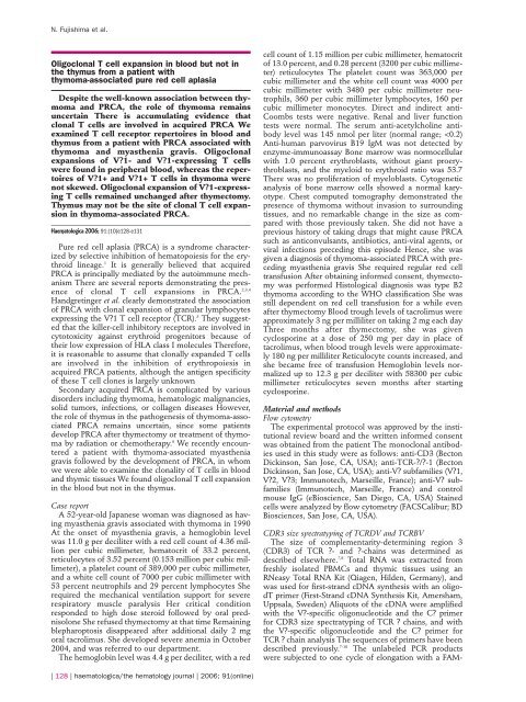
Figure 1. TCRBV and TCRDV repertoires before and after thymectomy<br />
and CyA <strong>the</strong>rapy. T <strong>cell</strong> repertoires were analyzed by flow<br />
cytometry. V?1+??T <strong>cell</strong>, V?1+ and V?9+ ??T (arrows) <strong>in</strong> peripheral<br />
<strong>blood</strong> were expanded more than that <strong>in</strong> normal <strong>in</strong>dividuals at <strong>the</strong><br />
onset of PRCA. Frequencies of V?1-, V?1- and V?9- express<strong>in</strong>g<br />
peripheral <strong>blood</strong> T <strong>cell</strong>s <strong>in</strong> healthy <strong>in</strong>dividuals were 1.2 ± 0.5 percent<br />
(19 ± 14 per cubic millimeter), 3.9 ± 1.0 percent (64 ± 35<br />
per cubic millimeter), and 3.4 ± 1.1 percent (51 ± 6 per cubic millimeter)<br />
(mean ± SD of 3 samples), respectively. The first flow<br />
cytometric analysis of TCR repertoires was performed one month<br />
after <strong>the</strong> onset of PRCA.<br />
labeled C? or C? primer The labeled PCR products were<br />
electrophoresed on acrylamide sequenc<strong>in</strong>g gels <strong>in</strong> an<br />
automated DNA sequencer (ABI 377, Perk<strong>in</strong>-Elmer<br />
Applied Biosystems, Foster, CA, USA), followed by<br />
analysis us<strong>in</strong>g GeneScan software (Perk<strong>in</strong>-Elmer)<br />
Sequenc<strong>in</strong>g of CDR3 regions<br />
PCR products were cloned <strong>in</strong>to <strong>the</strong> PCR2.1 TA clon<strong>in</strong>g<br />
vector (Invitrogen, Carlsbad, CA) and were sequenced<br />
us<strong>in</strong>g a Big-Dye Term<strong>in</strong>ator Cycle Sequenc<strong>in</strong>g Kit<br />
(Perk<strong>in</strong>-Elmer) Sequence analysis was performed us<strong>in</strong>g an<br />
automated DNA sequencer.<br />
Results<br />
TCRBV and TCRDV repertoires of T lymphocytes <strong>in</strong> <strong>blood</strong> and<br />
thymoma<br />
We first exam<strong>in</strong>ed <strong>the</strong> usage of TCRBV and TCRDV<br />
subfamilies by peripheral <strong>blood</strong> T lymphocytes to determ<strong>in</strong>e<br />
whe<strong>the</strong>r <strong>the</strong>re was a skew of TCR variable region<br />
repertoires As compared to those of healthy <strong>in</strong>dividuals,<br />
<strong>the</strong> usages of V?1, V?1 and V?9 segments were <strong>in</strong>creased<br />
(Figure 1A and 1B) 11,12,13 V?1+ ??T <strong>cell</strong>s accounted for 18.8<br />
Figure 2. CDR3 size distri<strong>but</strong>ion patterns of T lymphocytes <strong>in</strong><br />
peripheral <strong>blood</strong> and thymoma. V?1- and V?1- express<strong>in</strong>g T <strong>cell</strong>s<br />
were skewed <strong>in</strong> <strong>blood</strong> <strong>but</strong> <strong>not</strong> <strong>in</strong> <strong>the</strong> thymoma (2A). Skewed pattern<br />
of <strong>the</strong> V?1 TCR repertoire were persist<strong>in</strong>g even after successful<br />
treatment with thymectomy and cyclospor<strong>in</strong>e (2B).<br />
percent of <strong>the</strong> CD3 + T <strong>cell</strong> population with an absolute<br />
number of 487 per cubic millimeter Whereas V?2+ ??T<br />
<strong>cell</strong>s, that are usually <strong>the</strong> major subtype of circulat<strong>in</strong>g ??T<br />
<strong>cell</strong>s, accounted for only 0.5 percent with an absolute<br />
number of 15 per cubic millimeter V?1+ and V?9+ ??T<br />
<strong>cell</strong>s accounted for 19.6 percent (506 per cubic millimeter),<br />
and 12.8 percent (332 per cubic millimeter) respectively<br />
CDR3 size distri<strong>but</strong>ion analysis demonstrated that<br />
V?1+ and V?1+ T <strong>cell</strong>s were comprised of oligoclonally<br />
expanded clones (Figure 2A) In contrast, <strong>the</strong> diversity of<br />
V?1+ and V?1+ T <strong>cell</strong>s <strong>in</strong> <strong>the</strong> <strong>thymus</strong> were polyclonal<br />
<strong>Oligoclonal</strong> <strong>expansion</strong> of T <strong>cell</strong>s <strong>in</strong> <strong>blood</strong> <strong>but</strong> <strong>not</strong> <strong>in</strong> <strong>the</strong><br />
thymoma was also confirmed by sequenc<strong>in</strong>g analysis of<br />
<strong>the</strong> CDR3 region of TCR ?- and ?-cha<strong>in</strong>s (Table 1).<br />
Longitud<strong>in</strong>al analysis of TCR repertoires after thymectomy and<br />
CyA <strong>the</strong>rapy<br />
We studied <strong>the</strong> longitud<strong>in</strong>al k<strong>in</strong>etics of T <strong>cell</strong> repertoires<br />
before and after thymectomy and cyclospor<strong>in</strong>e<br />
<strong>the</strong>rapy The numbers of V?1+, V?1+ and V?9+ T <strong>cell</strong>s,<br />
which were expanded <strong>in</strong> <strong>the</strong> present case, decreased<br />
along with treatment (Figure 1A) Although <strong>the</strong> absolute<br />
numbers of expanded V?1+ and V?1+ T <strong>cell</strong>s were<br />
reduced after thymectomy and cyclospor<strong>in</strong>e <strong>the</strong>rapy,<br />
<strong>the</strong>se subsets rema<strong>in</strong>ed predom<strong>in</strong>ant <strong>in</strong> circulat<strong>in</strong>g <strong>blood</strong><br />
(Figure 1B) Moreover, <strong>the</strong> skewed diversity of V?1+ T<br />
<strong>cell</strong>s rema<strong>in</strong>ed <strong>the</strong> same (Figure 2B), <strong>in</strong>dicat<strong>in</strong>g that oligoclonally<br />
expanded T <strong>cell</strong>s were still present dur<strong>in</strong>g hematological<br />
remission.<br />
haematologica/<strong>the</strong> hematology journal | 2006; 91(onl<strong>in</strong>e) | 129 |
N. Fujishima et al.<br />
Table 1. <strong>Oligoclonal</strong>ly expanded T <strong>cell</strong>s <strong>in</strong> <strong>blood</strong> were <strong>not</strong> detected <strong>in</strong> <strong>the</strong><br />
<strong>thymus</strong>.<br />
TCR V segment n-D-n J segment ColonyFrequency<br />
Blood Thymoma<br />
TCRDV1 YFCALGE LGPYGTGGS YTDKLIFGKG 7/32 0/31<br />
YFCALGE LGPGGYQIP YTDKLIFGKG 3/32 0/31<br />
YFCALGE LVGIPSPTGG YTDKLIFGKG 3/32 0/31<br />
TCRBV1 LYFCASSV GGEA GELFFGEG 11/13 0/13<br />
Naohito Fujishima*, Makoto Hirokawa*, Masumi Fujishima*, Chizu<br />
Wada**, Itaru Toyoshima**, Sumio Watanabe** and Ken-ichi Sawada*<br />
Division of *Hematology and Oncology, and Division of **Neurology,<br />
Department of Medic<strong>in</strong>e, Akita University School of Medic<strong>in</strong>e<br />
Key words; V?1+??T <strong>cell</strong>s, thymoma, pure red <strong>cell</strong> aplasia (PRCA)<br />
Correspondence: Naohito Fujishima, M.D., Division of Hematology and<br />
Oncology, Department of Medic<strong>in</strong>e, Akita University School of Medic<strong>in</strong>e,<br />
1-1-1 Hondo, Akita Japan 010-8543<br />
Tel: +81-18-884-6116 Fax: +81-18-836-2613<br />
E-mail: naofuji@doc.med.akita-u.ac.jp<br />
Discussion<br />
We presented a case of thymoma-associated PRCA<br />
with oligoclonal <strong>expansion</strong>s of TCR?? and TCR?? T lymphocytes<br />
<strong>in</strong> <strong>blood</strong> <strong>but</strong> <strong>not</strong> <strong>in</strong> <strong>the</strong> <strong>thymus</strong> Mamiya et al.<br />
previously reported that 17 of 150 patients were complicated<br />
with thymoma, and that 10 patients developed<br />
PRCA after thymectomy or treatment of thymoma<br />
<strong>in</strong>clud<strong>in</strong>g radiation or chemo<strong>the</strong>rapy. 6 O<strong>the</strong>r groups also<br />
reported that 4 patients with myas<strong>the</strong>nia gravis developed<br />
PRCA after thymectomy. 14 These f<strong>in</strong>d<strong>in</strong>gs raise <strong>the</strong><br />
question of whe<strong>the</strong>r thymoma is <strong>in</strong>deed <strong>in</strong>volved <strong>in</strong> <strong>the</strong><br />
pathogenesis of PRCA. To date, <strong>the</strong>re are few reports<br />
describ<strong>in</strong>g <strong>the</strong> T <strong>cell</strong> repertoire <strong>in</strong> thymoma-associated<br />
PRCA We found a report describ<strong>in</strong>g <strong>the</strong> oligoclonal T <strong>cell</strong><br />
<strong>expansion</strong> <strong>in</strong> thymoma <strong>but</strong> <strong>not</strong> <strong>in</strong> peripheral <strong>blood</strong> <strong>in</strong><br />
contrast to <strong>the</strong> present case 15 Type B2 thymoma has been<br />
previously reported to be associated with myas<strong>the</strong>nia<br />
gravis, <strong>but</strong> PRCA is <strong>not</strong> a common complication. 16<br />
Although we expected that oligoclonally expanded T<br />
<strong>cell</strong>s would be also detected <strong>in</strong> <strong>the</strong> thymoma, our f<strong>in</strong>d<strong>in</strong>gs<br />
suggest that oligoclonal T <strong>cell</strong>s <strong>in</strong> <strong>the</strong> present case<br />
were selected and expanded <strong>in</strong> peripheral tissues.<br />
There are some reports suggest<strong>in</strong>g <strong>the</strong> pathological<br />
roles of V?1+ ??T <strong>cell</strong>s <strong>in</strong> GLPD-associated PRCA 4,5,17 Hara<br />
et al. have also demonstrated that ??T <strong>cell</strong>s mediate an<br />
<strong>in</strong>hibition of erythropoiesis <strong>in</strong> type I autoimmune polyglandular<br />
syndrome. 18 This case also showed <strong>the</strong> oligoclonal<br />
<strong>expansion</strong> of V?1+ ??T <strong>cell</strong>s <strong>in</strong> peripheral <strong>blood</strong> At<br />
present, we do <strong>not</strong> have direct evidence for this T <strong>cell</strong><br />
subset <strong>in</strong> <strong>the</strong> <strong>in</strong>hibition of erythropoiesis No obvious<br />
improvement of anemia was observed dur<strong>in</strong>g three<br />
months after thymectomy, while a global reduction of T<br />
lymphocytes <strong>in</strong> <strong>blood</strong> was seen after thymectomy and<br />
absolute numbers of oligoclonally expanded V?1 and V?1<br />
T <strong>cell</strong>s also decreased However, oligoclonal <strong>expansion</strong>s of<br />
T <strong>cell</strong>s are still present even after achiev<strong>in</strong>g complete<br />
hematological response S<strong>in</strong>ce cyclospor<strong>in</strong>e is a functional<br />
<strong>in</strong>hibitor of T <strong>cell</strong>s, cyclospor<strong>in</strong>e can <strong>in</strong>hibit <strong>the</strong> function<br />
of pathogenic T lymphocytes <strong>but</strong> <strong>not</strong> eradicate those<br />
<strong>cell</strong>s.<br />
It would be an <strong>in</strong>terest<strong>in</strong>g to address why this patient<br />
developed PRCA dur<strong>in</strong>g immunosuppressive <strong>the</strong>rapy<br />
with tacrolimus and prednisolone, <strong>the</strong>n showed amelioration<br />
after tak<strong>in</strong>g cyclospor<strong>in</strong>e There is a report describ<strong>in</strong>g<br />
<strong>the</strong> patient develop<strong>in</strong>g PRCA on tacrolimus after liver<br />
transplantation. 19 However, <strong>the</strong>re is also a report that<br />
tacrolimus was effective for PRCA. 20 In any case, it is possible<br />
that a sufficient dose of cyclospor<strong>in</strong>e was given to<br />
control <strong>the</strong> disease <strong>in</strong> <strong>the</strong> present case <strong>but</strong> <strong>the</strong> dose of<br />
tacrolimus might <strong>not</strong> have been sufficient.<br />
In summary, <strong>the</strong> present case implies that <strong>the</strong> <strong>thymus</strong><br />
may <strong>not</strong> be <strong>the</strong> site of clonal T <strong>cell</strong> <strong>expansion</strong> <strong>in</strong> thymoma-associated<br />
PRCA, and fur<strong>the</strong>r <strong>in</strong>vestigations are necessary<br />
to address this issue Clonal <strong>expansion</strong> of T <strong>cell</strong>s <strong>in</strong><br />
<strong>the</strong> marrow and erythroid suppressive effects of autologous<br />
T <strong>cell</strong>s need to <strong>the</strong> studied <strong>in</strong> <strong>the</strong> future.<br />
References<br />
1. Dessypris EN, et al: In: Greer JP, et al (eds): W<strong>in</strong>trobe’s Cl<strong>in</strong>ical<br />
Hematology, 11th ed, Lipp<strong>in</strong>cott Williams & Wilk<strong>in</strong>s,<br />
Philadelphia, London, p1421-1427, 2004.<br />
2. Masuda M, Arai Y, Okamura T, Mizoguchi H. Pure red <strong>cell</strong><br />
aplasia with thymona: evidence of T-<strong>cell</strong> clonal disorder. Am J<br />
Hematol. 1997;54:324-328.<br />
3. Masuda M, Saitoh H, Mizoguchi H. Clonality of acquired primary<br />
pure red <strong>cell</strong> aplasia. Am J Hematol. 1999;62:193-195.<br />
4. Masuda M, Teramura M, Matsuda A, Bessho M, Shimamoto<br />
T, Ohyashiki K, et al. Clonal T <strong>cell</strong>s of pure red-<strong>cell</strong> aplasia.<br />
Am J Hematol. 2005;79:332-333.<br />
5. Handgret<strong>in</strong>ger R, Geiselhart A, Moris A, Grau R, Teuffel O,<br />
Bethge W, et al. Pure red-<strong>cell</strong> aplasia associated with clonal<br />
<strong>expansion</strong> of granular lymphocytes express<strong>in</strong>g killer-<strong>cell</strong><br />
<strong>in</strong>hibitory receptors. N Engl J Med. 1999;340:278-284.<br />
6. Mamiya S, Itoh T, Miura AB. Acquired pure red <strong>cell</strong> aplasia <strong>in</strong><br />
Japan. Eur J Haematol. 1997;59:199-205.<br />
7. Horiuchi T, Hirokawa M, Satoh K, Kitabayashi A, Muira AB.<br />
Clonal <strong>expansion</strong> of gammadelta-T lymphocytes <strong>in</strong> an HTLV-<br />
I carrier, associated with chronic neutropenia and rheumatoid<br />
arthritis. Ann Hematol. 1999;78:101-104.<br />
8. Hirokawa M, Horiuchi T, Kawabata Y, Kitabayashi A, Miura<br />
AB. Reconstitution of gammadelta T <strong>cell</strong> repertoire diversity<br />
after human allogeneic hematopoietic <strong>cell</strong> transplantation and<br />
<strong>the</strong> role of peripheral <strong>expansion</strong> of mature T <strong>cell</strong> population <strong>in</strong><br />
<strong>the</strong> graft. Bone Marrow Transplant. 2000;26:177-185.<br />
9. Saitoh H, Hirokawa M, Fujishima N, Ichikawa Y, Kawabata Y,<br />
Miura I, et al. The presence and longevity of peripherally<br />
expanded donor-derived TCRalphabeta+ mature T lymphocyte<br />
clones after allogeneic bone marrow transplantation for<br />
adult myeloid leukemias. Leukemia. 2003;17:1626-1635.<br />
10. Hirokawa M, Horiuchi T, Kitabayashi A, Kawabata Y,<br />
Matsutani T, Suzuki R, et al. Delayed recovery of CDR3 complexity<br />
of <strong>the</strong> T-<strong>cell</strong> receptor-beta cha<strong>in</strong> <strong>in</strong> recipients of allogeneic<br />
bone marrow transplants who had virus-associated<br />
<strong>in</strong>terstitial pneumonia: monitor of T-<strong>cell</strong> function by CDR3<br />
spectratyp<strong>in</strong>g. J Allergy Cl<strong>in</strong> Immunol. 2000;106:S32-39.<br />
11. Matsutani T, Yoshioka T, Tsuruta Y, Iwagami S, Toyosaki-<br />
Maeda T, Horiuchi T, et al. Restricted usage of T-<strong>cell</strong> receptor<br />
alpha-cha<strong>in</strong> variable region (TCRAV) and T-<strong>cell</strong> receptor betacha<strong>in</strong><br />
variable region (TCRBV) repertoires after human allogeneic<br />
haematopoietic transplantation. Br J Haematol.<br />
2000;109:759-769.<br />
12. Parker CM, Groh V, Band H, Por<strong>cell</strong>i SA, Morita C, Fabbi M,<br />
et al. Evidence for extrathymic changes <strong>in</strong> <strong>the</strong> T <strong>cell</strong> receptor<br />
gamma/delta repertoire. J Exp Med. 1990;171:1597-1612.<br />
13. Dechanet J, Merville P, Berge F, Bone-Mane G, Taup<strong>in</strong> JL,<br />
Michel P, et al. Major <strong>expansion</strong> of gammadelta T lymphocytes<br />
follow<strong>in</strong>g cytomegalovirus <strong>in</strong>fection <strong>in</strong> kidney allograft<br />
recipients. J Infect Dis. 1999;179:1-8.<br />
14. Suzuki S, Nogawa S, Tanaka K, Koto A, Fukuuchi Y, Kuwana<br />
M. Initial predictors of development of pure red <strong>cell</strong> aplasia <strong>in</strong><br />
myas<strong>the</strong>nia gravis after thymectomy. Cl<strong>in</strong> Neurol Neurosurg.<br />
2003;106:16-18.<br />
15. Handa SI, Schofield KP, Sivakumaran M, Short M, Pumphrey<br />
RS. Pure red <strong>cell</strong> aplasia associated with malignant thymoma,<br />
myas<strong>the</strong>nia gravis, polyclonal large granular lymphocytosis<br />
and clonal thymic T <strong>cell</strong> <strong>expansion</strong>. J Cl<strong>in</strong> Pathol. 1994;47:676-<br />
679.<br />
16. Kuo T, Shih LY. Histologic types of thymoma associated with<br />
pure red <strong>cell</strong> aplasia: a study of five cases <strong>in</strong>clud<strong>in</strong>g a composite<br />
tumor of organoid thymoma associated with an unusual<br />
lipofibroadenoma. Int J Surg Pathol. 2001;9:29-35.<br />
17. Kondo H, Uematsu M, Watanabe J, Takahashi Y, Hayashi K,<br />
Iwasaki H. CD3+, CD4-, CD8-, TCR alpha beta-, TCR gamma<br />
delta+ granular lymphocyte proliferative disorder without<br />
| 130 | haematologica/<strong>the</strong> hematology journal | 2006; 91(onl<strong>in</strong>e)
haematologica onl<strong>in</strong>e 2006<br />
lymphocytosis and cl<strong>in</strong>ical symptoms. Acta Haematol.<br />
2000;104:54-56.<br />
18. Hara T, Mizuno Y, Nagata M, Okabe Y, Taniguchi S, Harada<br />
M, et al. Human gamma delta T-<strong>cell</strong> receptor-positive <strong>cell</strong>mediated<br />
<strong>in</strong>hibition of erythropoiesis <strong>in</strong> vitro <strong>in</strong> a patient with<br />
type I autoimmune polyglandular syndrome and pure red<br />
<strong>blood</strong> <strong>cell</strong> aplasia. Blood. 1990;75:941-950.<br />
19. Misra S, Moore TB, Ament ME, Busuttil RW, McDiarmid SV.<br />
Red <strong>cell</strong> aplasia <strong>in</strong> children on tacrolimus after liver transplantation.<br />
Transplantation. 1998;65:575-577.<br />
20. Yoshida S, Konishi T, Nishizawa T, Yoshida Y. Effect of<br />
tacrolimus <strong>in</strong> a patient with pure red-<strong>cell</strong> aplasia. Cl<strong>in</strong> Lab<br />
Haematol. 2005;27:67-69.<br />
haematologica/<strong>the</strong> hematology journal | 2006; 91(onl<strong>in</strong>e) | 131 |