Sudden Death in Addison's Disease - Healthcare Bulletin
Sudden Death in Addison's Disease - Healthcare Bulletin
Sudden Death in Addison's Disease - Healthcare Bulletin

Create successful ePaper yourself
Turn your PDF publications into a flip-book with our unique Google optimized e-Paper software.
SUDDEN DEATH IN ADDISON’S DISEASE: LEAD POISONING-LIKE GUM APPEARANCE<br />
The autopsy was performed on 3 November 2009 (21 hours after<br />
the pronounced death). The body was of a middle aged, poor nourished<br />
female, 153 cm <strong>in</strong> length with short black hair. Axillary and<br />
pubic hair was completely absent. Her sk<strong>in</strong> looked generally dark<br />
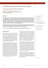
especially on the palmar creases, all jo<strong>in</strong>t areas, and lips. The upper<br />
gum showed bluish-black patches, which looked like a lead l<strong>in</strong>e <strong>in</strong><br />
lead poison<strong>in</strong>g (Figure 1).<br />
Figure 1: Hyperpigmentation of the gum mimick<strong>in</strong>g the<br />
lead l<strong>in</strong>e <strong>in</strong> lead poison<strong>in</strong>g<br />
The toxicological analysis was negative. Lead <strong>in</strong> the blood sample<br />
was negative. Postmortem blood cortisol was 0.86 µg/dL (range<br />
5-25 AM, 2.5-12.5 PM).<br />
Unfortunately, anti-adrenal and anti-thyroid antibodies could not<br />
be analysed <strong>in</strong> Thailand. Bra<strong>in</strong>, thyroid gland, heart, lung and kidney<br />
tissues were submitted for microscopic exam<strong>in</strong>ation: The cardiac<br />
muscles showed hypertrophy with a small focal area of fibrosis.<br />
Some focal haemorrhages were present <strong>in</strong> the subepicardium and<br />
the myocardium. There were random contraction band necroses.<br />
The thyroid gland showed Hashimoto thyroditis. There was pulmonary<br />
oedema with some focal hemorrhages <strong>in</strong> the lungs. The bra<strong>in</strong><br />
tissue was unremarkable.<br />
DISCUSSION<br />
Fatal Addison’s disease is rarely found <strong>in</strong> forensic practice, especially<br />
<strong>in</strong> Northern Thailand. <strong>Sudden</strong> death from Addison’s disease<br />
has been reported (9-12) but mostly <strong>in</strong> Caucasians. Several studies<br />
showed that Addison’s disease could only be diagnosed dur<strong>in</strong>g autopsy<br />
(9-12) . The most specific sign of primary adrenal <strong>in</strong>sufficiency<br />
is hyperpigmentation of the sk<strong>in</strong> and mucosal surfaces which is due<br />
to the high plasma corticotroph<strong>in</strong> concentrations that occur as a result<br />
of a decrease of cortisol feedback (4) . Malaise, hyperpigmented<br />
sk<strong>in</strong>, hypotension and hyponatremia <strong>in</strong> our case were clues for diagnosis<br />
of chronic primary adrenal <strong>in</strong>sufficiency. However, the dark<br />
l<strong>in</strong>e on the gum may be mistaken as the lead l<strong>in</strong>e <strong>in</strong> lead poison<strong>in</strong>g.<br />
Eur J Cardiovasc Med © <strong>Healthcare</strong> Bullet<strong>in</strong> 2011<br />
There was no wound or <strong>in</strong>jection mark on the sk<strong>in</strong>. The <strong>in</strong>ternal exam<strong>in</strong>ation<br />
showed no evidence of vital organs <strong>in</strong>jury. The bra<strong>in</strong> had<br />
no pathological lesion. The pituitary fossa had no abnormal mass.<br />
The thyroid gland was normal shape and weighed 15 g. The airways<br />
showed no edema or foreign body obstruction. Both lungs showed<br />
mild edema with left upper lung consolidation. There was no pulmonary<br />
thromboembolism. The right and the left lung weighed<br />
370 g and 470 g, respectively. There were some petechiae on the<br />
anterior external surface of the heart. The left anterior descend<strong>in</strong>g<br />
coronary artery showed 10% stenosis. The right ma<strong>in</strong> coronary artery<br />
was widely patent. The left ventricular free wall thickness was<br />
12 mm. Neither valvular abnormality nor congenital anomaly was<br />
observed. The heart weighed 290 g and had a normal shape.<br />
There was no evidence of peritonitis. The liver, spleen, small bowel,<br />
large bowel and pancreas had no significant gross pathologic abnormality.<br />
Both kidneys showed a diffuse micronodular surface.<br />
No evidence of acute pyelonephritis was detected. The adrenal<br />
gland was searched for <strong>in</strong> the suprarenal areas and <strong>in</strong> other areas<br />
<strong>in</strong>clud<strong>in</strong>g chest wall, but could not be identified grossly. There was<br />
no fibrosis at the suprarenal areas. The retroperitoneal region had<br />
no blood collection. The pelvic organs showed no significant gross<br />
lesion. There were 10 millilitres of light green mucous-mixed liquid<br />
<strong>in</strong> the stomach. The mucosa of the stomach showed generalised<br />
gastritis. The femoral blood, heart blood and gastric contents were<br />
submitted for toxicological analysis at the Regional Medical Science<br />
Center, Chiang Mai prov<strong>in</strong>ce.<br />
Corticotrop<strong>in</strong> stimulation is the most commonly used test for the<br />
diagnosis of primary adrenal <strong>in</strong>sufficiency (4) , but it cannot be performed<br />
postmortem. However, a very low level of plasma cortisol (3<br />
or less µg/dL) confirmed adrenal <strong>in</strong>sufficiency (4) . A previous study<br />
showed that serum cortisol rema<strong>in</strong>s constant dur<strong>in</strong>g the early postmortem<br />
period (13) . This supports that the very low level of postmortem<br />
blood cortisol is due to severe adrenal <strong>in</strong>sufficiency <strong>in</strong> this<br />
case. The absence of the adrenal gland <strong>in</strong> our case may be caused<br />
by severe atrophy.<br />
However, the detection of blood cortisol <strong>in</strong>dicates some rema<strong>in</strong><strong>in</strong>g<br />
cortisol secret<strong>in</strong>g tissue. Autoimmune adrenalitis is the ma<strong>in</strong> cause<br />
of Addison’s disease and may occur alone or as a component of<br />
type I or II autoimmune polyglandular syndrome (2, 4, 6) . The Hashimoto<br />
thyroiditis of this case <strong>in</strong>dicated that the autoimmune disease<br />
was the probable cause of adrenocortical <strong>in</strong>sufficiency.<br />
Autoantibodies aga<strong>in</strong>st 21-hydroxylase, one of the enzymes <strong>in</strong><br />
steroid biosynthesis <strong>in</strong>side the adrenal glands, can be found <strong>in</strong> approximately<br />
80% of the Addisonian persons. These autoantibodies<br />
thus clearly correlate with the disease (14) and are useful for its diagnosis.<br />
Approximately 21% of persons positive for adrenal cortex<br />
autoantibodies (ACA) developed overt Addison’s disease with<strong>in</strong><br />
5.2 years, while negative ACA persons ma<strong>in</strong>ta<strong>in</strong>ed normal adrenal<br />
function dur<strong>in</strong>g the observation period (15) . ACA is also an additional<br />
marker to predict Addison’s disease. In conclusion, cl<strong>in</strong>icians<br />
should not overlook hyperpigmentation of the sk<strong>in</strong> comb<strong>in</strong>ed with<br />
other significant cl<strong>in</strong>ical signs and basic laboratory tests for the correct<br />
diagnose of adrenal <strong>in</strong>sufficiency which is potentially fatal if<br />
not recognised and promptly treated.<br />
EUROPEAN JOURNAL OF CARDIOVASCULAR MEDICINE<br />
VOL I ISSUE III<br />
39