Crystal-Induced Arthritis - HealthPlexus.net
Crystal-Induced Arthritis - HealthPlexus.net
Crystal-Induced Arthritis - HealthPlexus.net

Create successful ePaper yourself
Turn your PDF publications into a flip-book with our unique Google optimized e-Paper software.
<strong>Arthritis</strong><br />
<strong>Crystal</strong>-<strong>Induced</strong><br />
<strong>Arthritis</strong><br />
Simon H.K. Huang, MD, FRCPC, Clinical Associate Professor, Division of<br />
Rheumatology, Faculty of Medicine, University of British Columbia,Vancouver, BC.<br />
Ian K.Tsang, MB, FRCPC, Clinical Professor Emeritus, Division of Rheumatology, Faculty<br />
of Medicine, University of British Columbia,Vancouver, BC.<br />
The two most common forms of crystal-induced arthritis among older adults are gout<br />
and calcium pyrophosphate dihydrate (CPPD) deposition disease. Gout in older adults<br />
has unique clinical features. The new case incidence is the same in males and females<br />
over age 60. Upper limb and polyarticular involvement are not unusual. CPPD deposition<br />
disease may present as asymptomatic chondrocalcinosis on radiographs and symptomatically<br />
as pseudogout, pseudo–rheumatoid arthritis, or pseudo-osteoarthritis. Other<br />
crystals may cause periarthritis or arthritis. Management of crystal-induced arthritis<br />
among older adults requires special considerations due to comorbid conditions and concomitant<br />
medications. Nonsteroidal anti-inflammatory drugs may be contraindicated.<br />
Steroids taken either orally or intra-articularly are often an alternative.<br />
Key words: gout, chondrocalcinosis, pseudogout, pseudo–rheumatoid arthritis, pseudoosteoarthritis<br />
Introduction<br />
The most common forms of crystalinduced<br />
arthritis are caused by deposition<br />
of monosodium urate (MSU), as in<br />
gout, and calcium pyrophosphate dihydrate<br />
(CPPD), as in pseudogout, pseudo–rheumatoid<br />
arthritis, and<br />
pseudo-osteoarthritis). Other crystals,<br />
such as basic calcium phosphate, primarily<br />
hydroxyapatite, and calcium oxalate,<br />
may induce an inflammatory reaction in<br />
and around the joint. This article discusses<br />
the clinical characteristics of gout,<br />
CPPD deposition disease, and other crystal-induced<br />
arthritis/periarthritis among<br />
older adults.<br />
Gout<br />
Incidence and Prevalence<br />
The prevalence of gout in the US population<br />
is
<strong>Crystal</strong>-<strong>Induced</strong> <strong>Arthritis</strong><br />
osteoarthritis is almost universally present<br />
in older persons. Third, the presence<br />
or absence of hyperuricemia does not<br />
establish or exclude the diagnosis of gout.<br />
Management<br />
The management of gout can be divided<br />
into the management of acute gout, intercritical<br />
gout, and tophaceous gout, and<br />
the management of risk factors associated<br />
with gout or hyperuricemia.<br />
Acute Gout<br />
Colchicine and nonsteroidal anti-inflammatory<br />
drugs (NSAIDs) are the standard<br />
pharmacological treatment for acute<br />
gout. However, due to comorbid conditions<br />
experienced by older adults, such<br />
as renal insufficiency, hypertension, and<br />
cardiovascular disease, and the use of<br />
concomitant medications such as antihypertensives<br />
and anticoagulants, NSAIDs<br />
should be avoided or used with great<br />
care. If NSAIDs cannot be used, an intraarticular<br />
steroid or a short course of<br />
tapering oral prednisone may be an alternative.<br />
While there are many dosage<br />
schedules, in our experience, a short<br />
course of prednisone starting at 30 mg<br />
tapering by 5 mg every 2 days is usually<br />
very effective. On the rare occasion when<br />
a steroid is contraindicated, such as when<br />
labile diabetes is present, analgesics<br />
including opioids may be used until the<br />
acute inflammation settles.<br />
Intercritical Gout<br />
The management of acute episodes of<br />
intercritical gout is the same as that for<br />
acute gout. However, if the frequency of<br />
acute episodes exceeds three over a 12-<br />
month period, efforts should be taken to<br />
lower the serum uric acid to prevent further<br />
attacks as the uric acid level correlates<br />
with the frequency of acute<br />
episodes. We have found that the optimal<br />
serum uric acid level to be achieved is<br />
between 250 and 300 µmol/L. To lower<br />
the serum uric acid level, attention<br />
should be directed toward eliminating or<br />
avoiding risk factors. As most of these<br />
factors are difficult to modify, especially<br />
among older adults, pharmacotherapy to<br />
lower the serum uric acid level is often<br />
Table 1: 1997 Criteria for the Classification of Acute <strong>Arthritis</strong> of Primary<br />
Gout<br />
1. More than one attack of acute arthritis<br />
2. Maximum inflammation developed within 1 day<br />
3. Monoarthritis attack<br />
4. Redness observed over joints<br />
5. First metatarsophalangeal joint painful or swollen<br />
6. Unilateral first metatarsophalangeal joint attack<br />
7. Unilateral tarsal joint attack<br />
8. Tophus (proven or suspected)<br />
9. Hyperuricemia<br />
10. Asymmetrical swelling within a joint on radiograph*<br />
11. Subcortical cysts without erosions on radiograph<br />
12. Monosodium urate monohydrate microcrystals in joint fluid during attack<br />
13. Joint fluid culture negative for organisms during attack<br />
*This criterion could logically be found on examination as well as on radiographs. However, the protocol did<br />
not request this information in regard to examination.<br />
Source: Adapted from Wallace SL et al., 1997. 5<br />
necessary.<br />
Two classes of medications are currently<br />
available to accomplish this—uricosuric<br />
agents and allopurinol, a<br />
xanthine oxidase inhibitor. Uricosuric<br />
agents are most effective for those with<br />
normal renal function with a high urine<br />
output, and in the absence of renal calculi;<br />
they are therefore difficult to use,<br />
especially among older adults. Thus,<br />
allopurinol becomes the hypouricemic<br />
drug of choice. As a rapid change in the<br />
serum uric acid level may precipitate an<br />
acute attack, this drug should be started<br />
at a low dose, 50–100 mg daily, titrating<br />
upwards every 3–4 weeks by 50–100 mg<br />
until the optimal uric acid level is<br />
achieved (between 250 and 300 µmol/L).<br />
As the serum uric acid level and the<br />
metabolism of allopurinol are dependent<br />
on the glomerular filtration rate of the<br />
kidney, periodic assessments of renal<br />
function and uric acid are required. For<br />
those who are allergic to allopurinol, the<br />
use of uricosuric agents or a trial of allopurinol<br />
desensitization may be appropriate.<br />
7 Newer hypouricemic drugs, for<br />
example, febuxostat, are currently under<br />
study but not yet available. Finally,<br />
agents to lower the serum uric acid level<br />
should only be initiated or altered after<br />
the acute attack has settled as lowering<br />
the uric acid during an acute phase may<br />
prolong the attack.<br />
Tophaceous Gout<br />
The management goals in tophaceous<br />
gout are to control the ongoing or recurrent<br />
joint inflammation and to lower the<br />
serum uric acid level to prevent acute<br />
episodes, decrease the chance of renal<br />
dysfunction, and reduce or eliminate the<br />
tophi. The management of tophaceous<br />
gout is the same as that of intercritical<br />
gout. However, due to the ongoing<br />
inflammation, it is often necessary to<br />
keep the patients on low-dose colchicines<br />
such as 0.6 mg once or twice per day or<br />
less, depending on the renal function;<br />
an optimal dose of NSAID (if not con-<br />
442 GERIATRICS & AGING • September 2008 • Volume 11, Number 8
<strong>Crystal</strong>-<strong>Induced</strong> <strong>Arthritis</strong><br />
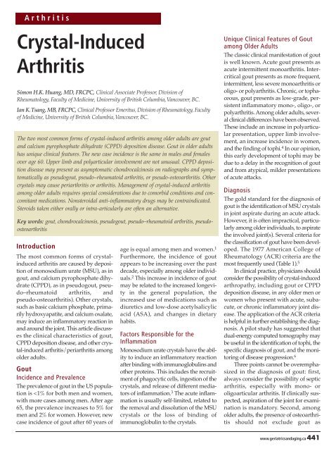
Figure 1:<br />
CPPD Deposition Disease<br />
Healthy Joint<br />
Joint with <strong>Crystal</strong> Deposits<br />
Bone<br />
Joint capsule<br />
Swollen<br />
joint<br />
Synovium<br />
Synovial<br />
fluid<br />
Inflamed<br />
synovium<br />
<strong>Crystal</strong> deposits<br />
within cartilage<br />
Cartilage<br />
Common Sites of the Disease<br />
Femur<br />
Phalanges<br />
Knee joint<br />
Tibia<br />
Fibula<br />
Carpals<br />
Ulna<br />
Metacarpals<br />
Radius<br />
www.geriatricsandaging.ca 443
<strong>Crystal</strong>-<strong>Induced</strong> <strong>Arthritis</strong><br />
Key Points<br />
Septic arthritis should always be considered, with acute mono- or oligoarticular arthritis.<br />
Physicians should consider the possibility of crystal-induced arthropathy, including gout<br />
or CPPD deposition disease, in any older men or women who present with acute, subacute,<br />
or chronic inflammatory joint disease.<br />
Pharmacological treatment for crystal-induced arthritis in older adults deserves special<br />
attention due to comorbid conditions and the use of concomitant medications.<br />
traindicated); or low-dose prednisone<br />
such as 5 mg/d. If steroid use is anticipated<br />
to be >12 weeks, osteoporosis prevention<br />
should be initiated.<br />
Risk Factors Associated with Gout<br />
or Hyperuricemia<br />
Risk factors such as age, gender, and ethnicity<br />
are not modifiable. Modifiable factors<br />
include obesity, diet, alcohol<br />
consumption, and medications. 8 A highprotein,<br />
purine-rich diet and alcohol consumption<br />
are associated with<br />
hyperuricemia and gout. Fruit, vegetables,<br />
nuts, and legumes are neutral, while<br />
plant oils and multiple vitamins may<br />
decrease the uric acid level. Common<br />
medications that increase serum uric acid<br />
include diuretics and low-dose ASA. As<br />
fluctuating uric acid levels often induce<br />
acute gouty arthritis, a steady serum uric<br />
acid level should be maintained.<br />
Calcium Pyrophosphate<br />
Dihydrate Deposition Disease<br />
The hallmark of CPPD deposition disease<br />
is the deposition of CPPD crystals<br />
within hyaline or fibrocartilage within<br />
joints (chondrocalcinosis) (Figure 1). It<br />
may present as acute arthritis as in gout<br />
(pseudo-gout), chronic low-grade inflammation<br />
resembling rheumatoid arthritis<br />
(pseudo–rheumatoid arthritis), or chronic<br />
noninflammatory arthritis resembling<br />
osteoarthritis (pseudo-osteoarthritis), or<br />
as asymptomatic articular cartilage deposition<br />
found incidentally on radiographs.<br />
Incidence and Prevalence<br />
As the presentation of CPPD deposition<br />
disease is variable, the true incidence and<br />
prevalence are not available. However,<br />
the prevalence of knee chondrocalcinosis<br />
on radiographs in a US population was<br />
3% for those 85 years of age. 9<br />
Factors Responsible for the<br />
Inflammation<br />
Calcium-containing crystals have been<br />
shown to induce collagenase and metalloproteases,<br />
leading to the generation of<br />
proinflammatory prostaglandins and<br />
other cytokines. 10 It has been suggested<br />
that CPPD deposition disease may play<br />
a role in an advanced form of osteoarthritis.<br />
11<br />
Clinical Features<br />
As mentioned, the prevalence of chondrocalcinosis<br />
increases with increasing<br />
age; thus, CPPD deposition disease primarily<br />
occurs among older adults. It<br />
may be asymptomatic or present with<br />
clinical features resembling gout,<br />
rheumatoid arthritis, or osteoarthritis;<br />
however, subtle differences may distinguish<br />
CPPD-induced arthritis from true<br />
gout, rheumatoid arthritis, and<br />
osteoarthritis. 12<br />
Pseudogout<br />
Unlike gout, the most common sites of<br />
acute arthritis are the knees, wrists, and,<br />
to a lesser degree, shoulders and hips.<br />
Pseudo–rheumatoid <strong>Arthritis</strong><br />
Unlike classic rheumatoid arthritis, the<br />
most common sites of chronic lowgrade<br />
inflammatory arthritis from<br />
CPPD deposition are the wrists and<br />
metacarpal phalangeal joints, particularly<br />
the second and third, usually sparing<br />
the proximal interphalangeal and<br />
metatarsal phalangeal joints, which are<br />
commonly affected in classic rheumatoid<br />
arthritis.<br />
Pseudo-osteoarthritis<br />
Unlike typical osteoarthritis, pseudoosteoarthritis<br />
associated with CPPD deposition<br />
often presents with low-grade<br />
synovial inflammation. Calcium<br />
pyrophosphate dihydrate deposition disease<br />
is more commonly seen in a number<br />
of metabolic conditions, including<br />
hemochromatosis, hyperparathyroidism,<br />
and possibly gout and hypothyroidism.<br />
Diagnosis<br />
Asymptomatic CPPD deposition disease<br />
is usually discovered incidentally<br />
by the finding of chondrocalcinosis on<br />
radiographs. Clinically, because CPPDinduced<br />
arthritis may present with different<br />
clinical features, a high index of<br />
suspicion is the key to establish the<br />
diagnosis, especially for those who have<br />
metabolic conditions associated with<br />
CPPD deposition. The gold standard<br />
for the diagnosis of CPPD-induced<br />
arthritis is the finding of CPPD crystal<br />
in the joint aspirate. However, it may<br />
not be practical to perform joint aspiration<br />
in all patients. The diagnosis may<br />
therefore rely on the clinical presentations,<br />
together with the finding of chondrocalcinosis<br />
on radiographs. It should<br />
be pointed out that the acute and chronic<br />
inflammatory arthritis associated<br />
with CPPD deposition disease may<br />
occur without the radiographic finding<br />
of chondrocalcinosis as the deposit may<br />
not be visible on radiographs. Finally, it<br />
must be emphasized again that the possibility<br />
of septic arthritis should be considered,<br />
especially with mono- or<br />
oligoarticular arthritis. If infection is<br />
clinically suspected, aspiration of the<br />
joint for examination is mandatory.<br />
Management<br />
Management of the arthritis associated<br />
with CPPD deposition disease is directed<br />
to controlling pain and inflammation<br />
and treating any underlying metabolic<br />
diseases. Nonsteroidal anti-inflammatory<br />
drugs are the standard pharmacological<br />
treatment for the pain and<br />
444 GERIATRICS & AGING • September 2008 • Volume 11, Number 8
<strong>Crystal</strong>-<strong>Induced</strong> <strong>Arthritis</strong><br />
inflammation. Colchicines may be considered in acute<br />
pseudogout and to prevent flare up. When NSAIDs and<br />
colchicines are contraindicated, an intra-articular steroid or a<br />
short course of tapering oral prednisone can be an alternative,<br />
as in the management of gout. Among individuals with<br />
CPPD-associated chronic arthritis, hydroxychloroquine can<br />
be considered, particularly in those for whom NSAIDs and<br />
steroids are contraindicated. 13<br />
Other <strong>Crystal</strong> Deposition<br />
Diseases<br />
A number of other crystals, predominantly hydroxyl apatite<br />
crystal (also known as basic calcium phosphate hydroxyl<br />
apatite) may cause periarticular or articular pathology. Two clinical<br />
syndromes, painful shoulder and Milwaukee shoulder, are<br />
briefly discussed.<br />
Painful Shoulder<br />
Calcification within the rotator cuffs is commonly seen. Occasionally,<br />
this condition may present with acute or subacute<br />
shoulder pain. Treatment should be primarily directed toward<br />
maintenance and improvement of range by active exercises and<br />
reduction of pain with nonpharmacological and/or pharmacological<br />
modalities, including steroid injection.<br />
1. Lawrence RC, Helmick CG, Ar<strong>net</strong>t FC, et al. Estimates of the<br />
prevalence of arthritis and selected musculoskeletal disorders in the<br />
United States. <strong>Arthritis</strong> Rheum 1998;41:778–99.<br />
2. Wallace KL, Riedel AA, Joseph-Ridge N, et al. Increasing prevalence of<br />
gout and hyperuricemia over 10 years among older adults in a managed<br />
care population. J Rheumatol 2004;31:1582–7.<br />
3. Schiltz C, Lioté F, Prudhommeaux F, et al. Monosodium urate monohydrate<br />
crystal-induced inflammation in vitro: quantitative histomorphometric<br />
analysis of cellular events. <strong>Arthritis</strong> Rheum<br />
2002;46:1643–50.<br />
4. Ter Borg E, Rasker JJ. Gout in the elderly, a separate entity? Ann<br />
Rheum Dis 1987;46:72.<br />
5. Wallace SL, Robinson H, Masi AT, et al. Preliminary criteria for the<br />
classification of the acute arthritis of primary gout. <strong>Arthritis</strong> Rheum<br />
1997;20:895–900.<br />
6. Nicolaou S. Utilization of the dual source CT for musculoskeletal<br />
applications: confirming the pattern of distribution of gout. Paper presented<br />
at the 10th International Symposium Multidetector Row CT;<br />
2008 May 13–16; Las Vegas, NV.<br />
7. Fam AG, Dunne SM, Iazzetta J, et al. Efficacy and safety of desensitization<br />
to allopurinol following cutaneous reaction. <strong>Arthritis</strong> Rheum<br />
2001;44:231–8.<br />
8. Choi H. Epidemiology of crystal arthropathy. Rheum Dis Clin North<br />
Am 2006;32:255–73.<br />
9. Felson DT, Anderson JJ, Naimark A, et al. The prevalence of chondrocalcinosis<br />
in the elderly and its association with knee osteoarthritis:<br />
the Framingham study. J Rheumatol 1989;16:1241–5.<br />
10. Morgan MP, McCarthy GM. Signalling mechanisms involved in crystal-induced<br />
tissue damage. Curr Opin Rheumatol 2002;14:292–7.<br />
11. Nalbant S, Martinez JA, Kitumnuaypong T, et al. Synovial fluid<br />
features and their relations to osteoarthritis severity: new findings<br />
from sequential studies. Osteoarthritis Cartilage 2003;11:50–4.<br />
12. Doherty M. Hochberg, et al. Rheumatology, 3rd edition. Toronto, ON:<br />
Mosby; 2003:1939.<br />
13. Wu WC, Terkeltaub R, Kalunian KC. Calcium-containing crystals and<br />
osteoarthritis: implication for the clinician. Curr Rheumatol Rep<br />
2005;7:213–9.<br />
Milwaukee Shoulder<br />
Occasionally, severe osteoarthritis of the glenohumeral joint<br />
with limited mobility and instability is associated with hydroxyl<br />
apatite deposition around the rotator cuff tendons: the socalled<br />
Milwaukee shoulder. Surprisingly, pain is not a<br />
prominent feature. No specific treatment is required outside of<br />
the maintenance of shoulder function.<br />
Conclusion<br />
<strong>Crystal</strong>-induced arthritis, especially gout and CPPD deposition<br />
disease, are more common in the older population. Clinically,<br />
crystal-induced arthritis may present as acute, subacute, or<br />
chronic mono, oligo, or polyarthritis. Therefore, physicians<br />
should consider the possibility of crystal-induced arthropathy<br />
in any older men or women who present with joint disease.<br />
Because of the frequent presence of comorbid conditions and<br />
the use of concomitant medications, pharmacological treatment<br />
for crystal-induced arthritis in older adults deserves special<br />
attention and care.<br />
No competing financial interests declared.<br />
References<br />
www.geriatricsandaging.ca 445