Pregnancy Summary - VA/DoD Clinical Practice Guidelines Home
Pregnancy Summary - VA/DoD Clinical Practice Guidelines Home
Pregnancy Summary - VA/DoD Clinical Practice Guidelines Home

Create successful ePaper yourself
Turn your PDF publications into a flip-book with our unique Google optimized e-Paper software.
Department of Veterans Affairs<br />
Department of Defense<br />
<strong>VA</strong>/<strong>DoD</strong> <strong>Clinical</strong> <strong>Practice</strong> Guideline<br />
for<br />
<strong>Pregnancy</strong> Management<br />
Guideline <strong>Summary</strong><br />
Prepared by:<br />
The <strong>Pregnancy</strong> Management<br />
Working Group<br />
With support from:<br />
The Office of Quality and Safety, <strong>VA</strong>, Washington, DC<br />
and<br />
Quality Management Directorate, United States Army MEDCOM<br />
Full guideline available at:<br />
http://www.healthquality.va.gov or https://www.qmo.amedd.army.mil<br />
QUALIFYING STATEMENTS<br />
The Department of Veterans Affairs (<strong>VA</strong>) and The Department of Defense (<strong>DoD</strong>) guidelines are based on the best information<br />
available at the time of publication. They are designed to provide information and assist decision-making. They are not<br />
intended to define a standard of care and should not be construed as one. Neither should they be interpreted as prescribing<br />
an exclusive course of management.<br />
Variations in practice will inevitably and appropriately occur when providers take into account the needs of individual<br />
patients, available resources, and limitations unique to an institution or type of practice. Every healthcare professional<br />
making use of these guidelines is responsible for evaluating the appropriateness of applying them in the setting of any<br />
particular clinical situation.<br />
Version 2.0 — 2009
2 | Guideline <strong>Summary</strong> – 2009<br />
THIS PAGE INTENTIONALLY LEFT BLANK
Table of Contents<br />
INTRODUCTION. . . . . . . . . . . . . . . . . . . . . . . . . . . . . . . . . . . . . . . . . . . . . . . . . . . . . . . . . . . . . . . . .7<br />
Algorithm . . . . . . . . . . . . . . . . . . . . . . . . . . . . . . . . . . . . . . . . . . . . . . . . . . . . . . . . . . . . . . . . . . 11<br />
ANNOTATIONS . . . . . . . . . . . . . . . . . . . . . . . . . . . . . . . . . . . . . . . . . . . . . . . . . . . . . . . . . . . . . . . . 12<br />
A–0. Organization of Prenatal Care. . . . . . . . . . . . . . . . . . . . . . . . . . . . . . . . . . . . . . . . . . . . . . . . . 12<br />
A–1. Confirmed <strong>Pregnancy</strong>. . . . . . . . . . . . . . . . . . . . . . . . . . . . . . . . . . . . . . . . . . . . . . . . . . . . . . 13<br />
A–2. First Visit with Nurse . . . . . . . . . . . . . . . . . . . . . . . . . . . . . . . . . . . . . . . . . . . . . . . . 13<br />
Table 1. Prenatal Risk Assessment by Nurse . . . . . . . . . . . . . . . . . . . . . . . . . . . . . . . . . . . . . . . . 14<br />
A–3. The First Provider Visit . . . . . . . . . . . . . . . . . . . . . . . . . . . . . . . . . . . . . . . . . . . . . . . 16<br />
Table 2. Conditions Requiring Supplemental Care . . . . . . . . . . . . . . . . . . . . . . . . . . . . . . . . . . . . 18<br />
A–4. Assessment of Risk Factors for Preterm Birth . . . . . . . . . . . . . . . . . . . . . . . . . . . . . . . . . 20<br />
Table 3. Risk Factors for Preterm Birth . . . . . . . . . . . . . . . . . . . . . . . . . . . . . . . . . . . . . . . . . . . . 21<br />
A–5. Routine Visits: Weeks 16–27 . . . . . . . . . . . . . . . . . . . . . . . . . . . . . . . . . . . . . . . . . . . . . . . . . 22<br />
A–6. Routine Visits: Weeks 28–41 . . . . . . . . . . . . . . . . . . . . . . . . . . . . . . . . . . . . . . . . . . . . . . . . . 22<br />
A–7. Postpartum Visit . . . . . . . . . . . . . . . . . . . . . . . . . . . . . . . . . . . . . . . . . . . . . . . . . . 22<br />
Interventions . . . . . . . . . . . . . . . . . . . . . . . . . . . . . . . . . . . . . . . . . . . . . . . . . . . . . . . . . . . . . . . 23<br />
<strong>Summary</strong> Table. Prenatal Care Interventions . . . . . . . . . . . . . . . . . . . . . . . . . . . . . . . . . . . . . . . . . . . 24<br />
Interventions at All Visits . . . . . . . . . . . . . . . . . . . . . . . . . . . . . . . . . . . . . . . . . . . . . . . . . . . . . . . . . 27<br />
I–1. Screening for Hypertensive Disorders of <strong>Pregnancy</strong> . . . . . . . . . . . . . . . . . . . . . . . . . . . 27<br />
I–2. Breastfeeding Education . . . . . . . . . . . . . . . . . . . . . . . . . . . . . . . . . . . . . . . . . . . . . . . . . . . . 27<br />
I–3. Exercise During <strong>Pregnancy</strong> . . . . . . . . . . . . . . . . . . . . . . . . . . . . . . . . . . . . . . . . . . . . 28<br />
I–4. Influenza Vaccine (Season-Related) . . . . . . . . . . . . . . . . . . . . . . . . . . . . . . . . . . . . . . . . . . . . . 28<br />
First Visit with Nurse (6-8 Weeks) . . . . . . . . . . . . . . . . . . . . . . . . . . . . . . . . . . . . . . . . . . . . . . . . . . . . 29<br />
I–5. Screening for Tobacco Use — Offer Cessation . . . . . . . . . . . . . . . . . . . . . . . . . . . . . . . 29<br />
I–6. Screening for Alcohol Use — Offer Cessation. . . . . . . . . . . . . . . . . . . . . . . . . . . . . . . . . . . . . . . 29<br />
I–7. Screening for Drug Use — Offer Treatment . . . . . . . . . . . . . . . . . . . . . . . . . . . . . . . . . . . . . . . . 29<br />
I–8. Screening for Blood Type (ABO, Rh) and Antibody Status . . . . . . . . . . . . . . . . . . . . . . . . . . . . . . . 30<br />
I–9. Screening for Rubella . . . . . . . . . . . . . . . . . . . . . . . . . . . . . . . . . . . . . . . . . . . . . . . . . . . . . . 30<br />
I–10. Screening for Varicella . . . . . . . . . . . . . . . . . . . . . . . . . . . . . . . . . . . . . . . . . . . . . . . . . . . . . 30<br />
I–11. Screening for Hepatitis B Virus (HBV) . . . . . . . . . . . . . . . . . . . . . . . . . . . . . . . . . . . . . 31<br />
I–12. Treatment for Hepatitis B Infection (Week 36) . . . . . . . . . . . . . . . . . . . . . . . . . . . . . . . 31<br />
I–13. Screening for Syphilis Rapid Plasma Reagin (RPR) . . . . . . . . . . . . . . . . . . . . . . . . . . . . . . . . . . . . 32<br />
I–14. Screening for Asymptomatic Bacteriuria . . . . . . . . . . . . . . . . . . . . . . . . . . . . . . . . . . . 32<br />
I–15. Screening for Tuberculosis . . . . . . . . . . . . . . . . . . . . . . . . . . . . . . . . . . . . . . . . . . . . 33<br />
I–16. Screening for HIV — Counsel . . . . . . . . . . . . . . . . . . . . . . . . . . . . . . . . . . . . . . . . . . . . . . . . . 33<br />
I–17. Screening for Td and Tdap Booster . . . . . . . . . . . . . . . . . . . . . . . . . . . . . . . . . . . . . . . . . . . . . 34<br />
Management of <strong>Pregnancy</strong> | Contents | 3
Table of Contents<br />
I–18. Screening for Anemia . . . . . . . . . . . . . . . . . . . . . . . . . . . . . . . . . . . . . . . . . . . . . . . . 35<br />
I–19. Screening for Hemoglobinopathies . . . . . . . . . . . . . . . . . . . . . . . . . . . . . . . . . . . . . . 35<br />
I–20. Screening for Domestic Abuse . . . . . . . . . . . . . . . . . . . . . . . . . . . . . . . . . . . . . . . . . 36<br />
I–21. Screening for Depression . . . . . . . . . . . . . . . . . . . . . . . . . . . . . . . . . . . . . . . . . . . . . . 36<br />
First Visit With Provider (10-12 Weeks) . . . . . . . . . . . . . . . . . . . . . . . . . . . . . . . . . . . . . . . . . . . . . . . . 37<br />
I–22. Establishing the Gestational Age . . . . . . . . . . . . . . . . . . . . . . . . . . . . . . . . . . . . . . . . . 37<br />
Table 4. Accuracy of <strong>Pregnancy</strong> Dating Information/Modalities . . . . . . . . . . . . . . . . . . . . . . . . . . . . 38<br />
I–23. Auscultation Fetal Heart Tones . . . . . . . . . . . . . . . . . . . . . . . . . . . . . . . . . . . . . . . . . . . . . . . . 38<br />
I–24. Screening Fundal Height . . . . . . . . . . . . . . . . . . . . . . . . . . . . . . . . . . . . . . . . . . . . . . . . . . . 38<br />
I–25. Assessing (Inappropriate) Weight Gain . . . . . . . . . . . . . . . . . . . . . . . . . . . . . . . . . . . . . . . . . . . 39<br />
I–26. Nutritional Supplements . . . . . . . . . . . . . . . . . . . . . . . . . . . . . . . . . . . . . . . . . . . . . . 39<br />
I–27. Obesity . . . . . . . . . . . . . . . . . . . . . . . . . . . . . . . . . . . . . . . . . . . . . . . . . . . . . . . . . . 40<br />
I–28. History of Gastric Bypass/Bariatric Surgery . . . . . . . . . . . . . . . . . . . . . . . . . . . . . . . . . . . 40<br />
I–29. Screening for Gonorrhea. . . . . . . . . . . . . . . . . . . . . . . . . . . . . . . . . . . . . . . . . . . . . . . . . . . . 41<br />
I–30. Screening for Chlamydia . . . . . . . . . . . . . . . . . . . . . . . . . . . . . . . . . . . . . . . . . . . . . . . . . . . . 41<br />
I–31. Screening for and Prevention of Cervical Cancer . . . . . . . . . . . . . . . . . . . . . . . . . . . . . 42<br />
I–32. Screening for HSV . . . . . . . . . . . . . . . . . . . . . . . . . . . . . . . . . . . . . . . . . . . . . . . . . . . 42<br />
I–33. Counseling for Cystic Fibrosis Screening . . . . . . . . . . . . . . . . . . . . . . . . . . . . . . . . . . 43<br />
I–34. Management of Depression During <strong>Pregnancy</strong> . . . . . . . . . . . . . . . . . . . . . . . . . . . . . . . . 43<br />
I–35. Periodontal Disease and Dental Care . . . . . . . . . . . . . . . . . . . . . . . . . . . . . . . . . . . . . . 44<br />
I–36. Prenatal Screening for Fetal Chromosomal Abnormalities . . . . . . . . . . . . . . . . . . . . . . . . . 44<br />
Visits During Weeks: 16-27 . . . . . . . . . . . . . . . . . . . . . . . . . . . . . . . . . . . . . . . . . . . . . . . . . . . . . . . . 47<br />
I–37. Obstetric Ultrasound . . . . . . . . . . . . . . . . . . . . . . . . . . . . . . . . . . . . . . . . . . . . . . 47<br />
Table 5. Indications for Ultrasonography During <strong>Pregnancy</strong> . . . . . . . . . . . . . . . . . . . . . . . . . . . . . . 48<br />
I–38. Education About Symptoms of Preterm Labor . . . . . . . . . . . . . . . . . . . . . . . . . . . . . . . . . . . . . . 49<br />
I–39. Counseling for Trial of Labor . . . . . . . . . . . . . . . . . . . . . . . . . . . . . . . . . . . . . . . . . . 50<br />
Visits During Weeks: 28-37 . . . . . . . . . . . . . . . . . . . . . . . . . . . . . . . . . . . . . . . . . . . . . . . . . . . . . . . . 51<br />
I–40. Screening for Gestational Diabetes . . . . . . . . . . . . . . . . . . . . . . . . . . . . . . . . . . . . . 51<br />
I–41. Iron Supplement . . . . . . . . . . . . . . . . . . . . . . . . . . . . . . . . . . . . . . . . . . . . . . . . . 52<br />
I–42. Anti-D Prophylaxis for Rh-Negative Pregnant Women . . . . . . . . . . . . . . . . . . . . . . . . . 52<br />
I–43. Assess for Preterm Labor . . . . . . . . . . . . . . . . . . . . . . . . . . . . . . . . . . . . . . . . . . . . 52<br />
I–44. Daily Fetal Movements Counts . . . . . . . . . . . . . . . . . . . . . . . . . . . . . . . . . . . . . . . . . . . . . . . . 53<br />
I–45. Counseling for Family Planning . . . . . . . . . . . . . . . . . . . . . . . . . . . . . . . . . . . . . . . . . . . . . . . 54<br />
I–46. Screening for Group B Streptococcus (GBS) . . . . . . . . . . . . . . . . . . . . . . . . . . . . . . . . 54<br />
I–47. Assessment of Fetal Presentation . . . . . . . . . . . . . . . . . . . . . . . . . . . . . . . . . . . . . . . . . . . . . . 54<br />
4 | Guideline <strong>Summary</strong> – 2009
Visits During Weeks: 38-41 . . . . . . . . . . . . . . . . . . . . . . . . . . . . . . . . . . . . . . . . . . . . . . . . . . . . . . . . 55<br />
I–48. Consider Weekly Cervical Check/Stripping (Sweeping) . . . . . . . . . . . . . . . . . . . . . . . . . 55<br />
I–49. Term Management . . . . . . . . . . . . . . . . . . . . . . . . . . . . . . . . . . . . . . . . . . . . . . . . . . 55<br />
I–50. Immunization HPV Vaccine . . . . . . . . . . . . . . . . . . . . . . . . . . . . . . . . . . . . . . . . . . . . . 56<br />
I–51. Education — Shaken Baby Syndrome (SBS) . . . . . . . . . . . . . . . . . . . . . . . . . . . . . . . . . . 56<br />
Interventions Not Recommended in Prenatal Care . . . . . . . . . . . . . . . . . . . . . . . . . . . . . . . . . . . . . 57<br />
I–52. Routine Screening with Fetal Fibronectin . . . . . . . . . . . . . . . . . . . . . . . . . . . . . . . . . 57<br />
I–53. Routine Cervical Examination. . . . . . . . . . . . . . . . . . . . . . . . . . . . . . . . . . . . . . . . . . . . . . . . . 57<br />
I–54. Routine Antenatal Pelvimetry. . . . . . . . . . . . . . . . . . . . . . . . . . . . . . . . . . . . . . . . . . . . . . . . . 57<br />
I–55. Routine Urine Dipstick Test: . . . . . . . . . . . . . . . . . . . . . . . . . . . . . . . . . . . . . . . . . . . . . . . . . . 57<br />
I–56. Routine Edema Evaluation. . . . . . . . . . . . . . . . . . . . . . . . . . . . . . . . . . . . . . . . . . . . . . . . . . . 58<br />
I–57. Routine Screening for Cytomegalovirus (CMV). . . . . . . . . . . . . . . . . . . . . . . . . . . . . . . . . . . . . . 58<br />
I–58. Routine Screening for Parvovirus . . . . . . . . . . . . . . . . . . . . . . . . . . . . . . . . . . . . . . . . . . . . . . 58<br />
I–59. Routine Screening for Toxoplasmosis. . . . . . . . . . . . . . . . . . . . . . . . . . . . . . . . . . . . . . . . . . . . 58<br />
I–60. Routine Screening for Bacterial Vaginosis . . . . . . . . . . . . . . . . . . . . . . . . . . . . . . . . . 59<br />
I–61. Immunization — MMR (Measles/Mumps/Rubella) . . . . . . . . . . . . . . . . . . . . . . . . . . . . . . . . . . . 59<br />
I–62. Routine Immunization — Varicella . . . . . . . . . . . . . . . . . . . . . . . . . . . . . . . . . . . . . . . . . . . . . 59<br />
I–63. Routine Ultrasound Evaluation of Cervical Length . . . . . . . . . . . . . . . . . . . . . . . . . . . . 59<br />
I–64. Repeat Screening for Anemia, Syphilis, and Isoimmunization . . . . . . . . . . . . . . . . . . . . . . . . . . . . 60<br />
I–65. Routine Screening for Hypothyroidism. . . . . . . . . . . . . . . . . . . . . . . . . . . . . . . . . . . . . . . . . . . 60<br />
Appendices<br />
Appendix A<br />
Appendix B<br />
Guideline Development Process. . . . . . . . . . . . . . . . . . . . . . . . . . . See Full Guideline<br />
Screening Items for Self-Administered Questionnaire – First Visit . . . . . . . See Full Guideline<br />
Appendix C Hemoglobinopathies . . . . . . . . . . . . . . . . . . . . . . . . . . . . . . . . . See Full Guideline<br />
Appendix D<br />
Appendix E<br />
Appendix F<br />
Table of Contents<br />
Risk Factors – Preterm Birth . . . . . . . . . . . . . . . . . . . . . . . . . . . . . See Full Guideline<br />
Prenatal Screening for Fetal Chromosomal Abnormalities . . . . . . . . . . . See Full Guideline<br />
Questions for Literature Search . . . . . . . . . . . . . . . . . . . . . . . . . . . See Full Guideline<br />
Appendix G Acronym List . . . . . . . . . . . . . . . . . . . . . . . . . . . . . . . . . . . . . . . . . . . . . . . . . . 61<br />
The Recommendation is new in Version 2.0 (2009)<br />
Recommendation was included in Version 1.0 (2003) and is modified in verison 2.0 (2009)<br />
Management of <strong>Pregnancy</strong> | Contents | 5
6 | Guideline <strong>Summary</strong> – 2009<br />
THIS PAGE INTENTIONALLY LEFT BLANK
INTRODUCTION<br />
The <strong>Clinical</strong> <strong>Practice</strong> Guideline for <strong>Pregnancy</strong> Management was developed under the auspices of the Veterans Health<br />
Administration (VHA) and the Department of Defense (<strong>DoD</strong>) pursuant to directives from the Department of Veterans<br />
Affairs (<strong>VA</strong>). VHA and <strong>DoD</strong> define clinical practice guidelines as:<br />
“ Recommendations for the performance or exclusion of specific procedures or services<br />
derived through a rigorous methodological approach that includes:<br />
»»<br />
Determination of appropriate criteria such as effectiveness, efficacy,<br />
population benefit, or patient satisfaction; and<br />
»»<br />
Literature review to determine the strength of the evidence in relation to these criteria.”<br />
The intent of the guideline is to:<br />
• Reduce current practice variation and provide facilities with a structured framework to help improve patient<br />
outcomes<br />
• Provide evidence-based recommendations to assist providers and their patients in the decision-making process<br />
concerning pregnancy<br />
• Identify outcome measures to support the development of practice-based evidence that can ultimately be used<br />
to improve clinical guidelines.<br />
2009 UPDATE VERSION OF THE GUIDELINE<br />
In 2003, the first <strong>DoD</strong>/<strong>VA</strong> <strong>Clinical</strong> <strong>Practice</strong> Guideline for the Management of Uncomplicated <strong>Pregnancy</strong> was implemented.<br />
One of the key components of this CPG was changing from the traditional interval-based visit template (every four weeks<br />
in the first and second trimesters) towards a system in which an antenatal visit is planned for a specific gestational age,<br />
with each visit having specific well-defined goals and objectives.<br />
The first version of the <strong>VA</strong>/<strong>DoD</strong> pregnancy guideline limited its scope of care to women with uncomplicated pregnancies.<br />
No recommendations or guidance were given for providers caring for women with common or minor complications<br />
of pregnancy. Thus, the guideline was named as the <strong>VA</strong>/<strong>DoD</strong> <strong>Clinical</strong> <strong>Practice</strong> Guideline for the Management of<br />
Uncomplicated <strong>Pregnancy</strong> (UCP). Women who initially received care according to the guideline simply “exited the<br />
guideline” when complications arose. No guidance for even the basic care of these women was provided and the tools,<br />
including the medical record materials (flow sheets, mother’s handbook, etc.), were deemed non-applicable. However, the<br />
UCP guideline materials continued to be used in most institutions and care has been supplemented as needed.<br />
The goal-oriented prenatal care system, first outlined in the 2003 version of the guideline, should be applied to all pregnant<br />
women regardless of their risk factors. As such, this guideline encompasses the basic components of prenatal care that<br />
will be provided to all pregnant women by low-risk providers (such as Certified Nurse-Midwives, Nurse Practitioners, or<br />
Family <strong>Practice</strong> Care Providers) as well as evidence-based recommendations for advanced prenatal care that should be<br />
applied when complications emerge during pregnancy or an increased risk for complications is identified.<br />
However, rather than discard the recommendations for the basic components of prenatal care in women who have been<br />
identified with some risk, this version of the guideline includes evidence-based recommendations for routine prenatal<br />
care. It also includes additional recommendations suggesting specific and general actions to initiate the appropriate<br />
advanced prenatal care for many women with identified risks or complications. Women with specific risk factors, or who<br />
develop high-risk conditions complicating the pregnancy, may require additional surveillance (i.e., additional ultrasounds,<br />
lab studies, etc.) and/or consultation with advanced prenatal care providers such as Obstetrician/Gynecologists (OB/GYN)<br />
specialists or Maternal-Fetal Medicine (MFM) subspecialists.<br />
The recommendations in this guideline may be modified according to local practice conditions and updated scientific<br />
evidence. Except in very unusual circumstances, the recommendations outlined in this guideline should serve as a<br />
backbone to the supplemental prenatal care that is provided or recommended by advanced prenatal care providers.<br />
Management of <strong>Pregnancy</strong> | Introduction | 7
The guideline and algorithms are designed to be adapted by individual facilities, considering needs and resources. The<br />
algorithm will serve as a guide that providers can use to determine best interventions and timing of care to optimize<br />
quality of care and clinical outcomes for their patients. This should not prevent providers from using their own clinical<br />
expertise in the care of an individual patient. Guideline recommendations are intended to support clinical decision-making<br />
but should never replace sound clinical judgment.<br />
BACKGROUND<br />
Target Population<br />
Goals of the Guideline<br />
• The primary goal of the <strong>Pregnancy</strong> Guideline is to improve pregnant woman and provider satisfaction with<br />
antenatal care (also referred to in the literature as “prenatal” or “antepartum” care). Approaches include:<br />
»»<br />
Outlining antenatal visits for specific gestational ages, with each visit having specific well-defined<br />
goals and objectives.<br />
»»<br />
Helping ensure both pregnant women and providers are aware of the specific expectations for<br />
each visit, thus promoting a partnership with the common goal of a healthy infant and mother. Enhanced<br />
patient education will be a hallmark of this healthcare partnership and the goal-oriented<br />
prenatal care system.<br />
»»<br />
Presenting a standardized care plan in the <strong>Pregnancy</strong> Guideline that is expected to improve overall<br />
patient satisfaction and lessen inter-provider variability, which is often perceived by pregnant<br />
women in a negative manner and as a sign of clinical naiveté and uncertainty.<br />
»»<br />
Providing a scientific evidence-base for practice interventions and evaluations.<br />
• The guideline offers best practice advice for antenatal care of pregnant women.<br />
• The guideline will not address specific intra-partum or post-partum needs.<br />
Audiences<br />
The guideline is relevant to primary and secondary healthcare professionals who have direct contact with pregnant women,<br />
and make decisions concerning antenatal care.<br />
Development Process<br />
The development process of this guideline follows a systematic approach described in “Guideline-for-<strong>Guidelines</strong>,” an<br />
internal working document of the <strong>VA</strong>/<strong>DoD</strong> Evidence-Based <strong>Practice</strong> Guideline Working Group.<br />
The literature was critically analyzed and evidence was graded using a standardized format. The evidence rating system for<br />
this document is based on the system used by the U.S. Preventative Services Task Force.<br />
8 | Guideline <strong>Summary</strong> – 2009
Evidence Rating System<br />
Strength of Recommendation (SR)*<br />
A<br />
B<br />
C<br />
D<br />
I<br />
A strong recommendation that the clinicians provide the intervention to eligible patients.<br />
Good evidence was found that the intervention improves important health outcomes and concludes that benefits substantially outweigh harm.<br />
A recommendation that clinicians provide (the service) to eligible patients.<br />
At least fair evidence was found that the intervention improves health outcomes and concludes that benefits outweigh harm.<br />
No recommendation for or against the routine provision of the intervention is made.<br />
At least fair evidence was found that the intervention can improve health outcomes, but concludes that the balance of benefits and harms is too close to<br />
justify a general recommendation.<br />
Recommendation is made against routinely providing the intervention to asymptomatic patients.<br />
At least fair evidence was found that the intervention is ineffective or that harms outweigh benefits.<br />
The conclusion is that the evidence is insufficient to recommend for or against routinely providing the intervention.<br />
Evidence that the intervention is effective is lacking, or poor quality, or conflicting, and the balance of benefits and harms cannot be determined.<br />
* SR= Strength of Recommendation<br />
Lack of Evidence — Consensus of Experts<br />
Where existing literature was ambiguous or conflicting, or where scientific data were lacking on an issue, recommendations<br />
were based on the clinical experience of the Working Group. These recommendations are indicated in the evidence tables<br />
as based on “Working Group Consensus.”<br />
This Guideline is the product of many months of diligent effort and consensus-building among knowledgeable individuals<br />
from the <strong>VA</strong>, <strong>DoD</strong>, and academia. An experienced guideline-development moderator from the private sector facilitated the<br />
multidisciplinary Working Group. The draft document was discussed in two face-to-face group meetings. The content and<br />
validity of each section was thoroughly reviewed in a series of conference calls. The final document is the product of those<br />
discussions and has been approved by all members of the Working Group.<br />
Implementation<br />
The guideline and algorithms are designed to be adapted to individual facility needs and resources. The algorithm will<br />
serve as a guide that providers can use to determine best interventions and timing of care for their patients to optimize<br />
quality of care and clinical outcomes. This should not prevent providers from using their own clinical expertise in the care<br />
of an individual patient. Guideline recommendations are intended to support clinical decision-making but should never<br />
replace sound clinical judgment.<br />
Although this guideline represents the state-of-the-art practice at the time of its publication, medical practice is evolving<br />
and this evolution will require continuous updating of published information. New technology and more research will<br />
improve patient care in the future. The clinical practice guideline can assist in identifying priority areas for research and<br />
optimal allocation of resources. Future studies examining the impact of this clinical practice guideline may lead to the<br />
development of new practice-based evidence.<br />
Outcomes<br />
1. Complete initial screening and intake by the nurse or provider during the first trimester.<br />
2. Timely comprehensive screening for risk factors as outlined in the guideline.<br />
3. Timely prenatal counseling and education as outlined in the guideline.<br />
Management of <strong>Pregnancy</strong> | Introduction | 9
Content of the Guideline<br />
The guideline consists of an algorithm that describes the step-by-step process of the clinical decision-making and<br />
intervention that should occur, and a summary chart that describes the interventions that should take place throughout<br />
the goal-oriented prenatal visits during pregnancy. General and specific recommendations for each visit are included in an<br />
annotation section. The links to these recommendations are embedded in the relevant specific steps in the algorithm and<br />
the chart describing the overall visits throughout pregnancy. (<strong>Summary</strong> Table)<br />
Each annotation includes a brief background and specific recommendations. The Strength of Recommendation [SR],<br />
based on the level of the supporting evidence, is presented in brackets following each recommendation. Level [I],<br />
indicating insufficient evidence, follows recommendations that are based on Working Group Consensus of expert opinion.<br />
Guideline Update Working Group*<br />
Department of Veterans Affairs<br />
Gwen Garmon, MD, MS<br />
Connie La Rosa, RN, BSN, MSA<br />
Patricia M. Hayes, PhD<br />
Veterans Health Administration<br />
Office of Quality and Safety<br />
Carla Cassidy, RN, MSN, NP<br />
Department of Defense<br />
Susan C. Altenburg, RN, BSN, MS, CNM<br />
Susan Farrar, LCDR, MD, MPH, MC, USN<br />
Bardett Fausett, Lt Col, MC, USAF<br />
Trisha Farrell, CDR, CNM, WHNP, NC, USN<br />
Nancy Hughes, COL, CNM, USA<br />
Ann Hryshko-Mullen, Lt Col, Ph.D, ABPP, USAF<br />
Kathryn Kanzler, Capt, Psy. D.<br />
Mary Kreuger, MAJ, DO, MPH, USA<br />
Len Kuskowski, CDR, MD, FACOG, MC, USN<br />
Jason Pates, MAJ, MD, MC, USA<br />
Mary Wahl, Lt Col, CNM, MSN, NC, USAF<br />
US Army Medical Command<br />
Quality Management Division<br />
Ernest Degenhardt, MSN, RN, ANP-FNP<br />
Evelyn Patterson, RN, MSN, MBA<br />
Marjory Waterman, RN, MN<br />
Facilitator<br />
Oded Susskind, MPH<br />
Healthcare Quality Informatics, Inc.<br />
Rosalie Fishman, RN, MSN, CPHQ<br />
Joanne Marko, MS, SLP<br />
Sue Radcliff<br />
* Bolded names are Co-Chair of the Guideline Working Group.<br />
Additional contributor contact information is available in Appendix H of the full guideline.<br />
10 | Guideline <strong>Summary</strong> – 2009
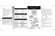
AlGORIThM<br />
2<br />
3<br />
5<br />
7<br />
8<br />
10<br />
1<br />
Confirmed <strong>Pregnancy</strong><br />
[A–1]<br />
First Visit with Nurse (Week 6 to 8)<br />
Complete self-questionnaire<br />
Assess for risk factors [A–2]<br />
Was any high risk<br />
identified that requires<br />
immediate referral to<br />
advanced prenatal care?<br />
(See Sidebar 1) [A–2]<br />
n<br />
Was any risk<br />
identified that requires<br />
follow-up with<br />
advanced prenatal care<br />
at 10-12 weeks? [A–2]<br />
n<br />
Visit with Provider (Week 10-12)<br />
(See <strong>Summary</strong> Table p. 26) [A–3]<br />
Any conditions requiring<br />
consultation/referral to<br />
advanced obstetric care provider?<br />
(See Sidebar 2) [A–3, A–4]<br />
n<br />
Continue goal oriented visit<br />
(See <strong>Summary</strong> Table p. 26)<br />
4<br />
6<br />
9<br />
Y<br />
Refer to advanced<br />
prenatal care for<br />
follow-up and treatment<br />
Y<br />
Schedule follow-up with<br />
advanced prenatal care<br />
provider at 10-12 weeks<br />
Y<br />
Consult/Refer to<br />
advanced prenatal care<br />
for supplemental care<br />
Sidebar 1: Indication for Immediate Referral<br />
Recurrent pregnancy loss • Pregestational diabetes<br />
Ectopic pregnancy risk<br />
(Type 1 or 2)<br />
Vaginal bleeding<br />
Renal disorder<br />
Uncontrolled hypertension<br />
Transplant<br />
• Significant abdominal pain/<br />
Moderate-severe depression<br />
cramping<br />
Suicidal<br />
Severe pelvic infections<br />
HIV<br />
Cardiovascular diseases<br />
Known teratogen exposure<br />
• Cardiac abnormality • History of genetic disease<br />
Sidebar 2: Indications for Advanced Obstetric Care<br />
Following 10-12 Weeks<br />
Prior macrosomia or GDM<br />
Current Infection<br />
• Severe pelvic infections • Positive Gonorrhea<br />
Second trimester pregnancy loss<br />
• Cervical; surgery<br />
(LEEP, cone biopsy)<br />
• Bariatric surgery within the past<br />
18 months<br />
• Uterine abnormality<br />
Drug use/alcohol use/smoking<br />
Current cancer<br />
T ransplant on medication<br />
• Cardiac abnormality<br />
Positive Chlamydia<br />
Hepatitis B<br />
Genital Herpes<br />
• HIV<br />
Pre-Existing Conditions<br />
• Abnormal pap-smear<br />
Mild asthsma<br />
Controlled hypothyroidism<br />
Previous gastric bypass<br />
• Depression<br />
• At risk for diabetes<br />
OB/GYN Conditions<br />
• At risk for preterm birth<br />
• Prior caesarean section<br />
Sidebar 3: Interventions at ALL Routine Visits<br />
Screening for hypertensive disorders<br />
Breast feeding education<br />
Exercise during pregnancy<br />
• Influenza vaccine (season-related)<br />
11<br />
Routine Visits (Week 16-27)<br />
(See Sidebar 3, 4) [A–5]<br />
Sidebar 4:<br />
Routine Visit<br />
Week 16 –27<br />
Sidebar 5:<br />
Routine Visit<br />
Week 28 –41<br />
12<br />
Routine Visits (Week 28-41)<br />
(See Sidebar 3, 5) [A–6]<br />
Auscultate fetal heart tones<br />
Screen fundal height<br />
Assess weight gain<br />
• Educate about symptoms of<br />
preterm labor (week 20)<br />
Auscultate fetal heart tones<br />
Screen fundal height<br />
• Assess weight gain<br />
• Assess for symptoms of preterm<br />
labor (week 28)<br />
• Assess fetal kick counts<br />
14<br />
13<br />
Labor and Birth<br />
Postpartum Visit<br />
[A–7]<br />
For Specific Intervention<br />
See <strong>Summary</strong> Table same<br />
as above page 24<br />
Management of <strong>Pregnancy</strong> | AlGorItHm | 11
ANNOTATIONS<br />
A–0.<br />
Organization of Prenatal Care<br />
BACKGROUND<br />
Goal-oriented visits can be provided during individual encounters with OB providers, or can be accomplished in a group<br />
setting. Recommendations for reduced visit prenatal care have been instituted at some healthcare facilities. This first <strong>DoD</strong>/<br />
<strong>VA</strong> <strong>Clinical</strong> <strong>Practice</strong> Guideline for the Management of Uncomplicated <strong>Pregnancy</strong> was implemented in 2003. One of the key<br />
components of the clinical practice guideline was changing from the traditional interval-based visit template (every four<br />
weeks in the first and second trimesters) towards a system in which an antenatal visit is planned for a specific gestational<br />
age, with each visit having specific well-defined goals and objectives.<br />
Group prenatal care has been implemented in many clinical practices in the United States and abroad. Centering<br />
<strong>Pregnancy</strong>® is a group model of prenatal care which provides care in a group setting, integrating assessment support and<br />
education at each visit. Studies have shown group prenatal care results in equal or improved perinatal outcomes with no<br />
added cost.<br />
Level of Care Settings:<br />
Throughout this guideline, the term Routine Prenatal Care refers to prenatal care generally provided to pregnant women by<br />
Family Medicine Physicians, Women’s Health Nurse Practitioners, Certified Nurse-Midwives or Obstetrician/Gynecologists.<br />
The term Advanced Prenatal Care generally refers to care provided to women with complicated pregnancies provided by<br />
Obstetrician/Gynecologists and/or Maternal-Fetal Medicine specialists.<br />
Routine Prenatal Care Providers<br />
Individuals qualified to provide routine obstetric care include Family <strong>Practice</strong> Physicians, Certified Nurse-Midwives,<br />
Women’s Health Nurse Practitioners, and Obstetrician/Gynecologists. These providers may have varied experience in<br />
providing more advanced prenatal care.<br />
Advanced Prenatal Care Providers<br />
Obstetrician-Gynecologist: Physician qualified by training and experience to manage complicated pregnancies by<br />
virtue of having completed four years of Obstetrics and Gynecology residency training and maintaining currency in the<br />
profession.<br />
Maternal-Fetal Medicine (MFM) Specialist: Physician who has completed two to three years of Maternal-Fetal Medicine<br />
fellowship after completing four years of Obstetrics and Gynecology residency training. Fellowship training provides<br />
additional education and practical experience to gain special competence in managing various obstetrical, medical, and<br />
surgical complications of pregnancy.<br />
MFM specialists function in collaboration with Family Medicine physicians, Women’s Health Nurse Practitioners, Certified<br />
Nurse-Midwives and Obstetricians. The relationship and referral patterns between Obstetrician-Gynecologists and MFM<br />
specialists will depend on the acuity of the patient’s condition and local circumstances.<br />
RECOMMENDATIONS<br />
1. Goal-oriented prenatal care system can be delivered to all pregnant women. [B]<br />
2. Education should be a central component of prenatal care for all pregnant women. [B]<br />
3. Group model of prenatal care, such as the Centering <strong>Pregnancy</strong>® model, is an acceptable alternative to<br />
individual provider appointments. [A]<br />
12 | Guideline <strong>Summary</strong> – 2009
A–1.<br />
Confirmed <strong>Pregnancy</strong><br />
Confirmation of pregnancy is established by a confirmed positive urine or serum pregnancy test.<br />
A–2. First Visit with Nurse Weeks 6 – 8<br />
Complete Self-Questionnaire; Assess for Risk Factors<br />
BACKGROUND<br />
After confirmation of the pregnancy, the goal of the first prenatal contact is to exchange information and identify existing<br />
risk factors that may impact the pregnancy. This initial contact may be accomplished in a group setting or during a oneon-one<br />
visit. This encounter provides an opportunity early in the pregnancy to obtain general short-term risk stratification.<br />
In this visit, the nurse should identify women who: (1) Need immediate referral to an advanced prenatal care provider<br />
(e.g., high risk for ectopic pregnancy); (2) Need to see an advanced prenatal care provider at the first provider visit; or (3)<br />
Can have the first provider visit with a low-risk prenatal care provider. Table 1 contains a checklist of the data collected<br />
during the first visit with the nurse and/or obstetric healthcare provider. These data are required to appropriately triage<br />
women into one of the three categories noted above. In addition, all active duty pregnant women are required to have an<br />
occupational health screening. This referral/consultation with occupational health should be done at this initial encounter.<br />
RECOMMENDATIONS<br />
1. Initial assessment by nurse may include the following actions:<br />
a. Assure the patient completes the Self-Questionnaire (See Appendix B of the full guideline)<br />
b. Review the patient’s completed Self-Questionnaire for issues requiring immediate<br />
evaluation or intervention (See Appendix B of the full guideline)<br />
c. Obtain initial prenatal lab tests to be reviewed and documented at the following visit<br />
d. Consult with an advanced prenatal care provider regarding advice or instruction to the<br />
patient if there are immediate needs (See Table 1)<br />
e. Arrange immediate referral to advanced prenatal care for follow-up in cases needing<br />
short-term assessment or intervention (See Table 1)<br />
f. Provide brief information about options for screening for fetal chromosomal abnormalities<br />
and arrange for counseling (See I-36)<br />
g. Arrange follow-up with the appropriate provider at 10-12 weeks.<br />
Management of <strong>Pregnancy</strong> | Annotations | 13
Table 1.<br />
Prenatal Risk Assessment by Nurse — Checklist<br />
Risk Assessed by Nurse<br />
Nurse Assessment<br />
& Questionnaire<br />
Laboratory Tests<br />
Immediate Referral<br />
to Advanced Prenatal<br />
Care Provider<br />
Consult with<br />
Advanced Prenatal<br />
Care Provider<br />
Follow-up with<br />
Advanced Prenatal<br />
Care: Weeks 10-12<br />
Uncertain dating criteria ❑ ✓ Ultrasound ❑ ✓<br />
Late presentation ❑ ✓ Ultrasound ❑ ✓<br />
Past OB History<br />
Recurrent pregnancy loss ❑ ✓ ❑ ✓<br />
Ectopic pregnancy risk (prior hx of ectopic, prior tubal surgery, current<br />
IUD, hx of tubal infertility, hx PID) ❑ ✓ Quantitative<br />
HCG/ US<br />
❑ ✓<br />
Prior macrosomia or prior gestational diabetes mellitus (GDM) ❑✓ Glucola forGDM<br />
Preterm birth ❑ ✓ ❑ ✓<br />
Second-trimester pregnancy loss ❑ ✓ Ultrasound ❑ ✓<br />
Cervical surgery (LEEP, cone biopsy) ❑ ✓ ❑ ✓<br />
Bariatric surgery (less than 18 months) ❑ ✓ ❑ ✓<br />
Current Problems<br />
Vaginal bleeding (current) ❑ ✓ ❑ ✓<br />
Significant abdominal pain/cramping (current) ❑ ✓ ❑ ✓<br />
Prescription, over-the-counter, and herbal medications ❑ ✓ ❑ ✓<br />
Drug/alcohol use ❑ ✓ ❑ ✓<br />
Smoking ❑ ✓ ❑ ✓<br />
Medical Conditions<br />
Cardiovascular diseases ❑ ✓ ❑ ✓ ❑ ✓<br />
Cardiac abnormality ❑ ✓ ❑ ✓<br />
Diabetes mellitus (DM) – Type 1 or 2 ❑ ✓ Hgb A1c ❑ ✓ ❑ ✓<br />
Renal disorder (includes pyelonephritis) ❑ ✓ ❑ ✓<br />
Hypertension ❑ ✓ If not controlled ❑ ✓<br />
Thyroid disorders ❑ ✓ Thyroid function ❑ ✓<br />
Gastrointestinal disorders on medications ❑ ✓ ❑ ✓<br />
Pulmonary disease ❑ ✓ ❑ ✓<br />
Family history of DM in first relative ❑✓ Glucola for GDM<br />
Neurological disorder ❑ ✓ ❑ ✓<br />
Autoimmune disorder/Lupus ❑ ✓ ❑ ✓ ❑ ✓<br />
Major mental illness ❑ ✓ ❑ ✓<br />
Blood disorders ❑ ✓ ❑ ✓<br />
Hepatitis ❑ ✓ Hepatitis panel ❑ ✓<br />
Sexually transmitted disease (STD) ❑ ✓ ❑ ✓<br />
Tuberculosis ❑ ✓ ❑ ✓<br />
Human immunodeficiency virus (HIV) ❑ ✓ ❑ ✓ ❑ ✓ ❑ ✓<br />
14 | Guideline <strong>Summary</strong> – 2009<br />
❑ ✓ indicates action should be taken
Table 1.<br />
Prenatal Risk Assessment by Nurse — Checklist (continued)<br />
Risk Assessed by Nurse<br />
Nurse Assessment<br />
& Questionnaire<br />
Laboratory Tests<br />
Immediate Referral<br />
to Advanced Prenatal<br />
Care Provider<br />
Consult with<br />
Advanced Prenatal<br />
Care Provider<br />
Follow-up with<br />
Advanced Prenatal<br />
Care: Weeks 10-12<br />
Medical Conditions (continued)<br />
Rash or viral illness ❑ ✓ ❑ ✓<br />
Radiation/toxic chemical exposure since becoming pregnant ❑ ✓ ❑ ✓<br />
Cancer ❑ ✓ ❑ ✓<br />
Transplant ❑ ✓ ❑ ✓ ❑ ✓<br />
Hx of genetic disease or family history of genetic disease ❑ ✓ ❑ ✓<br />
Dental complaint ❑✓ To Dentistry<br />
Screen for Major Depressive Disorder<br />
❑ ✓ To Behavior Health if<br />
suicidal or moderate or<br />
severe MDD<br />
Occupational hazards ❑✓ To Public Health<br />
<strong>Home</strong>less ❑✓ To Social Services<br />
Domestic violence ❑✓ To Social Work if unsafe<br />
Hx of infertility ❑ ✓ Transvaginal US ❑ ✓<br />
Hx of mental illness on medications ❑ ✓ ❑ ✓<br />
Diet restriction ❑ ✓ To Nutrition Counseling ❑ ✓<br />
Eating disorder ❑ ✓ ❑ ✓<br />
Body mass index (BMI) > 30 kg/m2 ❑ ✓ Glucola for GDM ❑ ✓<br />
BMI < 20 kg/m2 ❑ ✓ ❑ ✓<br />
Age (34) ❑ ✓ ❑ ✓<br />
Routine Lab Tests<br />
Human immunodeficiency virus (HIV) ❑ ✓<br />
Complete blood count (CBC) ❑ ✓<br />
(ABO Rh) blood typing ❑ ✓<br />
Antibody screen ❑ ✓<br />
Rapid plasma reagent (RPR) ❑ ✓<br />
Hepatitis B surface antigen test ❑ ✓<br />
Rubella test ❑ ✓<br />
Urinalysis and culture ❑ ✓<br />
Additional Information<br />
Religion<br />
❑ ✓<br />
Language barrier<br />
❑ ✓<br />
Currently or previously deployed or family member<br />
❑ ✓<br />
Born outside the United States<br />
❑ ✓<br />
Lives with cats<br />
❑ ✓<br />
Wears seat belts ❑ ✓<br />
Planned pregnancy<br />
❑ ✓<br />
Highest level of education<br />
❑ ✓<br />
❑ ✓ indicates action should be taken<br />
Management of <strong>Pregnancy</strong> | Annotations | 15
A–3. The First Provider Visit Weeks 10-12<br />
BACKGROUND<br />
The first provider visit offers an opportunity for the provider to review the information obtained through the Self-<br />
Questionnaire and the results of the initial laboratory studies and to note any salient issues previously identified at the<br />
initial 6-8 week nurse’s visit. The provider also has an opportunity to further investigate notable issues, complete a physical<br />
examination, address/document fetal viability, confirm the gestational age, and address any complications that may have<br />
arisen in the interval since the initial nurse’s visit.<br />
The provider will outline the plan of care based on the information gathered from this and the initial nurse's visit. The plan<br />
for the ongoing prenatal care should be based on the backbone of routine prenatal care outlined in this guideline and<br />
then individualized by addressing any currently identifiable risks/complications and outlining any indicated supplemental<br />
prenatal interventions. The outline of care may involve referring the patient to, or consulting with, an advanced prenatal<br />
care provider. (See Annotation A-0 for Level of Care Settings)<br />
RECOMMENDATIONS<br />
1. At the first provider visit, a complete medical history and physical examination (including thyroid, breast<br />
and pelvic examination) should be obtained. Information from the previous visit(s) and laboratory studies<br />
should be reviewed and significant problems/risks should be assessed.<br />
2. At the first provider visit, the provider should outline an individualized plan of prenatal care that includes<br />
guideline-based routine prenatal care and consultation with advanced prenatal care providers or other<br />
medical specialty care services if needed.<br />
3. The following are conditions not addressed by this guideline that will require supplemental care that<br />
might be best provided by routine or advanced obstetric care providers and/or behavioral health<br />
providers depending on the individual circumstances and local conditions:<br />
»»<br />
Current mental illness requiring medical therapy<br />
»»<br />
Substance use disorders<br />
»»<br />
Eating disorders.<br />
4. The following are among conditions that require supplemental prenatal care or consultation with or<br />
referral to an advanced prenatal care provider (See Table 2):<br />
a. General<br />
»»<br />
Body mass index (BMI) 30<br />
»»<br />
Age ( 34 years at delivery)<br />
»»<br />
At risk for diabetes<br />
b. Infections:<br />
»»<br />
Hepatitis B or C (See I-11)<br />
»»<br />
Human immunodeficiency virus (HIV)<br />
»»<br />
Syphilis (positive RPR)<br />
»»<br />
Cytomegalovirus (CMV)<br />
»»<br />
Toxoplasmosis<br />
»»<br />
Primary herpes<br />
»»<br />
Rubella<br />
»»<br />
Parvovirus<br />
»»<br />
Positive gonorrhea (See I-29)<br />
»»<br />
Positive chlamydia (See I-30)<br />
»»<br />
Genital herpes (See I-32)<br />
»»<br />
Recurrent urinary tract infections/stones<br />
16 | Guideline <strong>Summary</strong> – 2009
c. Pre-existing Medical Conditions:<br />
»»<br />
Abnormal pap smear (See I-31)<br />
»»<br />
Controlled hypothyroidism<br />
»»<br />
Previous gastric bypass/bariatric surgery (See I-28)<br />
»»<br />
Mild depression (See I-21 & I-34)<br />
»»<br />
Cardiovascular disease<br />
»»<br />
High blood pressure<br />
»»<br />
Familial hyperlipidemia<br />
»»<br />
Pregestational diabetes<br />
»»<br />
Kidney disease (including pyelonephritis)<br />
»»<br />
Inflammatory bowel disease<br />
»»<br />
Pulmonary disease (including asthma)<br />
»»<br />
Autoimmune diseases (including Anticardiolipin Antibody Syndrome and Systemic Lupus Erythematosis)<br />
»»<br />
Thromboembolic disease, current or historical<br />
»»<br />
Cancer<br />
»»<br />
Seizure disorders<br />
»»<br />
Hematologic disorders (including anemia, thrombocytopenia)<br />
»»<br />
Genetic disease with known effect on pregnancy<br />
d. Obstetric Conditions:<br />
»»<br />
Vaginal bleeding<br />
»»<br />
Isoimmunization<br />
»»<br />
Placenta previa—symptomatic or present beyond 28 weeks<br />
»»<br />
Placental abruption<br />
»»<br />
At risk for preterm birth (See A-4)<br />
»»<br />
Prior cesarean section (See I-39)<br />
»»<br />
Previous uterine or cervical surgery<br />
»»<br />
Intrauterine fetal demise<br />
»»<br />
Preterm Labor<br />
»»<br />
Preterm ruptured membranes<br />
»»<br />
Recurrent pregnancy loss<br />
»»<br />
Suspected or documented fetal growth abnormalities (intrauterine growth<br />
restriction [IUGR] or macrosomia)<br />
»»<br />
Abnormalities of amniotic fluid including oligohydramnios, polyhydramnios<br />
»»<br />
Fetal anomaly(s)<br />
»»<br />
Multiple gestation<br />
»»<br />
Surgical condition during pregnancy (e.g., appendectomy, ovarian cystectomy, cerclage)<br />
Management of <strong>Pregnancy</strong> | Annotations | 17
Table 2. Conditions Requiring Supplemental Care<br />
Risk Assessed by Routine Prenatal Care Provider<br />
GENERAL CONDITIONS<br />
Referral/Consult<br />
with Advanced Prenatal<br />
Care Provider<br />
Genetic condition potentially affecting fetus ❑ ✓<br />
Consider Referral/Consult<br />
with Advanced Prenatal<br />
Care Provider<br />
Body Mass Index (BMI < 20 or > 30) ❑ ✓<br />
Age < 16 or > 34 ❑ ✓<br />
Genetic condition affecting patient or spouse ❑ ✓<br />
OBSTETRIC CONDITIONS (current or historical)<br />
Recurrent pregnancy loss ❑ ✓<br />
Ectopic pregnancy ❑ ✓<br />
Significant abdominal pain/cramping ❑ ✓<br />
Vaginal bleeding ❑ ✓<br />
Second-trimester pregnancy loss ❑ ✓<br />
Preterm labor (current) or birth (history) ❑ ✓<br />
Cervical surgery (LEEP, cone biopsy) ❑ ✓<br />
Uterine abnormality ❑ ✓<br />
Short ( 36 weeks) ❑ ✓<br />
Placenta Previa (symptomatic or beyond 28 weeks) ❑ ✓<br />
Abnormal amniotic fluid: oligo/poly hydramnios ❑ ✓<br />
Preterm ruptured membranes ❑ ✓<br />
Fetal growth abnormality (90 %tile) ❑ ✓<br />
Known or suspected fetal anomaly ❑ ✓<br />
Multiple gestation ❑ ✓<br />
Isoimmunization ❑ ✓<br />
Abnormal prenatal screening result (aneuploidy risk) ❑ ✓<br />
Abnormal prenatal screening result (ONTD risk) ❑ ✓<br />
Intrauterine fetal demise ❑ ✓<br />
Teratogenic exposure including drugs or radiation ❑ ✓<br />
Placental abruption ❑ ✓<br />
Prior cesarean section ❑ ✓<br />
Intrapartum complications ❑ ✓<br />
18 | Guideline <strong>Summary</strong> – 2009<br />
❑ ✓ indicates action should be taken
Table 2. Conditions Requiring Supplemental Care<br />
Risk Assessed by Routine Prenatal Care Provider<br />
GYNECOLOGIC, MEDICAL, SURGICAL CONDITIONS<br />
Referral/Consult<br />
with Advanced Prenatal<br />
Care Provider<br />
Current need for surgery ❑ ✓<br />
Bariatric surgery (< 18, > 36 months ago) ❑ ✓<br />
Diabetes mellitus (DM) – Type 1 or 2 ❑ ✓<br />
Hematalogic disorders (except mild anemia) ❑ ✓<br />
Gastrointestinal disorders on medication ❑ ✓<br />
Chronic hypertension ❑ ✓<br />
Cardiovascular disease ❑ ✓<br />
Pulmonary disease including asthma ❑ ✓<br />
Cancer (current or recent) ❑ ✓<br />
Neurological disorders including epilepsy ❑ ✓<br />
Renal, urinary tract disorder ❑ ✓<br />
Autoimmune disorder including Lupus ❑ ✓<br />
Antiphospholipid syndrome ❑ ✓<br />
Hyperlipidemia prior to pregnancy ❑ ✓<br />
Transplant ❑ ✓<br />
Consider Referral/Consult<br />
with Advanced Prenatal<br />
Care Provider<br />
Abnormal pap smear ❑ ✓<br />
Breast abnormality ❑ ✓<br />
Pelvic surgery for infertility or infection ❑ ✓<br />
Illicit drug, alcohol, or tobacco use ❑ ✓<br />
Thyroid disorders ❑ ✓<br />
INFECTIOUS DISEASES<br />
Severe pelvic infections ❑ ✓<br />
Hepatitis ❑ ✓<br />
Tuberculosis ❑ ✓<br />
HIV ❑ ✓<br />
TORCH infection ❑ ✓<br />
Sexually transmitted disease (STD) ❑ ✓<br />
PSYCHOSOCIAL CONDITIONS<br />
Major Depressive Disorder (MDD)<br />
To Mental Health if suicidal or<br />
moderate or severe MDD<br />
Domestic violence<br />
To Social Work if unsafe<br />
environment<br />
❑ ✓<br />
<strong>Home</strong>less To Social Service ❑ ✓<br />
❑ ✓<br />
❑ ✓ indicates action should be taken<br />
Management of <strong>Pregnancy</strong> | Annotations | 19
A–4.<br />
Assessment of Risk Factors for Preterm Birth<br />
BACKGROUND<br />
Preterm birth is the second leading cause of neonatal mortality in the United States. Although many preterm births are<br />
due to the development of obstetric complications, over 70 percent result from spontaneous preterm birth which includes<br />
deliveries related to idiopathic preterm labor, preterm rupture of membranes, and cervical insufficiency. Demographic or<br />
historical risk factors for spontaneous preterm birth delivery may be discovered at the initial nursing intake or provider visit.<br />
Other risk factors develop as a woman’s pregnancy progresses and include certain symptoms or physical examination and<br />
imaging findings. Although the prediction and prevention of spontaneous preterm birth remain challenging, continual<br />
surveillance for these risk factors may be beneficial as effective therapeutic options are developed. Some risk factors<br />
only require annotation in the obstetric record and routine surveillance as indicated in this pregnancy guideline. The<br />
identification of other risk factors should prompt increased surveillance in the form of consultation with an advanced<br />
prenatal care provider or ancillary testing and imaging studies.<br />
To date, no single test or sequence of tests has an optimal sensitivity or predictive value for preterm birth. Fetal fibronectin<br />
testing and cervical length measurement by transvaginal ultrasound appear to be useful in the management of some<br />
women meeting the criteria for increased surveillance. Most studies have shown that these tests have limited utility when<br />
used in the asymptomatic woman at low risk for preterm delivery. Importantly, modalities such as salivary estriol levels,<br />
bacterial vaginosis screening, and home uterine activity monitoring are generally not effective at predicting preterm birth<br />
regardless of risk status.<br />
Recent data suggest that the administration of progesterone intramuscularly or intravaginally beginning early in pregnancy<br />
in women at high risk for preterm birth significantly reduces the rate of preterm delivery. Specifically, women with a prior<br />
spontaneous birth at less than 37 weeks’ of gestation and asymptomatic women with a shortened cervical length in<br />
the 2 nd trimester appear to benefit from the administration of progesterone beginning early in pregnancy. Progesterone<br />
therapy is typically begun early in the 2 nd trimester and continued until approximately 36 weeks. Both intramuscular<br />
17 alpha hydroxyprogesterone caproate (250 mg, administered weekly) and vaginal progesterone suppositories (100 to<br />
200 mg, administered once daily) have been described in the literature.<br />
Despite the apparent benefits of progesterone in high-risk populations and its growing use, the ideal progesterone<br />
formulation and long-term safety of the drug must be confirmed by additional studies. Progesterone supplementation<br />
for the prevention of preterm birth should still be considered investigational.<br />
The identification of women at risk for preterm birth now increases in importance in light of the expanded availability of<br />
and indications for progesterone therapy for the prevention of preterm birth.<br />
RECOMMENDATIONS<br />
Assessment of Preterm Birth<br />
1. Women should be assessed for preterm birth risk as early as possible in the pregnancy in order to<br />
optimize maternal and newborn outcomes.<br />
2. Screening for preterm birth risk factors should continue up to 37 weeks estimated gestational age.<br />
3. Women at increased risk, but meeting the criteria for normal surveillance, should have the risk factor(s)<br />
documented in the medical record to increase awareness of the risk but may continue to be followed in<br />
accordance with the routine management of the pregnancy guideline.<br />
4. Routine care providers should consult with an advanced prenatal care provider whenever a woman meets<br />
the criteria for increased surveillance for preterm birth.<br />
5. Women requiring increased surveillance should be considered for ancillary studies and other additional<br />
intervention. Progesterone supplementation should be considered in these women.<br />
6. Routine screening of fetal fibronectin (fFN) in asymptomatic or low-risk women is not recommended<br />
(See I-52). fFN testing in symptomatic or high-risk women between 24 and 34 6/7 weeks’ gestation may be<br />
useful in guiding management.<br />
20 | Guideline <strong>Summary</strong> – 2009
7. The measurement of cervical length by transvaginal ultrasound may be useful in some patients requiring<br />
increased surveillance for preterm labor. Sonographic cervical length measurement is not recommended as<br />
a routine screening or prediction tool in women only requiring normal surveillance.<br />
8. The determination of salivary estriol levels, bacterial vaginosis screening, and home uterine activity<br />
monitoring are not recommended as a means to predict preterm birth.<br />
Progesterone Therapy<br />
9. It is reasonable to offer antenatal progesterone therapy to women at high-risk for preterm delivery and<br />
who meet the generally accepted inclusion criteria. [B]<br />
10. Progesterone may be administered intramuscularly on a weekly basis or intravaginally on a daily basis. [B]<br />
11. Progesterone therapy should only be initiated after consultation with an advanced prenatal care provider<br />
(obstetrician or maternal-fetal medicine specialist). [C]<br />
Table 3. Risk Factors for Preterm Birth Stratified by those Requiring either Normal or Increased Surveillance *<br />
Normal Surveillance<br />
• Nonwhite race<br />
• Age younger than 16 years or older<br />
than 34 years<br />
• Low socioeconomic status<br />
• Single parent<br />
• Smoking<br />
• History multiple first trimester<br />
spontaneous abortions<br />
• History of lower genital tract infection<br />
• Low pre-pregnancy weight/body<br />
mass index<br />
• Occupational stress or prolonged standing<br />
(greater than 3 hours)<br />
• Periodontal disease<br />
Increased Surveillance<br />
Risk factors known from prior history:<br />
• Prior cervical surgery<br />
• History of preterm delivery (less than 34 weeks)<br />
Findings that may be identified during the pregnancy:<br />
• Antepartum vaginal bleeding or persistent placenta previa<br />
• Uterine over-distension due to any cause<br />
(e.g., multiple gestation, polyhydramnios)<br />
• Abnormality of uterine cavity or architecture<br />
(e.g., septate uterus, uterine fibroids)<br />
• Uterine contractions, back ache, or pelvic pressure<br />
• Shortened cervical length<br />
• Rupture of membranes<br />
• Cervical dilation greater than or equal to 2 cm in 2nd trimester in<br />
symptomatic women (nulliparous women)<br />
• Soft cervical consistency in 2nd trimester in symptomatic women<br />
(nulliparous women)<br />
• Abdominal surgery during current pregnancy<br />
• Illicit drug use (e.g., methamphetamine, cocaine)<br />
• Use of assisted reproductive technology<br />
*Women with multiple risk factors in the Normal Surveillance category may require individualized assessment and warrant consultation<br />
with an advanced prenatal care provider.<br />
Management of <strong>Pregnancy</strong> | Annotations | 21
A–5. Routine Visits Weeks 16-27<br />
Visits during this period should include the following:<br />
»»<br />
Auscultation of fetal heart tones - if negative, elevate care<br />
»»<br />
Screening fundal height<br />
»»<br />
Screening for hypertensive disorders<br />
»»<br />
Assessing weight gain<br />
»»<br />
Education about symptoms of preterm labor (week 20)<br />
For specific interventions see Prenatal Care Interventions – Weeks 16-27<br />
A–6. Routine Visits Weeks 28-41<br />
Visits during this period should include the following:<br />
»»<br />
Auscultation of fetal heart tones - if negative, elevate care<br />
»»<br />
Screening fundal height<br />
»»<br />
Screening for hypertensive disorders<br />
»»<br />
Assessing weight gain<br />
»»<br />
Assessing for symptoms of preterm labor (week 28, 32)<br />
»»<br />
Assessing fetal kick counts<br />
For specific interventions see Prenatal Care Interventions – Weeks 28-41<br />
A–7.<br />
Postpartum Visit<br />
BACKGROUND<br />
The postpartum visit provides the opportunity for providers to interact with the new mother and her infant through<br />
interview, exam, and testing. The timing and the content of the postpartum visit have often been topics for debate. Recent<br />
literature helps the provider to answer these questions based on the evidence. The maternal postpartum visit should<br />
occur approximately eight weeks after delivery. Eight weeks is the optimal time to decrease the rate of false positive<br />
cervical smears.<br />
RECOMMENDATIONS<br />
1. The following should be included in the postpartum visit:<br />
»»<br />
Pelvic and breast examinations. [B]<br />
»»<br />
Cervical smear should be completed as indicated by cervical cancer screening guidelines<br />
(See I-31). [A]<br />
»»<br />
Initiate or continue the HPV vaccine series for women age < 26 years (See I-50). [C]<br />
»»<br />
Screening for postpartum depression (See I-21). [B]<br />
»»<br />
Screening for domestic violence (See I-20). [B]<br />
»»<br />
Diabetes testing for patients with pregnancies complicated by gestational diabetes.<br />
The two-hour 75g oral glucose tolerance test (GTT) is recommended but a fasting glucose<br />
can also be done. [B]<br />
»»<br />
Education about contraception, infant feeding method, sexual activity, weight, exercise and<br />
the woman’s assessment of her adaptation to motherhood. Pre-existing or chronic medical<br />
conditions should be addressed with referral for appropriate follow-up as indicated. [I]<br />
22 | Guideline <strong>Summary</strong> – 2009
Interventions<br />
Prenatal care for all pregnant women should include the interventions listed in the following <strong>Summary</strong> Table.<br />
Each intervention should be completed by the indicated week<br />
Management of <strong>Pregnancy</strong> | Interventions • All Visits | 23
<strong>Summary</strong> Table. Prenatal Care Interventions<br />
Intervention<br />
Trimester<br />
First Second Third<br />
WEEK<br />
Post<br />
Date †<br />
6-8 10-12 16-20 24 28 32 36 38-41 PP<br />
I-1 Screening for hypertensive disorders ❑ ✓ ❑ ✓ ❑ ✓ ❑ ✓ ❑ ✓ ❑ ✓ ❑ ✓ ❑ ✓<br />
I-2 Breastfeeding education ❑ ✓ ❑ ✓ ❑ ✓ ❑ ✓ ❑ ✓ ❑ ✓ ❑ ✓ ❑ ✓<br />
I-3 Exercise during pregnancy ❑ ✓ ❑ ✓ ❑ ✓ ❑ ✓ ❑ ✓ ❑ ✓ ❑ ✓ ❑ ✓<br />
I-4 Influenza vaccine (season-related) ❑ ✓ ❑ ✓ ❑ ✓ ❑ ✓ ❑ ✓ ❑ ✓ ❑ ✓ ❑ ✓<br />
I-5 Screening for tobacco use - offer cessation ❑ ✓<br />
I-6 Screening for alcohol use - offer cessation ❑ ✓<br />
I-7 Screening for drug abuse - offer cessation ❑ ✓<br />
I-8 Screening for Rh status ❑ ✓<br />
I-9 Screening for rubella ❑ ✓<br />
I-10 Screening for varicella ❑ ✓<br />
I-11 Screening for hepatitis B ❑ ✓<br />
I-12 Treatment for hepatitis B * ❑ ✓ ❑ ✓<br />
I-13 Screening for syphilis (Rapid Plasma Reagin) ❑ ✓<br />
I-14 Screening for asymptomatic bacteriuria ❑ ✓<br />
I-15 Screening for tuberculosis ❑ ✓<br />
I-16 Screening for HIV – counsel ❑ ✓<br />
I-17 Immunization – Td booster (first trimester) ❑ ✓<br />
I-18 Screening for anemia ❑ ✓<br />
I-19 Screening for hemoglobinopathies * ❑ ✓<br />
I-20 Screening for domestic abuse ❑ ✓ ❑ ✓ ❑ ✓<br />
I-21 Screening for depression ❑ ✓ ❑ ✓ ❑ ✓<br />
I-22 Establishing the gestational age ❑ ✓<br />
I-23 Auscultation fetal heart tones ❑ ✓ ❑ ✓ ❑ ✓ ❑ ✓ ❑ ✓ ❑ ✓ ❑ ✓<br />
I-24 Screening fundal height ❑ ✓ ❑ ✓ ❑ ✓ ❑ ✓ ❑ ✓ ❑ ✓ ❑ ✓<br />
I-25 Assessing weight gain (inappropriate) ❑ ✓ ❑ ✓ ❑ ✓ ❑ ✓ ❑ ✓ ❑ ✓ ❑ ✓<br />
I-26 Nutritional supplement ❑ ✓<br />
I-27 Management of obesity * ❑ ✓<br />
I-28 Gastric bypass consideration * ❑ ✓<br />
I-29 Screening for gonorrhea ❑ ✓<br />
I-30 Screening for chlamydia ❑ ✓<br />
I-31 Screening for cervical cancer ❑ ✓<br />
I-32 Screening for HSV and prophylaxis ❑ ✓<br />
I-33 Counseling for cystic fibrosis screening ❑ ✓<br />
I-34 Management of depression * ❑ ✓ ❑ ✓ ❑ ✓<br />
I-35 Assessment of periodontal disease ❑ ✓<br />
I-36<br />
Education about prenatal screening for fetal chromosonal<br />
abnormalities<br />
❑ ✓<br />
• Screening test 1st trimester<br />
❑ ✓<br />
• Counseling and test 2nd trimester<br />
❑ ✓<br />
Postpartum<br />
† Between weeks 38-41, weekly visits may be needed.<br />
* Intervention only applies if the risk/condition has been identified/diagnosed.<br />
❑ ✓ Intervention should be done<br />
Intervention should NOT be done<br />
24 | Guideline <strong>Summary</strong> – 2009
<strong>Summary</strong> Table. Prenatal Care Interventions<br />
Intervention<br />
Trimester<br />
First Second Third<br />
WEEK<br />
Post<br />
Date †<br />
6-8 10-12 16-20 24 28 32 36 38-41 PP<br />
I-37 Obstetric ultrasound ❑ ✓<br />
I-38 Education about preterm labor ❑ ✓<br />
I-39 Counseling for trial of labor * ❑ ✓<br />
I-40 Screening for gestational diabetes ❑ ✓<br />
I-41 Iron supplementation * ❑ ✓<br />
I-42 Anti-D prophylaxis for Rh-negative women * ❑ ✓<br />
I-43 Assess for preterm labor ❑ ✓ ❑ ✓<br />
I-44 Daily fetal movement counts ❑ ✓ ❑ ✓ ❑ ✓ ❑ ✓<br />
I-45 Counseling for family planning ❑ ✓<br />
I-46 Screening for Group B Streptococcus (GBS) ❑ ✓<br />
I-47 Assessment of fetal presentation ❑ ✓ ❑ ✓<br />
I-48 Consider weekly cervical check/stripping ❑ ✓<br />
I-49 Term management ❑ ✓<br />
I-50 Immunization HPV vaccine * ❑ ✓<br />
I-51 Education on Shaken Baby Syndrome ❑ ✓<br />
Postpartum<br />
Interventions NOT RECOMMENDED in Routine Prenatal Care<br />
I-52 Screening with fetal fibronectin<br />
I-53 Cervical examination<br />
I-54 Antenatal pelvimetry<br />
I-55 Urine dipstick test<br />
I-56 Edema evaluation<br />
I-57 Screening for cytomegalovirus (CMV)<br />
I-58 Screening for parvovirus<br />
I-59 Screening for toxoplasmosis<br />
I-60 Screening for bacterial vaginosis<br />
I-61 Immunization – MMR<br />
I-62 Immunization – Varicella<br />
I-63 Ultrasound (US) evaluation of cervical length at week 24<br />
I-64 Repeat screening for anemia, syphilis, and isoimmunization<br />
I-65 Screening for hypothyroidism<br />
† Between weeks 38-41, weekly visits may be needed.<br />
* Intervention only applies if the risk/condition has been identified/diagnosed.<br />
❑ ✓ Intervention should be done<br />
Intervention should NOT be done<br />
Management of <strong>Pregnancy</strong> | InterventIons • All VIsIts | 25
26 | Guideline <strong>Summary</strong> – 2009<br />
THIS PAGE INTENTIONALLY LEFT BLANK
INTERVENTIONS at All VISITS<br />
I–1. Screening for Hypertensive Disorders of <strong>Pregnancy</strong><br />
Weeks (All)<br />
BACKGROUND<br />
Hypertension in pregnancy can be defined as either a diastolic pressure greater than 90 mmHg or systolic pressure<br />
greater than 140 mmHg recorded on two separate occasions more than six hours apart, at any time during the gestation.<br />
Hypertension detected before the 20 th week of gestation in the absence of gestational trophoblastic disease or highorder<br />
multiple gestation is generally considered indicative of chronic hypertension. Gestational hypertension is defined<br />
as isolated hypertension in the absence of proteinuria occurring after 20 weeks’ gestation. Hypertension occurring in<br />
conjunction with proteinuria beyond 20 weeks’ gestation is classified as preeclampsia. Proteinuria is defined as >300 mg<br />
in a 24-hour urine collection in the absence of evidence of a urinary tract infection. Regardless of the etiology or specific<br />
diagnosis, all hypertensive disorders of pregnancy are associated with an increased risk for adverse perinatal outcome and<br />
require supplemental monitoring and care beyond the routine care outlined in this guideline.<br />
RECOMMENDATIONS<br />
1. Recommend measuring blood pressure of all pregnant women at each prenatal visit, following the<br />
guidelines of the National High Blood Pressure Education Program and the <strong>VA</strong>/<strong>DoD</strong> <strong>Clinical</strong> <strong>Practice</strong><br />
<strong>Guidelines</strong> for Hypertension. [B]<br />
2. Women diagnosed with hypertension during pregnancy should be managed by, or in consultation with,<br />
an advanced prenatal care provider. [C]<br />
3. Korotkoff 5 sound (disappearance of sound) will be used to determine the diastolic pressure. [C]<br />
I–2. Breastfeeding Education<br />
Weeks (All)<br />
BACKGROUND<br />
Between 50 and 90 percent of expectant mothers decide how they will feed their babies either before conceiving or very<br />
early in pregnancy (Bailey & Sheriff, 1992; Dix, 1991). Prenatal breastfeeding education is a key opportunity to educate<br />
expectant mothers on the benefits and methods associated with successful breastfeeding during the time they are<br />
making their decision on choice of infant feeding method.<br />
RECOMMENDATIONS<br />
1. Recommend offering breastfeeding education to all pregnant women during the first visit with<br />
the provider. [B]<br />
2. Recommend asking pregnant women, “What do you know about breastfeeding?” rather than,<br />
“Do you plan on breast or bottle feeding?” to provide an open opportunity for education. [B]<br />
3. Recommend continuing education throughout pregnancy for those pregnant women who express<br />
a desire to breastfeed or for those who are still undecided on feeding method. [C]<br />
4. Recommend including family/significant others in breastfeeding education. [B]<br />
Management of <strong>Pregnancy</strong> | Interventions • All Visits | 27
I–3. Exercise During <strong>Pregnancy</strong><br />
Weeks (All)<br />
BACKGROUND<br />
Attitudes toward exercise during pregnancy have changed markedly in recent decades. The underlying concern has<br />
revolved around fears that the exercise-induced increases in maternal body temperature, circulating stress hormones,<br />
and biomechanical stress, coupled with the decreased visceral blood flow, could have adverse effects on multiple aspects<br />
of the course and outcome of pregnancy. Only recently has a substantial amount of research been completed to support<br />
the idea that it is both safe and beneficial to exercise during pregnancy. Currently, there is no evidence to suggest that<br />
regular maternal exercise is associated with fetal compromise or unexplained fetal death. Furthermore, regular exercise<br />
improves maternal fitness, reduces the usual musculoskeletal complaints associated with pregnancy, enhances feelings of<br />
well being, improves body image, and decreases maternal weight gain and fat deposition in late pregnancy.<br />
RECOMMENDATIONS<br />
1. Strongly recommend all healthy pregnant women perform regular mild to moderate exercise sessions,<br />
three or more times per week. [A]<br />
2. Recommend individualized exercise programs for all pregnant women based on their pre-pregnancy<br />
activity level. [I]<br />
3. Recommend against high-altitude (>10,000 feet) activities, scuba diving, and contact sports during<br />
pregnancy. [I]<br />
I–4. Influenza Vaccine (Season-Related)<br />
Weeks (Any Week)<br />
BACKGROUND<br />
Women who acquire influenza during pregnancy may experience an increase in morbidity and mortality during an<br />
epidemic, with a possible increased abortion rate. Most recent CDC guidelines suggest that immunization of pregnant<br />
women for influenza has been found to be safe for both the mother and the fetus regardless of gestational age.<br />
RECOMMENDATIONS<br />
1. Recommend immunizing all pregnant women for influenza during the epidemic season. [B]<br />
28 | Guideline <strong>Summary</strong> – 2009
FIRST VISIT with NURSE (6-8 WEEks)<br />
I–5. Screening for Tobacco Use — Offer Cessation Weeks 6 - 8<br />
BACKGROUND<br />
Tobacco use in pregnancy is associated with decreased birth weight, as well as risk for spontaneous abortion and preterm<br />
labor. Newborns exposed to environmental tobacco smoke experience an increased incidence of upper respiratory<br />
infections and deaths from Sudden Infant Death Syndrome (SIDS). Behavioral and pharmacologic methods for smoking<br />
cessation are both safe and effective in pregnancy.<br />
RECOMMENDATIONS<br />
1. Strongly recommend routine screening for tobacco use in pregnancy at the initial prenatal visit. For<br />
patients who smoke, recommend assessment of smoking status at each subsequent prenatal visit.<br />
2. If the screening is positive, cessation should be strongly recommended. [A]<br />
3. There is insufficient data to recommend for or against pharmacologic therapy for tobacco cessation<br />
in pregnancy. [I]<br />
[A]<br />
I–6. Screening for Alcohol Use — Offer Cessation Weeks 6 - 8<br />
BACKGROUND<br />
Alcohol is a known teratogen with adverse effects on fetal facial and central nervous system development. Maternal<br />
alcohol consumption is a leading preventable cause of birth defects and childhood disabilities in the United States. While<br />
there is a clear dose-dependent effect, numerous observational studies have failed to delineate a threshold level for safe<br />
alcohol consumption during pregnancy.<br />
RECOMMENDATIONS<br />
1. Recommend routine screening for alcohol consumption using a standardized tool (See also <strong>VA</strong>/<strong>DoD</strong><br />
<strong>Clinical</strong> <strong>Practice</strong> Guideline for the Management of Substance Use Disorders). [B]<br />
2. If the screening is positive, cessation should be strongly recommended. [B]<br />
3. There is insufficient evidence regarding which cessation intervention tool is the most effective. [I]<br />
I–7. Screening for Drug Use — Offer Treatment Weeks 6 - 8<br />
BACKGROUND<br />
Up to one in ten babies may be exposed to illegal drugs during pregnancy. Use of these drugs may be harmful to the<br />
health and growth of the fetus, particularly early in pregnancy. Drug use later in pregnancy increases the risk for preterm<br />
delivery and fetal growth restriction. Risks to the mother include HIV, hepatitis, and addiction.<br />
RECOMMENDATIONS<br />
1. Recommend routine screening for illicit drug use using a self-report method. [C]<br />
2. Recommend pregnant women identified as abusing drugs be offered treatment and receive care in<br />
consultation with or referral to an advanced prenatal care provider. (See also <strong>VA</strong>/<strong>DoD</strong> <strong>Clinical</strong> <strong>Practice</strong><br />
Guideline for the Management of Substance Use Disorders.) [C]<br />
Management of <strong>Pregnancy</strong> | Interventions • week 6–8 | 29
I–8. Screening for Blood Type (ABO,Rh) and Antibody Status Weeks 6 to 8<br />
BACKGROUND<br />
Since the introduction of anti-D (Rhogam) immune globulin injections during and after pregnancy in women who are<br />
D antigen negative, the incidence of isoimmunization has fallen from 10 cases to 1.3 cases/1,000 live births. Testing and<br />
identification of pregnant women with non-anti-D antibodies allows for early treatment of infants, which may improve<br />
fetal outcomes.<br />
RECOMMENDATIONS<br />
1. Recommend evaluation of maternal ABO and Rh blood type and blood antibody status at the initial<br />
prenatal visit. [B]<br />
2. Pregnant women with positive antibody screens should be referred for consultation to assist with<br />
further management. [C] (See I-42)<br />
3. There is insufficient evidence to recommend for or against routine repeat testing at 28 weeks’<br />
gestation. [I]<br />
I–9. Screening for Rubella Weeks 6 - 8<br />
BACKGROUND<br />
Congenital Rubella Syndrome (CRS) is a constellation of findings in newborns exposed to the rubella virus prior to 16<br />
weeks’ gestation. The syndrome includes hearing loss, developmental delay, and ocular and cardiac defects. The incidence<br />
of CRS has declined dramatically since the advent of rubella vaccination in 1969. Identification of women lacking rubella<br />
immunity during the preconception period allows for immunization before pregnancy. Identification of non-immune<br />
women during pregnancy allows for risk counseling and immunization postpartum.<br />
RECOMMENDATIONS<br />
1. Recommend all pregnant women have a serum screen for rubella status at the initial prenatal visit. [B]<br />
2. Recommend seronegative pregnant women be counseled to avoid exposure. [B]<br />
3. Recommend seronegative pregnant women be vaccinated in the immediate postpartum period. [B]<br />
I–10. Screening for Varicella Weeks 6 to 8<br />
BACKGROUND<br />
Varicella infection during pregnancy may lead to poor outcomes for both mother and fetus. The incidence of varicella<br />
in pregnancy is less than one in 1,000. Most adults are immune to varicella due to previous exposure. In women who<br />
report no history of infection, 85 percent are found to have positive antibody titers. Identification of non-immune persons<br />
through screening with subsequent immunization may decrease the incidence of varicella.<br />
RECOMMENDATIONS<br />
1. Recommend routine screening for varicella through history. [B]<br />
2. If negative/unsure history, obtain a varicella titer. [B]<br />
3. Recommend offering vaccination postpartum, if varicella is non-immune. [B]<br />
30 | Guideline <strong>Summary</strong> – 2009
I–11. Screening for Hepatitis B Virus (HBV) Weeks 6 - 8<br />
BACKGROUND<br />
Each year in the United States an estimated 22,000 infants are born to women with chronic hepatitis B virus (HBV). The<br />
incidence of acute hepatitis B in pregnancy is 1–2/1,000 and the prevalence of chronic hepatitis B is 5 –15/1,000. Certain<br />
groups including Southeast Asians, Pacific Islanders, Alaskan Native Americans, drug addicts, transfusion recipients,<br />
women on dialysis, and those with tattoos have an increased prevalence of infection (Duff, 1998).<br />
However, these risk factors will only identify 60 percent of women with HBV infection. Perinatal transmission of hepatitis<br />
B virus occurs if the mother has an acute infection, particularly during late pregnancy or the early postpartum period, or if<br />
the mother is a chronic hepatitis B antigen carrier.<br />
The risk of vertical transmission (mother to infant) can be greatly reduced (>90 percent) if the infectious status of the<br />
mother is known and therapy is given to the baby shortly after delivery. The risk of vertical transmission may also be<br />
reduced by maternal therapy during the last month of pregnancy.<br />
RECOMMENDATIONS<br />
1. Recommend routine laboratory screening for hepatitis B surface antigen at the initial prenatal visit. [A]<br />
2. Repeat laboratory screening of pregnant women with identification of hepatitis risk factors during the<br />
pregnancy (e.g., healthcare worker, intravenous (IV) drug use, exposure to hepatitis, visit for evaluation or<br />
therapy for sexually transmitted infections, new tattoos, and blood transfusions). [C]<br />
3. Vaccinate pregnant women with hepatitis risk factors who have not been previously vaccinated. [B]<br />
4. Women at risk for HBV infection in pregnancy should be counseled concerning additional methods to<br />
prevent HBV infection. [C]<br />
I–12. Treatment for Hepatitis B Infection Week 36<br />
BACKGROUND<br />
Perinatal transmission of hepatitis B virus occurs if the mother has an acute infection, particularly during late pregnancy<br />
or the early postpartum period, or if the mother is a chronic hepatitis B antigen carrier.<br />
Vertical transmission of infection occurs in 90 percent of pregnancies where the mother is hepatitis B e antigen positive<br />
and in about 10 percent of surface antigen positive, e antigen negative mothers. Most (85 to 95 percent) of infected<br />
infants become chronic carriers. Infants born to infectious mothers treated by both hepatitis vaccination and hepatitis<br />
B-specific immunoglobulin are 90 percent less likely to become infected than untreated infants.<br />
There is also evidence that treating the mother in the last month of pregnancy with lamivudine or hepatitis B<br />
immunoglobulin may further reduce the transmission rate if she is highly infectious (HBV-DNA >1.2x10 9 geq/mL).<br />
RECOMMENDATIONS<br />
1. Treat all infants born to hepatitis B positive mothers with hepatitis B immunoglobulin and initiate hepatitis<br />
B vaccination within 12 hours of birth. [A]<br />
2. Strongly consider treating infants born to women at high risk for hepatitis B who have not been<br />
vaccinated or whose infectious status is unknown. [B]<br />
3. Consider treating women who have high copy numbers of HBV-DNA with lamivudine during the last<br />
month of pregnancy. [B]<br />
4. Women with HBV infection should be taught, and encouraged to implement, strategies to decrease<br />
transmission to non-infected intimate contacts. [B]<br />
Management of <strong>Pregnancy</strong> | Interventions • week 6–8 | 31
I–13. Screening for Syphilis Rapid Plasma Reagin (RPR) Weeks 6 - 8<br />
BACKGROUND<br />
Syphilis is a sexually transmitted disease that can cause significant mortality and morbidity in both the mother and<br />
fetus. The disease is acquired through either sexual or congenital transmission and can be effectively treated using<br />
broad-spectrum antibiotics. Screening for maternal syphilis, treating, and tracking all confirmed cases, can prevent<br />
congenital syphilis.<br />
RECOMMENDATIONS<br />
1. Recommend routine screening for syphilis using serologic testing (i.e., RPR or VDRL) at the<br />
initial prenatal visit. [B]<br />
2. Recommend a confirmatory test using a more specific treponemal assay (FTA-ABS, MHA-TP, HATTS)<br />
for pregnant women who test positive. [B]<br />
3. Strongly recommend therapy with penicillin G antibiotic for pregnant women who have confirmed<br />
syphilis, as recommended by other sexually transmitted disease (STD) guidelines. [A]<br />
4. Recommend appropriate medical and legal mandated follow-up and state/service branch reporting<br />
requirements for pregnant women screening positive. [I]<br />
I–14. Screening for Asymptomatic Bacteriuria Weeks 6 - 8<br />
BACKGROUND<br />
Bacteriuria occurs in two to seven percent of pregnant women. Asymptomatic bacteriuria (ASB) in pregnant women is an<br />
established risk factor for serious complications including pyelonephritis, preterm delivery, and low birth weight.<br />
RECOMMENDATIONS<br />
1. Strongly recommend screening for ASB at initial obstetrical visit via urine culture and sensitivity. [A]<br />
2. There is insufficient evidence to recommend for or against repeat screening throughout the remainder<br />
of pregnancy. [I]<br />
3. Strongly recommend a three to seven-day course of appropriate antibiotics based on positive culture and<br />
sensitivity, and woman’s history of medication allergies. [A]<br />
4. There is insufficient evidence to recommend for or against a test of cure (TOC) after completion of<br />
antibiotic therapy, except in pregnant women with ASB-Group B Strep. [I]<br />
32 | Guideline <strong>Summary</strong> – 2009
I–15. Screening for Tuberculosis Weeks 6-8<br />
BACKGROUND<br />
The incidence of tuberculosis has increased in the United States. Most new cases occur in cities with at least 500,000<br />
people and in women in higher risk groups. High-risk groups for tuberculosis include individuals who:<br />
»»<br />
Have HIV infection<br />
»»<br />
Live in close contact with individuals known or suspected to have tuberculosis<br />
»»<br />
Have medical risk factors known to increase risk of disease if infected<br />
»»<br />
Are born in a country with high tuberculosis prevalence<br />
»»<br />
Are medically underserved<br />
»»<br />
Have a low income<br />
»»<br />
Are alcoholics<br />
»»<br />
Are intravenous drug users<br />
»»<br />
Are residents of long-term care facilities (e.g., correctional institutions, mental institutions,<br />
nursing homes and facilities)<br />
»»<br />
Are healthcare professionals working in high-risk healthcare facilities.<br />
RECOMMENDATIONS<br />
1. All pregnant women from one or more high-risk groups should be screened for tuberculosis with a<br />
Mantoux test with purified protein derivative (PPD) soon after the pregnancy is diagnosed. [C]<br />
2. Pregnant women with a positive PPD with known conversion in the last two years and no clinical or X-ray<br />
evidence of disease should be treated with isoniazid (300 mg per day) starting after the first trimester and<br />
continuing for nine months. [C]<br />
3. For pregnant women with a positive PPD whose time of conversion is unknown and who have no clinical<br />
or X-ray evidence of disease present, consider delaying therapy until after the pregnancy. [C]<br />
4. Pregnant women with active tuberculosis should be treated with multi-drug therapy including isoniazid<br />
and rifampin, supplemented by ethambutol if isoniazid drug resistance is suspected. [C]<br />
I–16. Screening for HIV — Counsel Weeks 6 - 8<br />
BACKGROUND<br />
During the past decade, HIV infection became a leading cause of morbidity and mortality among women. As the incidence<br />
of HIV infection has increased among women of childbearing age, increasing numbers of children have become infected<br />
through perinatal transmission.<br />
RECOMMENDATIONS<br />
1. Strongly recommend routine testing for HIV infection at the initial prenatal visit. [A]<br />
2. Pregnant women who test positive for HIV should be referred for treatment and counseling. [I]<br />
3. Recommend retesting all high-risk pregnant women during the early third trimester and offer repeat<br />
testing for patients who refused the first test. [B]<br />
Management of <strong>Pregnancy</strong> | Interventions • week 6–8 | 33
I–17. Screening for Td and Tdap Booster Weeks 6 - 8<br />
BACKGROUND<br />
Tetanus and diphtheria were serious causes of infectious morbidity and mortality of people of all ages prior to the advent<br />
of widespread effective active immunization programs. The majority of cases of diphtheria and tetanus occur in adults<br />
who have not received adequate vaccination, and fatality rates for diphtheria are approximately 10 percent and 25 percent<br />
for tetanus. The tetanus-diphtheria (Td) vaccine is made up of bacterial toxins that cause the production of antibodies<br />
against the live bacterium when administered to an individual. Unfortunately, the immune response is not lifelong, thus<br />
periodic revaccination is required to ensure immunity. Since the vaccine is made up of inactive bacterial particles and<br />
not live bacteria, pregnancy is not a contraindication to providing indicated preventive services such as tetanus booster<br />
vaccination.<br />
RECOMMENDATIONS<br />
1. Strongly recommend routine screening for Tdap booster status at the initial prenatal visit. [A]<br />
2. If there is no documentation of Td booster within the last 10 years: [A]<br />
a. Provide Tdap in the immediate postpartum period before discharge from the hospital or<br />
birthing center<br />
b. May provide Tdap at an interval as short as two years since the most recent Td vaccine<br />
c. Provide Td during pregnancy for tetanus and diphtheria protection when indicated, or<br />
defer the Td vaccine indicated during pregnancy to substitute Tdap vaccine in the<br />
immediate postpartum period if the woman is likely to have sufficient protection against<br />
tetanus and diphtheria.<br />
3. Td booster should be provided if indicated. There are no contraindications other than a previous severe<br />
reaction to Td vaccination, such as anaphylaxis, generalized uriticaria, or angioedema. [A]<br />
4. If the pregnant woman is an immigrant and it is unclear that she ever received the primary vaccination series,<br />
she should be given a primary series with an initial dose, a second dose a month later, and a third dose 12<br />
months later. [B]<br />
34 | Guideline <strong>Summary</strong> – 2009
I–18. Screening for Anemia Weeks 6-8<br />
BACKGROUND<br />
Anemia occurs in two to four percent of pregnant women. It is defined as a hemoglobin or hematocrit concentration<br />
less than the fifth percentile of a healthy pregnant population and varies during the trimesters of pregnancy and in<br />
African versus non-African populations. Non-African women with a hematocrit less than 33, 32 and 33 percent in the first,<br />
second and third trimesters, respectively, are anemic. The threshold for anemia in the African-American population is two<br />
percent lower. Severe anemia, defined as a hemoglobin < 6 gm/dL is associated with adverse pregnancy outcomes due to<br />
inadequate fetal oxygenation. Iron deficiency and acute blood loss are the most common causes of anemia. Anemia can<br />
be categorized by the size of the red blood cell (microcytic, normocytic and macrocytic), the mechanism of the anemia,<br />
or by whether the anemia is acquired or inherited. Certain ethnic groups are at increased risk for inheritable causes of<br />
anemia and should be screened for such (See Appendix C of the full guideline). Iron deficiency anemia is usually microcytic<br />
(Mean Corpuscular Volume
I–20. Screening for Domestic Abuse Weeks 6 - 8<br />
BACKGROUND<br />
Domestic violence is an epidemic problem that may be first identified during pregnancy. Unfortunately, high-quality<br />
evidence-based documentation does not exist regarding the benefits of specific interventions to decrease domestic<br />
violence. Healthcare providers need to be aware that a woman’s decision to leave an abusive relationship may result in an<br />
escalation of violence.<br />
RECOMMENDATIONS<br />
1. Recommend routine screening for domestic abuse at the first visit, week 28, and the post partum visit,<br />
using the following three simple/direct questions: [B]<br />
»»<br />
Within the last year, have you been hit, slapped, kicked, or otherwise physically hurt by someone?<br />
»»<br />
Since you’ve been pregnant, have you been hit, slapped, kicked, or otherwise physically hurt<br />
by someone?<br />
»»<br />
Within the last year, has anyone forced you to engage in sexual activities?<br />
»»<br />
There is insufficient evidence to recommend for or against specific interventions for identified<br />
domestic abuse in pregnancy. [I]<br />
2. If the screening is positive, follow appropriate medical/legal mandates for reporting requirements for<br />
state/branch of service. [C]<br />
I–21. Screening for Depression<br />
Weeks 6-8, 28, Postpartum Visit<br />
BACKGROUND<br />
Depression in pregnancy in general, and in the peripartum period in particular, is a well-recognized problem. Although<br />
estimates vary, in the first three months after childbirth, 14.5 percent of women have a new episode of major or minor<br />
depression, and 10 percent to 20 percent of mothers are believed to suffer with depression sometime during their<br />
postpartum course, making postpartum depression the most common serious postpartum disorder. In addition, it is an<br />
under-recognized entity, with 50 percent of cases undetected. This rate of under-detection can be reduced by the use<br />
of a screening instrument administered during the course of pre- and postnatal visits. This detection can lead to further<br />
diagnostic interviews and to appropriate treatment, lessening the deleterious effects of depression on both the mother<br />
and child. When a woman is diagnosed with depression, treatment should follow (See I-34).<br />
RECOMMENDATIONS<br />
1. Women should be screened for depression during their first contact with obstetric healthcare services, at<br />
week 28, and at the postpartum visit. [B]<br />
2. Depression screening should be performed using a standardized screening tool such as the Edinburgh<br />
Postnatal Depression Scale (EDPS) or the PHQ-2. [B]<br />
3. Women should be asked early in pregnancy if they have had any previous psychiatric illnesses, and if they<br />
had a past history of serious psychiatric disorder they should be referred for a psychiatric assessment<br />
during the antenatal period. [B]<br />
36 | Guideline <strong>Summary</strong> – 2009
FIRST VISIT WITh PROVIDER (10-12 WEEks)<br />
I–22. Establishing the Gestational Age Weeks 10-12<br />
BACKGROUND<br />
Establishing accurate pregnancy dating impacts the management of normal and abnormal pregnancies and thus is one<br />
of the most important responsibilities of prenatal care providers. Accurate dating is essential for timing tests such as<br />
prenatal screening tests for aneuploidy, assessment of proper fetal growth and maturity, and management of the pregnancy<br />
past the due date. Common usage of the term gestational age refers to menstrual age that equals conceptional age<br />
plus 14 days.<br />
Currently, the gestational age is assessed by menstrual history, clinical examination, ultrasound, or by a known conceptional<br />
date. Gestational age is most accurately established by a certain conception date as occurs with reproductive technologies,<br />
single intercourse associated conceptions and basal body temperature records, each of which is highly predictive of<br />
conceptional age. The next most accurate assessment of menstrual age is by a six to eleven-week crown-rump length<br />
measurement by ultrasound, followed by a certain last menstrual period in women with regular cycles, then by early<br />
second-trimester sonographic examinations, and then first trimester followed by second-trimester physical examination.<br />
Dating the pregnancy by menstrual history or clinical examination is subject to considerable error. The mother’s initial<br />
detection of fetal movements and late pregnancy ultrasound are too unreliable to be useful for accurate assessment of the<br />
gestational age.<br />
RECOMMENDATIONS<br />
1. Establish the gestational age-based estimated delivery date (EDD) prior to 20 weeks’ gestational age. [B]<br />
2. Various information and methods for dating a pregnancy may be available for consideration. EDD should<br />
be based on the most accurate information/method available for the individual pregnancy (See Table 4.<br />
Accuracy of <strong>Pregnancy</strong> Dating Information/Modalities (Prioritized List). [B]<br />
3. Gestational age permitting, first-trimester ultrasound should be used to establish the gestational age and<br />
EDD if there is any uncertainty regarding the EDD due to: a pelvic examination discrepancy (> +/- two<br />
weeks), an unknown or uncertain last menstrual period (LMP), or irregular menstrual cycles. [B]<br />
4. When a first-trimester dating ultrasound has not been previously performed, a dating ultrasound at<br />
16 to 22 weeks should be obtained. This examination can be combined with a basic screening<br />
anatomy ultrasound. [B]<br />
5. Situations with abnormal fetal biometric ratios (e.g., head / abdominal circumference [HC/AC], biparietal<br />
diameter /femur length [BPD/FL]) limit the accuracy of biometric measurements for pregnancy dating<br />
and may signal fetal anomalies or karyotype abnormalities. Such circumstances require individualized<br />
assessment by an advanced prenatal care provider to establish dating and recommend ongoing<br />
assessment(s) and management. [C]<br />
6. When clinical decisions late in pregnancy necessitate gestational age information and the dates have<br />
not been established prior to the 29 th week, fetal maturity may be assumed when one of the following<br />
criteria are met: [C]<br />
a. 20 weeks of audible fetal heart tones by a non-electronic method<br />
b. 30 weeks of audible fetal heart tones by an electronic method<br />
c. 36 weeks from a positive pregnancy test in a reliable laboratory.<br />
Management of <strong>Pregnancy</strong> | Interventions • Weeks 10–12 | 37
Table 4. Accuracy of <strong>Pregnancy</strong> Dating Information/Modalities (Prioritized List)<br />
Priority<br />
Modality<br />
1 In vitro fertilization (+/- 1 day).<br />
2<br />
Ovulation induction, artificial insemination, a single intercourse record, ovulation predictor assay or basal<br />
body temperature measurement (+/- 3 days).<br />
3 First-trimester sonographic assessment (6-11 weeks) (+/- 8%).<br />
4 Reported LMP, if reliable.<br />
5<br />
12 to 22-week second-trimester sonographic examination (CRL or BPD, HC, AC and FL) if the LMP is unknown<br />
or uncertain or if the LMP is more than 8 percent discordant from the sonographic examination.<br />
6<br />
23 to 28-week second-trimester sonographic examination (BPD, HC, AC, FL) confirmed by a second<br />
examination 3–6 weeks later demonstrating normal interval growth (+/- 8%).<br />
7 Third-trimester sonographic evaluation (+/-8%).<br />
I–23. Auscultation Fetal Heart Tones<br />
Weeks 10-12; All Following Visits<br />
BACKGROUND<br />
No studies show improved perinatal outcome from identifying fetal heart tones, but expert opinion concurs that an<br />
occasional fetal demise may be found (with no other signs or symptoms) or an occasional cardiac anomaly might be<br />
detected. The primary indication for identifying fetal heart tones is the enormous psychological benefit to parents.<br />
RECOMMENDATIONS<br />
1. Recommend assessing fetal heart tones at each prenatal visit, starting at 10 to 12 weeks. [C]<br />
I–24. Screening Fundal Height<br />
Weeks 10-12; All Following Visits<br />
BACKGROUND<br />
Fundal height is commonly used as an indicator of fetal growth. A discrepancy between fundal height and gestational age<br />
in weeks, particularly between weeks 20 and 36, may indicate abnormal growth and/or abnormalities in amniotic fluid<br />
volume. Timely detection and treatment of these abnormalities may improve fetal outcomes.<br />
RECOMMENDATIONS<br />
1. Recommend measuring fundal height in all pregnant women at each visit during the second<br />
and third trimesters. [B]<br />
2. There is insufficient evidence to recommend for or against measuring fundal height after<br />
36 weeks’ gestation. [I]<br />
38 | Guideline <strong>Summary</strong> – 2009
I–25. Assessing (Inappropriate) Weight Gain<br />
Weeks 10-12; All Following Visits<br />
BACKGROUND<br />
Pregnant women who experience inappropriate weight gain may be at risk for a number of complications. Excessive<br />
weight gain may increase the risk for macrosomic infants, shoulder dystocia, operative delivery, and postpartum obesity.<br />
Inadequate weight gain is associated with preterm delivery, intrauterine growth restriction, and low birth weight. Screening<br />
for inappropriate weight gain allows for early intervention to prevent these complications.<br />
Obesity is defined as a BMI of 30 kg/m 2 or greater and affects approximately one-third of adult women. Obese women are<br />
at increased risk for several pregnancy complications (See I-27).<br />
RECOMMENDATIONS<br />
1. Recommend assessing and documenting body mass index (BMI) of all pregnant women at the initial visit. [B]<br />
2. Pregnant women found to have a BMI
Omega3<br />
7. There is insufficient evidence to support the use of Omega 3 supplements in the prevention of preterm<br />
birth, preeclampsia, and low birth weight. [I]<br />
Other Dietary Supplements<br />
8. Other dietary supplements should be used with caution and only after discussion with provider. [I]<br />
I–27. Obesity Weeks 10-12<br />
BACKGROUND<br />
Obesity is defined as a BMI of 30 kg/m 2 or greater and affects approximately one-third of adult women. Obese women are<br />
at increased risk for several pregnancy complications.<br />
RECOMMENDATIONS<br />
1. Recommend the following for obese pregnant women: [I]<br />
a. Provision of specific information concerning maternal and fetal risks of obesity<br />
b. Consideration of screening for gestational diabetes mellitus (GDM) on presentation or in the<br />
first trimester and repeated screening later in pregnancy if results are initially negative<br />
c. Assessment and possible supplementation of vitamin B12, folate, iron, and calcium for<br />
women who have undergone bariatric surgery<br />
d. Anesthesia consultation before labor<br />
e. Possible use of graduated compression stockings, hydration, and early mobilization during<br />
and after cesarean section<br />
f. Continuation of nutrition counseling and exercise program after delivery, and consultation<br />
with weight loss specialists before attempting another pregnancy.<br />
I–28. History of Gastric Bypass/Bariatric Surgery Weeks 10-12<br />
BACKGROUND<br />
The number of obese women of childbearing age undergoing bariatric surgery is increasing, resulting in questions<br />
regarding appropriate management of subsequent pregnancies. <strong>Pregnancy</strong> outcomes after bariatric surgery are consistent<br />
with general community outcomes. Nutritional and vitamin deficiencies, specifically iron, vitamin B12, folate and calcium<br />
are the most common complications.<br />
RECOMMENDATIONS<br />
1. Women with a gastric band should be monitored by their general surgeons during pregnancy because<br />
adjustment of the band may be necessary. [C]<br />
2. Women who have undergone bariatric surgery should be evaluated for nutritional deficiencies and need<br />
for nutritional supplementation where indicated (e.g., Vitamin B12, folate, iron, and calcium). [C]<br />
3. Women who experience dumping syndrome should NOT be screened for gestational diabetes with a<br />
glucose load but rather with fasting and two-hour postprandial glucose values. [C]<br />
40 | Guideline <strong>Summary</strong> – 2009
I–29. Screening for Gonorrhea Weeks 10-12<br />
BACKGROUND<br />
The CDC (1998) reports that there are approximately one million new cases of gonorrhea each year, and up to 80 percent<br />
of women infected with gonorrhea are asymptomatic. The reported prevalence among pregnant women varies from 0.4 to<br />
7.5 percent. In pregnancy, infection with this organism can be asymptomatic or cause cervicitis, endometritis, or systemic<br />
illness. It has also been associated with septic abortion, neonatal ophthalmic infections, and abscesses of Bartholin’s or<br />
Skene’s glands. Maternal infection with gonorrhea has been associated with adverse pregnancy outcomes such as preterm<br />
labor, premature rupture of membranes (PROM), and preterm delivery (McGregor et al., 1990).<br />
RECOMMENDATIONS<br />
1. Recommend screening for gonorrhea in all pregnant women. [B]<br />
2. Pregnant women with positive cultures should be treated with ceftriaxone, per the CDC guidelines. [B]<br />
3. Pregnant women with positive screens for gonorrhea should be screened for other sexually transmitted<br />
diseases (STDs) and follow local mandatory reporting requirements. [I]<br />
4. Recommend performing a test of cure (TOC) during pregnancy after completing antibiotic therapy. TOC<br />
in pregnant women, unlike non-pregnant women, is recommended due to risk of complications resulting<br />
from persistent or recurrent infections. [I]<br />
5. Recommend counseling to decrease rate of reinfection. [C]<br />
6. Recommend referring the partner for testing and treatment, as appropriate. [C]<br />
7. Infected pregnant women must abstain from intercourse pending TOC. [C]<br />
I–30. Screening for Chlamydia Weeks 10-12<br />
BACKGROUND<br />
Chlamydia trachomatis is one of the most common STDs in the United States. It is a leading cause of urethritis, cervicitis, PID,<br />
infertility, chronic pelvic pain, and ectopic pregnancy. In pregnancy, it can lead to preterm labor and delivery with resultant<br />
complications. Infection rates for neonatal conjunctivitis range between 15 and 25 percent and for neonatal pneumonitis<br />
between five and 15 percent. The morbidity and mortality rates for pregnant and nonpregnant women are equal.<br />
RECOMMENDATIONS<br />
1. Recommend screening all pregnant women for Chlamydia trachomatis at the initial physical<br />
examination. [B]<br />
2. Pregnant women with positive cultures should be treated with azithromycin or erythromycin, per the<br />
CDC guidelines. [A]<br />
3. Pregnant women with positive screens for Chlamydia should be screened for other sexually<br />
transmitted diseases (STDs). [I]<br />
4. Recommend performing a test of cure (TOC) during pregnancy after completing antibiotic therapy. TOC<br />
in pregnant women, unlike nonpregnant women, is recommended due to risk of complications resulting<br />
from persistent or recurrent infections. [C]<br />
5. Recommend counseling to decrease rate of re-infection. [C]<br />
6. Recommend referring partner for testing and treatment, as appropriate. [C]<br />
7. Infected pregnant women must abstain from intercourse pending TOC. [C]<br />
Management of <strong>Pregnancy</strong> | Interventions • Weeks 10–12 | 41
I–31. Screening for and Prevention of Cervical Cancer Weeks 10-12<br />
BACKGROUND<br />
Pregnant women who are exposed to oncogenic human papilloma virus are at risk for cervical cancer. Screening for<br />
cervical cancer usually begins within three years of a woman becoming sexually active or by age 21, whichever comes first.<br />
The screening is performed annually with conventional cervical cytology smears or every two years using liquid-based<br />
cytology in women with no history of dysplasia. <strong>Pregnancy</strong> presents an opportunity to detect disease in those women<br />
not previously screened and to initiate preventative measures in those who fit criteria for human papilloma virus (HPV)<br />
immunization.<br />
RECOMMENDATIONS<br />
1. Women current with routine screening for cervical cancer do not need to undergo additional testing. If the<br />
woman will come due for routine screening before the eight week postpartum visit, a screening test should<br />
be performed at the first prenatal visit. [B]<br />
2. For women who do not receive cervical cancer screening antenatally, screening should be considered<br />
at the eight-week postpartum visit to ensure compliance with routine cervical cancer screening<br />
guidelines. [B]<br />
3. Recommend performing cervical screening in pregnancy with a brush sampler and spatula. [A]<br />
4. Recommend women with abnormal cervical cytology during pregnancy be managed based on local<br />
algorithms, which may include repeat testing, observation, or colposcopy. [C]<br />
I–32. Screening for HSV<br />
Weeks 10-12 or Onset of Symptoms<br />
BACKGROUND<br />
Herpes Simplex Virus (HSV) is one of the most common sexually transmitted infections but it is not a reportable disease<br />
so the true incidence is not known. Approximately 45 million adolescent and adult Americans are infected with HSV-2<br />
(Fleming et al., 1997). However, HSV-1 can also cause genital disease.<br />
Approximately 10 percent of women who are HSV-2 seronegative have partners who are seropositive and are at risk of<br />
transmission during pregnancy (Gardella et al., 2005). Most new infections in pregnancy are asymptomatic (Brown et al.,<br />
1997). Approximately 80 percent of infected infants are born to mothers with no reported history of HSV infection (Whitley<br />
et al., 1988).<br />
RECOMMENDATIONS<br />
1. Routine HSV culture-based screening of pregnant patients is not recommended. [I]<br />
2. Symptomatic patients, those who are seropositive, or seronegative patients who have infected partners<br />
require further testing and counseling. [B]<br />
42 | Guideline <strong>Summary</strong> – 2009
I–33. Counseling for Cystic Fibrosis Screening Weeks 10-12<br />
BACKGROUND<br />
Cystic fibrosis (CF) is an autosomal recessive genetic condition. More than 1,300 mutations have been identified in the<br />
gene for cystic fibrosis, but the severity of disease (the phenotype) resulting from some of these mutations is not well<br />
characterized. Screening tests for common mutations are available and can reduce a couple’s risk for having a child with<br />
cystic fibrosis. The risk of being a carrier depends on an individual’s ethnicity and family history.<br />
RECOMMENDATIONS<br />
1. Information about cystic fibrosis (CF) should be provided to all couples. [I]<br />
2. For couples who desire screening at
I–35. Periodontal Disease and Dental Care Weeks 10-12<br />
BACKGROUND<br />
Prenatal care providers have a significant role in educating women concerning the importance of good oral health during<br />
pregnancy. Periodontal disease prevalence among women of reproductive age is estimated at 37 percent to 46 percent<br />
and can be as high as 30 percent among pregnant women. In addition, fewer than half of women receive dental care<br />
during pregnancy. The prenatal care team can encourage women to maintain a high level of oral hygiene, to visit an oral<br />
health professional, and promote the completion of all needed treatment during pregnancy. Instruction on oral health for<br />
pregnant women should include expected physiologic changes in the mouth and interventions to prevent threats to their<br />
oral health.<br />
Periodontal disease is a bacterial infection characterized by gingivitis (gum inflammation, bleeding, redness, tenderness<br />
and sensitivity) and periodontitis. If left untreated, periodontal disease can result in the formation of pockets around teeth<br />
caused by the destruction of the attachment of gums to teeth and teeth to the alveolar bone. Eventually it may lead to<br />
tooth loss. Periodontal disease is both preventable and curable. Treatment of periodontal disease in pregnant women is<br />
safe, and improves periodontal health.<br />
Treatment of periodontal disease during pregnancy and the risk of adverse outcomes to the fetus have been debated in<br />
the literature. The most recent research completed does not support a direct relationship between provision of periodontal<br />
treatment in pregnancy to reduce adverse outcomes in pregnancy<br />
RECOMMENDATIONS<br />
1. Assessment of oral health and instruction on maintaining a high level of oral hygiene should be offered to<br />
all pregnant women during their initial prenatal assessment to promote oral health and the general health<br />
of the woman. [C]<br />
2. Preventative dental treatment is safe and should be provided as early in pregnancy as possible. [B]<br />
3. Routine dental care, including x-rays and periodontal therapy, are effective and safe during pregnancy, and<br />
should be recommended. [B]<br />
4. There is insufficient evidence to recommend the routine treatment of periodontal disease in order to alter<br />
the rates of preterm delivery (PTD), low birth weight (LBW) or fetal growth restriction. [I]<br />
I–36. Prenatal Screening for Fetal Chromosomal Abnormalities Weeks 10-12; 16-20<br />
BACKGROUND<br />
There is a three to five percent chance for a pregnancy to be complicated by a fetal structural or karyotype abnormality. Many<br />
of these abnormalities are detectable prior to delivery. Prenatal detection of fetal abnormalities provides an opportunity<br />
for preparation and/or intervention that may optimize the desired pregnancy outcome. Normal findings on prenatal tests<br />
may also decrease parental anxiety and stress.<br />
Methods of prenatal evaluation broadly fall into screening and diagnostic categories. While screening modalities gather<br />
information used to calculate an individualized risk for the pregnancy, the results are not definite. These risks are usually<br />
reported both as a ratio of the likelihood that the baby(s) will be abnormal (e.g., 1:300) and more generally as “high risk” or<br />
“low risk”. High risk is usually defined as being a ratio of more than 1:100 to 1:270 where the cutoff value depends on the<br />
specific test selected. Diagnostic testing provides yes or no (i.e., affected or not affected) results.<br />
Screening modalities include measurement of maternal serum analytes, ultrasound evaluation, or a combination of<br />
maternal serum analyte and ultrasound evaluation. These modalities can variably be applied in each of the three trimesters<br />
of pregnancy but are typically performed in the first and second trimesters. The information obtained from these modalities<br />
can be combined to yield a complex array of screening strategies, each with its own inherent strengths and weaknesses<br />
including varied accuracy rates.<br />
Multiple screening and testing modalities are now defined and offered but the individual tests/strategies are not uniformly<br />
available. Due to the complex nature of the testing strategies, the potential harm of both screening and diagnostic testing,<br />
their varied local availability, and their inherent elective nature, it is imperative that detailed counseling be provided to the<br />
prospective mother prior to electing or declining a specific testing strategy.<br />
44 | Guideline <strong>Summary</strong> – 2009
Ultrasound can be diagnostic of certain fetal anomalies but a fetal tissue sample is necessary to diagnose or exclude fetal<br />
karyotype abnormalities. Fetal tissue samples are usually obtained by chorionic villus sampling (CVS), amniocentesis,<br />
or fetal cord blood sampling (cordocentesis). Each of these methods has limitations and inherent risks of provoking a<br />
pregnancy loss. Some large studies have shown amniocentesis to be significantly less likely to cause pregnancy loss<br />
than cordocentesis or CVS but the risk of CVS in experienced hands has been reported to be very low in other studies.<br />
Amniocentesis is also the most widely available of the diagnostic methods. Chorionic villus sampling provides an earlier<br />
result than either amniocentesis or cordocentesis.<br />
RECOMMENDATIONS<br />
1. All pregnant women, regardless of age, should be offered a prenatal screening test for the most common<br />
clinically significant fetal anomalies as a routine part of prenatal care. [B]<br />
2. Women presenting for care at appropriate gestational ages should have aneuploidy screening and<br />
diagnostic options available to them that provide first-trimester results as well as strategies that provide<br />
second-trimester results. The specific first-trimester screening strategy made available by or in the<br />
institution must be decided prior to embarking upon that strategy. [B]<br />
3. Initial limited and comprehensive prescreen/pretest counseling methods may include written or<br />
multimedia communication, one-on-one, or group counseling formats. Posttest and late entry counseling<br />
should be provided in an individualized one-on-one format. [B]<br />
4. Screening programs should show respect for the needs and quality of life of the woman and her family.<br />
Counseling should be nondirective and should respect a woman’s choice to accept or to refuse any or all of<br />
the testing or options offered at any point in the process. [I]<br />
5. The following modes of prenatal screening/diagnostic testing should be available for women receiving<br />
prenatal care in the <strong>DoD</strong>/<strong>VA</strong>: [B] (See Appendix E of the full guideline)<br />
a. No test at all<br />
b. Screening with results in first trimester<br />
c. Screening with results in second trimester<br />
d. Diagnostic/invasive test in first and second trimester.<br />
6. In order to make these screening and diagnostic options available, each institution providing prenatal<br />
care should provide locally or arrange for access to: genetic counseling, first- and second-trimester serum<br />
marker assessment, first-trimester nuchal translucency (NT) measurement, basic and comprehensive<br />
second-trimester ultrasound assessment, first-trimester chorionic villus sampling and second-trimester<br />
amniocentesis. [I]<br />
7. All women considered high-risk, due to maternal age, personal or family history, or the result of a previous<br />
test, should be offered the choice of a first- or second-trimester screening strategy and the choice of<br />
first- or second-trimester diagnostic testing including appropriate comprehensive pre- and post-test<br />
genetic counseling. [I]<br />
8. A comprehensive ultrasound may be offered as a primary or follow-on screening test. [B]<br />
9. First-trimester NT should be interpreted for risk assessment only when performed by a trained sonographer<br />
who is accredited to provide this service [B] and when offered together with biochemical markers. [A]<br />
10. For women who undertake first-trimester screening (FTS), second-trimester serum alpha fetoprotein<br />
(AFP) screening and/or ultrasound examination should be offered to screen for open neural tube<br />
defects (ONTD). [B]<br />
11. Pregnant women with persistent unexplained elevations of maternal serum alphafetoprotein (MSAFP)<br />
are at increased risk for adverse perinatal outcome and should receive specialized prenatal care. [B]<br />
12. The Quad Marker Screen should be used rather than the Triple Marker Screen when second-trimester<br />
serum screening is undertaken. [B]<br />
Management of <strong>Pregnancy</strong> | Interventions • Weeks 10–12 | 45
Counseling<br />
Comprehensive Counseling should include:<br />
• Initial brief counseling/information. This counseling/information, ideally provided in the first trimester, seeks<br />
to provide summary information and identify women who desire to forgo any screening or diagnostic testing<br />
for fetal aneuploidy/anomalies and to provide an opportunity for women to begin to consider screening/testing<br />
options.<br />
• Comprehensive prescreen/pretest counseling. This counseling should be comprehensive in nature and should<br />
be provided to all women who are considering undertaking a screening strategy or diagnostic testing for fetal<br />
aneuploidy/anomalies. Comprehensive counseling should include information regarding the elective nature of<br />
the testing, the various available screening strategies, the potential benefits and limitations of screening tests,<br />
the potential risks and benefits of diagnostic testing, the locally available diagnostic testing strategies, and the<br />
financial and institutional limitations of pregnancy termination in the <strong>DoD</strong>/<strong>VA</strong>.<br />
• Posttest counseling. This counseling should be provided to all women who have undergone screening or<br />
diagnostic testing when the result of the testing is abnormal or “high risk.” This posttest counseling should<br />
include a discussion of the significance of the result, including its limitations such as the false positive rate and an<br />
outline of further options and management strategies for the woman and her family.<br />
• Late entry counseling. This counseling should be provided to women presenting for prenatal care when the<br />
gestational age of her pregnancy limits options for screening strategies or diagnostic testing. The counseling<br />
should be based on the individual circumstances including the gestational age and patient desires.<br />
46 | Guideline <strong>Summary</strong> – 2009
VISITS DURING WEEks: 16-27<br />
I–37. Obstetric Ultrasound Week 16-20<br />
BACKGROUND<br />
Ultrasound is commonly performed for a host of reasons to include earlier detection of severe anomalies, confirmation<br />
of dating, general assessment of fetal well-being, and maternal reassurance. It is not possible to completely separate<br />
aneuploidy screening from the above when ultrasounds are performed.<br />
Fetal assessment by a comprehensive sonographic survey has been proven to be a useful means of ascertaining fetal<br />
health and establishing an accurate gestational age in, pregnant women.<br />
Women with specific risk factors, or who develop high-risk conditions that may complicate the pregnancy, require additional<br />
surveillance including ultrasound(s) to assist in decision making.<br />
None-the-less, the routine use of screening ultrasound in low risk women has not been conclusively demonstrated to<br />
improve long-term outcome in the offspring of these women. Thus, from a cost effectiveness standpoint, the routine use of<br />
screening ultrasound in well-dated pregnancies remains controversial.<br />
A single screening ultrasound examination at 18-20 weeks for all pregnant women who desire the examination after having<br />
been counseled regarding the limitations and safety of the exam is supported by ACOG.<br />
RECOMMENDATIONS<br />
1. Recommend counseling and educating all pregnant women prior to scheduling sonographic studies about<br />
the potential benefits, limitations, and safety of prenatal ultrasound. Documentation of education and<br />
counseling is recommended; however, written informed consent is not deemed necessary. [C]<br />
2. A complete obstetric sonographic examination should be recommended and available to women<br />
considering an invasive test on the basis of age or other risk factors; when a more accurate gestational age<br />
is required for decision-making regarding medical or antenatal routine care interventions; or for predicting<br />
actual date of delivery. [A]<br />
3. A complete obstetric sonographic examination should be recommended and available to women or who<br />
are at increased risk for a sonographically detectable maternal or fetal abnormality where an intervention<br />
may improve the outcome. (See Table 5 for list of indications) [A]<br />
4. There is insufficient evidence to recommend for or against complete obstetric sonographic examination in<br />
the second trimester to all low-risk asymptomatic consenting pregnant women [I]<br />
5. All complete obstetric sonographic studies should be performed and interpreted by qualified healthcare<br />
providers. [A]<br />
(See Standard for Performance of Antepartum Obstetrical Ultrasound Examination at www.aium.org)<br />
Management of <strong>Pregnancy</strong> | Interventions • Weeks 16–27 | 47
Table 5. Indications for Ultrasonography During <strong>Pregnancy</strong><br />
Category<br />
Indications<br />
A<br />
Evaluation of Known or Suspected<br />
Complications of <strong>Pregnancy</strong><br />
• confirm intrauterine pregnancy<br />
• suspected ectopic pregnancy<br />
• vaginal bleeding<br />
• abdominal and pelvic pain<br />
• maternal pelvic or adnexal masses<br />
• uterine abnormalities<br />
• cervical insufficiency<br />
• suspected amniotic fluid abnormalities<br />
• suspected placental abruption<br />
• premature rupture of membranes<br />
• premature labor<br />
• suspected placenta previa<br />
• suspected hydatidiform mole<br />
• evaluate abdominal / pelvic pain or mass<br />
B <strong>Pregnancy</strong> Dating • uncertain gestational age<br />
• assigned gestational age and clinical size discrepancy<br />
• evaluation of suspected multiples<br />
C<br />
As Component of Screening for<br />
Fetal Aneuploidy<br />
• abnormal biochemical markers<br />
• history of previous congenital anomaly<br />
• to assess findings that may increase or decrease the risk of aneuploidy<br />
• family, environmental or maternal history increasing the risk for<br />
fetal anomalies<br />
• to screen for fetal anomalies<br />
D Evaluation of Fetal Growth and<br />
Well Being<br />
• confirm cardiac activity<br />
• suspected fetal death<br />
• determine fetal presentation<br />
• fetal condition in late registrants for prenatal care<br />
• medical conditions posing high risk of fetal growth abnormalities<br />
• signs of fetal growth abnormality<br />
• follow up of known fetal anomaly<br />
E As Adjunct for Procedures • amniocentesis, chorionic villus sampling or fetal surgery<br />
• external cephalic version<br />
• cervical cerclage placement/evaluation<br />
• embryo transfer, or localization and removal of an intrauterine device<br />
48 | Guideline <strong>Summary</strong> – 2009
I–38. Education about Symptoms of Preterm Labor Week 24<br />
BACKGROUND<br />
The majority of women who are admitted for the treatment of preterm labor, often in the advanced stages of labor and<br />
delivering within 12 hours of admission, recognized that there was something “different” about their pregnancy for hours<br />
or even days prior to seeking medical attention.<br />
True preterm labor is defined as progressive cervical effacement and dilation in the presence of regular uterine<br />
contractions at a gestational age of at least 20 weeks, but no more than 37 weeks. A growing body of evidence suggests<br />
that progesterone administered to women at high risk for preterm birth significantly prolongs gestation. Additionally,<br />
many experts assert that the tocolysis of acute preterm labor allows the administration of antenatal corticosteroids and<br />
optimizes neonatal outcome. Thus, in addition to early risk factor assessment for preterm birth (See A–4), comprehensive<br />
patient education regarding the symptoms of preterm labor may maximize the opportunity for early evaluation and<br />
intervention to prevent delivery.<br />
RECOMMENDATIONS<br />
1. Pregnant women should be educated about the most common symptoms of preterm labor:<br />
a. Low, dull backache<br />
b. Four or more uterine contractions per hour. Uterine contractions may be perceived by<br />
the patient as:<br />
Menstrual-like cramps<br />
Sensation of the “baby rolling up in a ball”<br />
Increased uterine activity compared to previous patterns<br />
»»<br />
Abdominal cramping (may be associated with diarrhea)<br />
c. Increased pelvic pressure (may be associated with thigh cramps)<br />
d. Change in vaginal discharge such as change in color of mucus, leaking of clear fluid,<br />
spotting or bleeding or discharge associated with itching or fish-like odor immediately<br />
after intercourse<br />
e. General sensation that “something feels different” (e.g., agitation, flu-like syndrome, and<br />
sensation that baby has “dropped”).<br />
2. A pregnant woman who experiences any of the above symptoms or is unsure about the presence of any<br />
of the above, should lie down on her side with one of her hands on her lower abdomen to palpate for<br />
uterine contractions for an additional hour. If symptoms persist and/or she palpates four or more uterine<br />
contractions in the hour, she should seek immediate medical care. The exception to this is the pregnant<br />
woman who notes the presence of vaginal bleeding, leaking of clear fluid from the vagina, or a vaginal<br />
discharge with a fish-like odor immediately after intercourse, all of which should prompt immediate<br />
medical attention. [I]<br />
3. Re-emphasize to the pregnant woman that she is the most important link in the early diagnosis of<br />
preterm labor, and that early diagnosis and treatment of preterm labor increases the chances for a<br />
healthy infant.<br />
4. Educate the pregnant woman that she can safely continue moderate exercise and activity during her<br />
pregnancy so long as she does not notice any of the symptoms of preterm labor. The exception to this is<br />
that she may notice some increase in uterine cramping with moderate exercise or activity. This is of no<br />
consequence so long as the cramping ceases when she stops her activity. She should limit her activity to<br />
no more than two hours per session. [B]<br />
5. Women with uncomplicated pregnancies may continue a standard work schedule throughout their<br />
pregnancy. If their work is strenuous or they spend long periods of time on their feet they should limit<br />
their work week to 40 hours and workday to eight hours during the last trimester (beginning at 28 weeks)<br />
or sooner if they frequently experience symptoms of preterm labor while at work. Pregnant women<br />
should attempt to limit periods of time on their feet to three hours. [B]<br />
Management of <strong>Pregnancy</strong> | Interventions • Weeks 16–27 | 49
6. There is no evidence that sexual intercourse increases the probability of preterm labor in women with<br />
uncomplicated pregnancy. They may experience some uterine contractions following orgasm; however,<br />
this is a normal response and she only needs to seek medical attention if they persist at four or more per<br />
hour for at least three hours, or if vaginal bleeding or spotting is noted.<br />
I–39. Counseling for Trial of Labor Week 24<br />
BACKGROUND<br />
A trial of labor after previous cesarean delivery has been accepted as a way to reduce the overall cesarean rate. Although<br />
vaginal birth after cesarean delivery (VBAC) is appropriate for many women with a prior low-transverse cesarean delivery,<br />
several factors increase the likelihood of a failed trial of labor which ultimately leads to increased maternal and perinatal<br />
morbidity and mortality. The rate of uterine rupture for women in spontaneous labor after one prior cesarean delivery is<br />
approximately 0.5 percent.<br />
Cesarean delivery on maternal request is defined as a cesarean delivery for a singleton pregnancy on maternal request<br />
at term in the absence of any medical or obstetric indicators. The overall U.S. cesarean rate rose to 29.1 percent in 2004,<br />
and limited evidence suggests that cesarean delivery on maternal request is also increasing for unclear reasons. Cesarean<br />
delivery on maternal request should be guided by the best possible information regarding potential health outcomes for<br />
both mother and baby.<br />
RECOMMENDATIONS<br />
1. Appropriate candidates for a trial of labor include women with one prior low transverse cesarean and no<br />
other contraindications to labor or vaginal delivery. Women with two prior low transverse cesareans are<br />
candidates provided they have undergone a previous vaginal delivery. [B]<br />
2. Women who meet the criteria for a possible trial of labor should be counseled regarding the risks and<br />
benefits of VBAC versus repeat low transverse cesarean delivery. Ideally, informed consent should be<br />
documented in the antepartum period after 24 weeks, and again at the time of admission for delivery.<br />
3. There is insufficient evidence to recommend for or against cesarean delivery on maternal request. [I]<br />
50 | Guideline <strong>Summary</strong> – 2009
VISITS DURING WEEks: 28-37<br />
I–40. Screening for Gestational Diabetes Week 28<br />
BACKGROUND<br />
Routine screening of all pregnant women for gestational diabetes mellitus (GDM) should be performed at 24 to 28 weeks’<br />
gestation. GDM is defined as marked impairment of glucose metabolism initially identified during pregnancy, and has also<br />
been associated with childhood obesity. Pregnant women with GDM are at increased risk for developing fetal macrosomia<br />
and requiring operative delivery. Uncontrolled or poorly controlled gestational diabetes may also lead to neonatal morbidity,<br />
such as hypoglycemia, polycythemia, and hyperbilirubinemia. Treatment aimed at normalizing glucose metabolism has<br />
been shown to reduce these risks. Therefore, any pregnant woman with GDM should have additional surveillance and<br />
management beyond the scope outlined in this guideline.<br />
RECOMMENDATIONS<br />
1. Recommend screening all pregnant women for GDM at 24 to 28 weeks’ gestation. [B]<br />
2. Screening for GDM should be performed by randomly administering a 50 gram oral glucose tolerance test<br />
(GTT) followed by a blood draw one hour later. Generally accepted threshold values of the 1-hour screen<br />
are between 130 mg/dL and 140 mg/dL. Pregnant women who are positive require the diagnostic threehour<br />
GTT. [B]<br />
3. In the three-hour GTT a 100-gram glucose load is administered to a woman who has fasted overnight<br />
(minimum eight hours). Blood draws are performed fasting and at one, two and three hours after the oral<br />
glucose load. No special diet is required before this test. [C]<br />
4. Two acceptable sets of threshold values for the three-hour 100-gram GTT can be used to diagnose<br />
gestational diabetes: the National Diabetes Data Group (NDDG) criteria and the Carpenter/Coustan<br />
conversion criteria. Institutions should adopt one of these two criteria sets based upon their<br />
population demographics. There should NOT be variance within the facility itself, though variance<br />
may occur between facilities. [B]<br />
5. For patients with only one abnormal value, consider one of the following: [C]<br />
a. Undergo a repeat three-hour 100-gram glucose challenge test approximately one month<br />
following the initial test<br />
b. Have dietary management and intermittent postprandial glucose testing performed in a<br />
manner similar to women with gestational diabetes.<br />
6. Patients with a history of gastric bypass surgery may experience a “dumping” syndrome following<br />
ingestion of large quantities of simple sugar. An alternative to the 50-gram glucose tolerance test in these<br />
patients includes a fasting and two-hour postprandial finger sticks for one week. Target ranges are 90 mg/<br />
dL or lower fasting and 120 mg/dL or lower for postprandial. [C]<br />
Management of <strong>Pregnancy</strong> | Interventions • Weeks 28–37 | 51
I–41. Iron Supplement Week 28<br />
BACKGROUND<br />
Iron supplementation in pregnancy is commonly practiced and generally expected by women in the United States. This<br />
tradition is based on the assumption that women have increased nutritional requirements during pregnancy that cannot<br />
be met by diet alone.<br />
RECOMMENDATIONS<br />
1. There is insufficient evidence to recommend for or against routinely supplementing iron for all<br />
pregnant women. [I]<br />
2. Women exhibiting signs or symptoms of anemia at any time during their pregnancy should be evaluated<br />
upon presentation. [I]<br />
3. Obtain a serum ferritin if iron deficiency anemia is suspected. Recommend supplementing with at least<br />
50 mg elemental iron (325 mg ferrous sulfate) twice a day (bid) in all pregnant women diagnosed with iron<br />
deficiency anemia (abnormal ferritin). [B]<br />
I–42. Anti-D Prophylaxis for Rh-Negative Pregnant Women Week 28<br />
BACKGROUND<br />
The incidence of Rh incompatibility varies by race and ethnicity. Approximately 15 percent of whites are Rh negative,<br />
compared with five to eight percent of African Americans, and one to two percent of Asians and Native Americans. Among<br />
whites, an Rh negative woman has an approximate 85 percent chance of mating with an Rh positive man, 60 percent of<br />
whom are heterozygous and 40 percent of whom are homozygous at the D locus. Pregnant women who have had D antigen<br />
isoimmunization in a previous pregnancy have an increased risk for development of fetal anemia and hydrops in future<br />
pregnancies. Since the introduction of anti-D (Rhogam) immune globulin injections during and after pregnancy in women<br />
who are D-antigen negative, the incidence of isoimmunization has fallen from 10 cases to 1.3 cases/1,000 live births.<br />
RECOMMENDATIONS<br />
1. Recommend determination of paternal erythrocyte antigen status for screen-positive women. [I]<br />
2. Recommend administering anti-D prophylaxis to all unsensitized D-negative pregnant women. [B]<br />
3. Recommend using either 300 mcg of anti-D immunoglobulin at 28 weeks or 100 mcg of<br />
anti-D-immunoglobulin at 28 and 34 weeks’ gestation. [I]<br />
4. Pregnant women who have had isoimmunization in a previous pregnancy or who are screened positive for<br />
antibody screen should be referred to a Maternal Fetal Medicine specialist for care. [A]<br />
I–43. Assess for Preterm Labor Weeks 28, 32<br />
BACKGROUND<br />
The assessment of risk for various adverse perinatal outcomes has become a routine component of prenatal care. One of the<br />
principal adverse outcomes that has been subjected to such risk assessment and profiling is preterm labor and subsequent<br />
preterm delivery. Preterm delivery, defined as delivery prior to 37 weeks’ gestation, occurs in approximately 11 percent of<br />
all pregnancies in the United States. Efforts to identify and prevent preterm delivery have been hampered by the lack of<br />
an effective preventive method and treatment modalities that are only effective in delaying preterm births for a few days.<br />
Early efforts at lowering the preterm delivery rate focused on the use of risk factor profiling. Unfortunately, subsequent<br />
analysis of such risk profiles demonstrated that only approximately 50 percent of women who delivered prematurely were<br />
identified by the risk profile system. Thus, all pregnant women must be considered at risk for preterm labor until they reach<br />
37 weeks’ gestation. This risk spans a wide spectrum and the approach of the practice guideline will be as follows:<br />
• Screen each pregnant woman for clinically substantive risk factors (See A-4)<br />
52 | Guideline <strong>Summary</strong> – 2009
• Provide patient education regarding early clinical signs and symptoms of preterm labor and<br />
appropriate responses (See I-38)<br />
• Inquire about the presence of clinical signs or symptoms of preterm labor at each visit between<br />
24 and 36 weeks’ gestation.<br />
RECOMMENDATIONS<br />
1. Pregnant women should be educated about the most common symptoms of preterm labor:<br />
a. Low, dull backache<br />
b. Four or more uterine contractions per hour. Uterine contractions may be perceived by<br />
the patient as:<br />
»»<br />
Menstrual-like cramps<br />
»»<br />
Sensation of the “baby rolling up in a ball”<br />
»»<br />
Increased uterine activity compared to previous patterns<br />
»»<br />
Abdominal cramping (may be associated with diarrhea)<br />
c. Increased pelvic pressure (may be associated with thigh cramps)<br />
d. Change in vaginal discharge such as change in color of mucus, leaking of clear fluid,<br />
spotting or bleeding or discharge associated with itching or fish-like odor immediately<br />
after intercourse.<br />
e. Sensation that “something feels different” (e.g., agitation, flu-like syndrome, and<br />
sensation that baby has “dropped”).<br />
2. A pregnant woman who experiences any of the above symptoms or is unsure about the presence of any of<br />
the above, should lie down on her side with one of her hands on her lower abdomen to palpate for uterine<br />
contractions for an additional hour. If symptoms persist or she palpates four or more uterine contractions<br />
in the hour, she should seek immediate medical care. The exception to this is the pregnant woman who<br />
notes the presence of vaginal bleeding, leaking of clear fluid from the vagina or a vaginal discharge with a<br />
fish-like odor immediately after intercourse, all of which should prompt immediate medical attention.<br />
3. If no diagnosis of preterm labor is established, continuation in the guideline is appropriate.<br />
I–44. Daily Fetal Movements Counts<br />
Weeks 28; All Following Visits<br />
BACKGROUND<br />
Nearly one-half of all fetal deaths occur in pregnancies of low-risk women. Since fetal movement is a sign of fetal wellbeing,<br />
it may be beneficial for all women to learn to assess fetal movement during the third trimester. One hundred percent<br />
of fetuses between 30 to 39 weeks’ gestation and 98 percent of fetuses 24 to 27 weeks’ gestation, move by the 75 th minute<br />
of observation, so maternal perception of movement should occur within 1½ hours (Patrick et al., 1982). A decrease in fetal<br />
movement may indicate fetal jeopardy and should immediately prompt the pregnant woman to seek further evaluation<br />
of fetal well-being.<br />
RECOMMENDATIONS<br />
1. Recommend instructing all pregnant women about the importance of assessing fetal movement on a<br />
daily basis beginning in the third trimester. [B]<br />
2. Recommend instructing all pregnant women as to the course of action they should take if they<br />
do not perceive the minimum fetal movement counts within the time frame specific to their<br />
healthcare facility. [B]<br />
Management of <strong>Pregnancy</strong> | Interventions • Weeks 28–37 | 53
I–45. Counseling for Family Planning Week 32<br />
BACKGROUND<br />
Antepartum counseling for family planning allows the pregnant woman and provider ample time for discussion<br />
and informed decision-making. The different options for birth control discussed during pregnancy, including permanent<br />
sterilization, may enable the woman to consider the pros and cons of each method and choose the one that best<br />
fits her lifestyle.<br />
RECOMMENDATIONS<br />
1. Recommend antepartum counseling and educating all pregnant women regarding family planning, to<br />
include various temporary contraceptive means and/or permanent sterilization. [C]<br />
I–46. Screening for Group B Streptococcus (GBS) Week 36<br />
BACKGROUND<br />
Group B streptococcus (GBS) infections are the leading cause of serious neonatal infections (i.e., sepsis, meningitis, and<br />
pneumonia) within the first seven days of life (early-onset infection). A preventive strategy using intrapartum antibiotics<br />
for prophylaxis (IAP) in women who have been identified as GBS carriers has been proven to decrease the incidence of<br />
early-onset GBS infections of the newborn.<br />
RECOMMENDATIONS<br />
1. Recommend screening all pregnant women for Group B streptococcus (GBS) at 35 to 37 weeks’ gestation,<br />
using a rectovaginal culture and selective broth media to identify colonized women. [B]<br />
2. Screening should be repeated every four weeks until delivery. [C]<br />
3. Pregnant women with positive rectovaginal cultures should be treated with intrapartum IV<br />
chemoprophylaxis with either penicillin or ampicillin (if no contraindications) (a) . [A]<br />
4. Pregnant women who have had a previous child with early-onset GBS infection or have GBS bacteruria in<br />
the current pregnancy should receive intrapartum antibiotics and do not need screening cultures. [A]<br />
(a) <br />
Management of the GBS-colonized parturient with a history of an allergic reaction to penicillin agents: due to emerging resistance to<br />
previous second-line antimicrobial agents, clindamycin and erythromycin (10 to 15 percent resistant strains in most centers), alternative<br />
second-line agents for women with a history of allergic reactions to penicillin or ampicillin are listed below:<br />
a. Administer cefazolin 2gm IV load, followed by 1 gm IV every eight hours, for allergic reaction other than immediate hypersensitivity<br />
b. Administer vancomycin 1 gm IV load, followed by 1 gm IV every 12 hours, for immediate hypersensitivity reaction (anaphylaxis,<br />
dyspnea, rapid onset of urticarial rash).<br />
I–47. Assessment of Fetal Presentation Weeks 36, 38-41<br />
BACKGROUND<br />
Fetal non-cephalic presentation at term can result in cesarean section delivery. Examination at 36 weeks can identify<br />
non-cephalic presentation. External version of the fetus to the vertex position can allow a trial of labor for vaginal delivery.<br />
Vaginal delivery is associated with less morbidity and mortality than cesarean section delivery.<br />
RECOMMENDATIONS<br />
1. Recommend screening for non-cephalic presentation for all patients at 36 weeks’ gestation. [B]<br />
2. There is insufficient evidence to recommend for or against Leopolds versus cervical exam as the best<br />
screening method to determine fetal presentation. [I]<br />
3. Recommend ultrasound for confirmation, if non-cephalic presentation is suspected. [B]<br />
4. If non-cephalic presentation is confirmed and there are no contraindications, recommend external<br />
cephalic version at 37 weeks or beyond and referral to an advanced prenatal care provider. [B]<br />
54 | Guideline <strong>Summary</strong> – 2009
VISITS DURING WEEks: 38-41<br />
I–48. Consider Weekly Cervical Check/Stripping (Sweeping) Weeks 38-41<br />
BACKGROUND<br />
Post-dates pregnancies (over 42 weeks) occur in 10 percent of uncomplicated pregnant women. Post-dates pregnancies<br />
have a higher incidence of induction of labor, operative delivery, post-partum hemorrhage and shoulder dystocia.<br />
Routine membrane stripping, in low-risk pregnant women with accurate dating criteria, has been proposed as a method<br />
of encouraging earlier delivery to prevent post-dates pregnancy.<br />
RECOMMENDATIONS<br />
1. Consider offering routine membrane sweeping to all pregnant women every visit beginning<br />
at 38 weeks. [C]<br />
2. There is insufficient data to encourage or discourage this practice in women known to be<br />
GBS-colonized. [I]<br />
I–49. Term Management Weeks 38-41<br />
BACKGROUND<br />
Intrapartum fetal distress, meconium staining, postmaturity syndrome, and primary cesarean section rates all increase<br />
after the 40 th week of gestation (Devoe, 1983). Pregnancies continuing past the 41 st week carry additional risk of<br />
oligohydramnios as well as perinatal morbidity and mortality (Sims & Walther, 1989). The goal of antepartum fetal testing is<br />
to prevent adverse fetal and maternal outcomes to include fetal death. The success of antenatal fetal testing at predicting<br />
these outcomes, as well as the appropriate time to initiate antenatal fetal testing, have both been topics of debate in the<br />
medical community.<br />
RECOMMENDATIONS<br />
1. In the absence of contraindications, labor induction should be offered to women who reach<br />
41 and 0/7 weeks undelivered. [A]<br />
2. In those patients with a favorable cervix (Bishop score > 6), induction after 39 weeks may be<br />
considered. [B]<br />
3. When labor induction is offered or planned, women should be educated on the risks of induction,<br />
including length of induction, discomfort involved, and the process in determining appropriate<br />
timing of induction. [B]<br />
4. Antepartum fetal testing should begin as soon as possible after 41 and 0/7 weeks if not scheduled for<br />
induction at this time. [C]<br />
5. Testing should consist of weekly amniotic fluid assessment and twice weekly non-stress testing (NST). [C]<br />
6. Inadequate amniotic fluid index should prompt further evaluation to determine the need for delivery. [B]<br />
Management of <strong>Pregnancy</strong> | Interventions • Weeks 38–41 | 55
I–50. Immunization HPV Vaccine<br />
Prior To Discharge; Postpartum Visit<br />
BACKGROUND<br />
Cervical cancer is currently the 13 th most frequently diagnosed cancer among American women (Saslow et al, 2002). Over<br />
70 percent of cervical cancers result from infection with high-risk human papilloma virus (HPV) types 16 and 18 (Wright<br />
et al., 2007). The U.S. Food and Drug Administration has approved a quadrivalent HPV vaccine for use in women between<br />
nine and 26 years of age. This vaccine is given in a series of three injections: an initial injection followed by subsequent<br />
injections at two and six months from date of initial injection. Pregnant women are members of the population at risk<br />
for cervical cancer due to exposure to oncogenic human papilloma virus. <strong>Pregnancy</strong> presents an opportunity to initiate<br />
preventative measures in those who fit criteria for HPV immunization. For women who do not receive cervical cancer<br />
screening antenatally, screening should be considered at the eight-week postpartum visit to ensure compliance with<br />
routine cervical cancer screening guidelines.<br />
RECOMMENDATIONS<br />
1. Offer vaccination before postpartum discharge to all women < 26 years of age who have not previously<br />
completed HPV vaccination series. [B]<br />
2. Women who begin their HPV vaccination series in the immediate postpartum period should complete<br />
the series with subsequent vaccinations at two months and six months following the first injection in the<br />
series. The eight-week postpartum visit provides an opportunity for the second injection. [C]<br />
3. Vaccination to protect against HPV in individuals with a history of dysplasia is controversial and the<br />
decision to proceed in this situation should be made between a patient and her provider. [I]<br />
4. Women who have initiated the HPV vaccine series before becoming pregnant should halt the series<br />
during pregnancy, and resume after delivery. [I]<br />
5. HPV vaccination may be given to lactating women. [I]<br />
I–51. Education — Shaken Baby Syndrome (SBS)<br />
At Discharge; Postpartum Visit<br />
BACKGROUND<br />
Shaken Baby Syndrome (SBS) is a preventable cause of injury and death. Thousands of cases occur each year in the United<br />
States. Unfortunately, evidenced-based documentation does not exist regarding prevention strategies.<br />
The SBS toolkits are derived from educational components of the National Center on SBS but are customized to military<br />
families. The kits were developed in consultation with a steering committee and were tested with several focus groups.<br />
RECOMMENDATIONS<br />
1. All pregnant women and fathers should receive education about Shaken Baby Syndrome prior to<br />
discharge from the hospital. [I]<br />
56 | Guideline <strong>Summary</strong> – 2009
INTERVENTIONS<br />
NOT RECOMMENDED IN PRENATAl CARE<br />
I–52. Routine Screening with Fetal Fibronectin<br />
Not Recommended<br />
BACKGROUND<br />
Fetal fibronectin levels can identify pregnant women at risk for preterm delivery. Routine fetal fibronectin screening of<br />
cervical vaginal fluid has been suggested by some experts as a means of reducing preterm delivery among low-risk/<br />
asymptomatic pregnancies. However, there is insufficient data to support fetal fibronectin screening in all pregnant women.<br />
RECOMMENDATIONS<br />
1. Recommend against routine screening for preterm birth with fetal fibronectin (fFN) test. [D]<br />
2. Utilization of fFN testing in symptomatic women between 24 and 34 6/7 weeks’ gestation may be useful in<br />
guiding management of women with signs and symptoms of preterm labor. [B]<br />
I–53. Routine Cervical Examination<br />
Not Recommended<br />
BACKGROUND<br />
Digital cervical examination can identify pregnant women at risk for preterm delivery. Universal screening of cervical<br />
dilation and effacement has been suggested as a means of reducing preterm delivery among low-risk/asymptomatic<br />
pregnancies. However, there is insufficient data to justify routine digital cervical examination in all pregnant women.<br />
RECOMMENDATIONS<br />
1. Recommend against performing cervical examination to screen for preterm birth prevention in low-risk<br />
asymptomatic pregnant women. [D]<br />
I–54. Routine Antenatal Pelvimetry<br />
Not Recommended<br />
BACKGROUND<br />
Traditionally, all pregnant women underwent clinical pelvimetry during the course of their pregnancy to detect pelvic<br />
diameters that would preclude a trial of labor or place a woman at increased risk of dystocia.<br />
RECOMMENDATIONS<br />
1. Recommend against the use of antenatal pelvimetry (clinical or radiographic) in routine prenatal care. [D]<br />
2. There is fair evidence that clinical pelvimetry is not effective in predicting the actual occurrence of<br />
cephalopelvic disproportion (CPD) and its performance is associated with significant increase in cesarean<br />
section rates. [D]<br />
I–55. Routine Urine Dipstick Test:<br />
Not Recommended<br />
BACKGROUND<br />
Random urine dipstick testing for protein and glucose has been traditionally done at each prenatal visit. Concerns have<br />
been raised about the efficacy of the urine dipstick in detecting protein elevation that may indicate preeclampsia.<br />
RECOMMENDATIONS<br />
1. Recommend against the use of urine dipstick testing for protein and glucose during prenatal visits<br />
(the appropriate screening test for gestational diabetes is the one-hour glucola). [D]<br />
2. Recommend the use of selective laboratory urinalysis for pregnant women with signs or<br />
symptoms of preeclampsia. [B]<br />
Management of <strong>Pregnancy</strong> | InterventIons • not reCommended | 57
I–56. Routine Edema Evaluation<br />
Not Recommended<br />
BACKGROUND<br />
Routine clinical evaluation of edema has been performed to screen for preeclampsia. Dependent edema (DE) is a common<br />
occurrence in normal pregnancies, thus limiting its usefulness as a screening tool for preeclampsia. According to the NIH<br />
consensus, “Edema occurs in too many normal pregnant women to be discriminant and has been abandoned as a marker<br />
in this and other classification schemes (for preeclampsia)” (NIH, 2000).<br />
RECOMMENDATIONS<br />
1. Recommend against routine evaluation for edema in pregnancy. [D]<br />
I–57. Routine Screening for Cytomegalovirus (CMV)<br />
Not Recommended<br />
BACKGROUND<br />
Cytomegalovirus (CMV) is the most common congenitally acquired infection (0.2 to two percent of all infants) and may<br />
result in significant poor perinatal outcome. Some have suggested routine screening for CMV antibody status to identify<br />
women at risk for primary CMV infection during pregnancy.<br />
RECOMMENDATIONS<br />
1. The evidence is insufficient to recommend for or against routine screening for cytomegalovirus (CMV). [I]<br />
2. Recommend counseling pregnant women about methods to prevent acquisition of CMV<br />
during pregnancy. [C]<br />
I–58. Routine Screening for Parvovirus<br />
Not Recommended<br />
BACKGROUND<br />
Acute parvovirus B19 infection in pregnancy has been rarely associated with the development of fetal anemia and hydrops.<br />
It has been suggested that early detection of this infection may improve fetal outcomes. There is no immunization or<br />
treatment for parvovirus B19.<br />
RECOMMENDATIONS<br />
1. Recommend against routine testing for parvovirus in pregnancy. [D]<br />
I–59. Routine Screening for Toxoplasmosis<br />
Not Recommended<br />
BACKGROUND<br />
Toxoplasmosis infection has been rarely associated with fetal morbidity and mortality. Common sources for infection<br />
include the handling of contaminated meats and cat feces. It has been suggested that early detection and subsequent<br />
treatment of this infection may improve fetal outcomes.<br />
RECOMMENDATIONS<br />
1. Recommend against routine testing for toxoplasmosis in pregnancy. [D]<br />
2. Recommend counseling pregnant women about methods to prevent acquisition of toxoplasmosis<br />
during pregnancy. [C]<br />
58 | Guideline <strong>Summary</strong> – 2009
I–60. Routine Screening for Bacterial Vaginosis<br />
Not Recommended<br />
BACKGROUND<br />
Bacterial vaginosis is found in approximately 10 to 20 percent of normal pregnancies and is a common condition in<br />
pregnancy that has been associated with an increased risk for preterm delivery. It has been suggested that screening for<br />
bacterial vaginosis may improve fetal outcomes through reduction of preterm labor.<br />
RECOMMENDATIONS<br />
1. Recommend against routine screening for bacterial vaginosis in asymptomatic pregnant women. [D]<br />
I–61. Immunization — MMR (Measles/Mumps/Rubella)<br />
Not Recommended<br />
BACKGROUND<br />
Rubella in the first 16 weeks of pregnancy causes miscarriage, abortion, stillbirth, and Congenital Rubella Syndrome<br />
(CRS). The most common manifestations of CRS are hearing loss, developmental delay, growth retardation, and cardiac<br />
and ocular defects. Since 1969, when the vaccine was made available in the United States and childhood immunization<br />
was initiated, no major periodic rubella epidemics have occurred. Adults accounted for 25 percent of the measles cases<br />
reported in 1994 (Baughman et al., 1994). Complications of measles, including pneumonia and encephalitis, are more<br />
common among adults than among school-aged children. In 1994, measles was reported in 232 American adults, age 20<br />
or older (Centers for Disease Control, 1994).<br />
RECOMMENDATIONS<br />
1. Recommend against routine measles/mumps/rubella (MMR) immunization during pregnancy. [D]<br />
I–62. Routine Immunization — Varicella<br />
Not Recommended<br />
BACKGROUND<br />
The CDC recommends that all adults be immunized for varicella, if seronegative. Immunization prevents over 90 percent<br />
of varicella infections. Congenital varicella syndrome, while rare, can cause significant neonatal morbidity and mortality.<br />
There are theoretical concerns regarding administration of an attenuated virus during pregnancy. These include potential<br />
alterations in fetal immunity and inducement of a congenital varicella-like syndrome in the fetus.<br />
RECOMMENDATIONS<br />
1. Recommend against routine varicella vaccination in pregnancy. [D]<br />
2. Recommend serological testing early in pregnancy for all pregnant women with a negative<br />
or uncertain history. [B]<br />
3. Recommend offering vaccination postpartum to pregnant women who are non-immune. [B]<br />
I–63. Routine Ultrasound Evaluation of Cervical Length<br />
Not Recommended<br />
BACKGROUND<br />
Preterm delivery remains one of the principal causes of adverse perinatal outcomes. Multiple interventions to identify<br />
pregnant women at risk for preterm delivery have been studied in the recent past. It has been determined that cervical<br />
length, as measured by transvaginal sonography, correlates with the incidence of preterm delivery. Observational studies<br />
have found a linear relationship between cervical length and the rate of preterm delivery as well as the gestational<br />
age of delivery. This finding has prompted questions regarding the usefulness of routine screening of cervical length<br />
in pregnant women.<br />
RECOMMENDATIONS<br />
1. Recommend against routine cervical length screening at 24 weeks’ gestation. [D]<br />
Management of <strong>Pregnancy</strong> | Interventions • Not Recommended | 59
I–64. Repeat Screening for Anemia, Syphilis, and Isoimmunization<br />
Not Recommended<br />
BACKGROUND<br />
Traditional maternal care often requires repeat testing of all women for anemia, syphilis, and anti-D and non-anti-D antigen<br />
antibody development in the mother at 24 to 28 weeks’ gestation. This testing was done to identify correctable causes of<br />
potential morbidity and mortality in the mother and fetus. Pregnant women with anemia may respond to vitamin and iron<br />
supplementation and those with syphilis can be treated with antibiotics. The unborn fetus with D isoimmunization may be<br />
helped by in utero transfusion or early delivery.<br />
RECOMMENDATIONS<br />
1. Recommend against routine repeat screening for blood group antibodies. [D]<br />
2. Recommend against routine repeat screening for anemia and syphilis. [D]<br />
3. Recommend providers consider repeat testing for anemia or syphilis at 24 to 28 weeks for women<br />
who are at higher risk for these conditions. [C]<br />
I–65. Routine Screening for Hypothyroidism<br />
Not Recommended<br />
BACKGROUND<br />
Recent publications have drawn attention to the role of thyroid hormone status of the mother on the future<br />
neuropsychological development of the child. Screening all pregnant women for thyroid hormone status has been<br />
suggested. To date, however, there are no evidence-based studies to provide meaningful and clinically relevant data to<br />
guide the practitioner.<br />
RECOMMENDATIONS<br />
1. Recommend against routine screening for thyroid hormone status of the mother. [D]<br />
2. Recommend ensuring adequate iodine intake during pregnancy for pregnant women in areas of the<br />
country with questionable levels of dietary iodine. [C]<br />
60 | Guideline <strong>Summary</strong> – 2009
APPENDIx G ACRONym List<br />
AAP<br />
ACOG<br />
AFP<br />
AIUM<br />
ASB<br />
bid<br />
BMI<br />
BPD<br />
CBC<br />
CBT<br />
CDC<br />
CF<br />
CI<br />
CMV<br />
CNM<br />
CPD<br />
CPG<br />
CPS<br />
CRS<br />
DE<br />
DM<br />
ECT<br />
EDC<br />
EDD<br />
EDPS<br />
EGA<br />
fFN<br />
FL<br />
GBS<br />
GDM<br />
GTT<br />
HBIG<br />
HC/AC<br />
HCG<br />
HIV<br />
HPV<br />
HSV<br />
IAP<br />
IOM<br />
IPT<br />
IV<br />
LBW<br />
American Academy of Pediatrics<br />
American College of Obstetricians and Gynecologists<br />
Alphafetoprotein<br />
American Institute of Ultrasound in Medicine<br />
Asymptomatic Bacteriuria<br />
Twice a Day<br />
Body Mass Index<br />
BiParietal Diameter<br />
Complete Blood Count<br />
Cognitive Behavioral Therapy<br />
Centers for Disease Control<br />
Cystic Fibrosis<br />
Confidence Interval<br />
Cytomegalovirus<br />
Certified Nurse Midwife<br />
Cephalopelvic Disproportion<br />
<strong>Clinical</strong> <strong>Practice</strong> Guideline<br />
<strong>Clinical</strong> Preventive Services<br />
Congenital Rubella Syndrome<br />
Dependent Edema<br />
Diabetes Mellitus<br />
Electroconvulsive Therapy<br />
Estimated Date of Confinement<br />
Estimated Date of Delivery<br />
Edinburgh Postnatal Depression Scale<br />
Estimated Gestational Age<br />
Fetal Fibronectin<br />
Femur Length<br />
Group B Streptococcus<br />
Gestational Diabetes Mellitus<br />
Glucose Tolerance Test<br />
Hepatitis B Immune Globulin<br />
Head/Abdominal circumference<br />
Human Chorionic Gonodatropin<br />
Human Immunodeficiency Virus<br />
Human Papillomavirus<br />
Herpes Simplex Virus<br />
Intrapartum Antibiotics for Prophylaxis<br />
Institute of Medicine<br />
Interpersonal Therapy<br />
Intravenous<br />
Low Birth Weight<br />
LEEP<br />
LMP<br />
MDD<br />
MFM<br />
MMR<br />
MSAFP<br />
NDDG<br />
HBV<br />
HC/AC<br />
NIH<br />
NNT<br />
NRT<br />
NST<br />
NT<br />
NTD<br />
OB/GYN<br />
ONTD<br />
OR<br />
PCR<br />
PID<br />
PPD<br />
PROM<br />
PTB<br />
PTD<br />
RCT<br />
RPR<br />
RR<br />
SBS<br />
SIDS<br />
SOGC<br />
SSRI<br />
STD<br />
Td<br />
Tdap<br />
TOC<br />
TORCH<br />
TSH<br />
US<br />
USPSTF<br />
VBAC<br />
VDRL<br />
Loop Electrosurgical Excisional Procedure<br />
Last Menstrual Period<br />
Major Depressive Disorder<br />
Maternal-Fetal Medicine Physician<br />
Measles/Mumps/Rubella<br />
Maternal Serum Alphafetoprotein<br />
National Diabetes Data Group<br />
Hepatitis B Virus<br />
Head/Abdominal Circumference<br />
National Institute of Health<br />
Number-Needed-To-Treat<br />
Nicotine Replacement Therapy<br />
Non-Stress Testing<br />
Nuchal Translucency<br />
Neural Tube Defect<br />
Obstetrician/Gynecologist or Obstetrical/Gynecological<br />
Open Neural Tube Defects<br />
Odds Ratio<br />
Polymerase Chain Reaction<br />
Pelvic Inflammatory Disease<br />
Purified Protein Derivative<br />
Premature Rupture of Membranes<br />
Preterm Birth<br />
Preterm Delivery<br />
Randomized Controlled Trials<br />
Rapid Plasma Reagin<br />
Relative Risks<br />
Shaken Baby Syndrome<br />
Sudden Infant Death Syndrome<br />
Society of Obstetricians and Gynaecologists of Canada<br />
Selective Serotonin Reuptake Inhibitors<br />
Sexually Transmitted Disease<br />
Tetanus-diphtheria<br />
diphtheria, and acellular pertussis<br />
Test of Cure<br />
Toxoplasmosis, Other infections, Rubella,<br />
Cytomegalovirus, Herpes simplex virus<br />
Thyroid Stimulating Hormone<br />
Ultrasound<br />
United States Preventive Services Task Force<br />
Vaginal Birth After Cesarean Delivery<br />
Venereal Disease Research Laboratory<br />
Management of <strong>Pregnancy</strong> | Appendix G | 61
62 | Guideline <strong>Summary</strong> – 2009<br />
Notes
Notes<br />
2010 | Guideline <strong>Summary</strong> | 63
64 | Guideline <strong>Summary</strong> – 2009<br />
THIS PAGE INTENTIONALLY LEFT BLANK
<strong>VA</strong>/<strong>DoD</strong> Evidence-Based <strong>Practice</strong><br />
http://www.healthquality.va.gov<br />
https://www.qmo.amedd.army.mil