Download PDF - Howard Hughes Medical Institute
Download PDF - Howard Hughes Medical Institute
Download PDF - Howard Hughes Medical Institute

You also want an ePaper? Increase the reach of your titles
YUMPU automatically turns print PDFs into web optimized ePapers that Google loves.
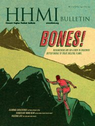
F a l l ’12 V o l.25 • No. 03<br />
perils of fatty liver<br />
vosshall on the scent<br />
über cameras
Observations<br />
the training pipeline<br />
Owen Gatley<br />
To prepare and retain a topnotch, diverse scientific workforce,<br />
trainees should be introduced to multiple career paths, according to<br />
a June report by the NIH Biomedical Workforce Working Group.<br />
One of those options—the staff scientist—could use a boost. Co-chaired<br />
by Princeton University President Shirley Tilghman (see “The Future<br />
of Science,” HHMI Bulletin, May 2011) and Sally Rockey, NIH deputy<br />
director for extramural research, the working group notes a decline in<br />
biomedical Ph.D.s in tenure-track faculty positions, and suggests<br />
modifying training programs to include diverse experiences relevant<br />
to the careers they are likely to find. HHMI investigators on the<br />
14-member committee included Leemor Joshua-Torr and Richard Lifton.<br />
The typical academic laboratory consists of a PI and one or a small<br />
number of permanent technical staff, with the majority of the research<br />
carried out by trainees. This creates a system in which a large number of<br />
future scientists are being produced each year, well in excess of the<br />
number of research-oriented jobs in academia, government and industry.<br />
The working group believes that even a modest change in the ratio of<br />
permanent staff to trainees could have a beneficial effect on the system<br />
without reducing the productivity of the research enterprise. Staff<br />
scientists—individuals with master’s or Ph.D. degrees—could play a<br />
more important role in biomedical research (one that may become<br />
increasingly necessary if the market for biomedical researchers<br />
strengthens outside of the United States in coming years).<br />
Today, these scientists bring stability to many labs and provide<br />
important functions as part of institutional core facilities, but have a<br />
wide variety of titles and employment conditions. As an example,<br />
staff scientists constitute an essential part of the NIH intramural<br />
research program. In the extramural program, these scientists do not<br />
apply for their own grants, but are supported by research project,<br />
Center and Program Project grants. They should be differentiated from<br />
“soft money” scientists, whose employment depends upon their<br />
successful competition for research funds, a category that has been<br />
increasing over the last few years.<br />
The working group encourages NIH study sections to be receptive to grant<br />
applications that include staff scientists and urges institutions to create<br />
position categories that reflect the value and stature of these researchers.<br />
Excerpted from Biomedical Workforce Working Group Report,<br />
A Working Group of the Advisory Committee to the Director, National<br />
<strong>Institute</strong>s of Health, June 14, 2012.
fall ’12 vol. 25 · no. o3<br />
12<br />
18<br />
Web-Only Content<br />
Meet some of the behind-the-scenes specialists<br />
who help modern science get done.<br />
Turtles and snakes and frogs, oh my! View the<br />
critters discovered during a herpetological<br />
survey at Janelia Farm.<br />
Read a Q&A with computer scientist<br />
Hanchuan Peng on developing sophisticated<br />
ways to make sense of biological images.<br />
Watch a fruit fly embryo develop and follow a<br />
dragonfly as it captures its prey, thanks to the<br />
latest developments in visualization techniques.<br />
Learn about the technology that lets<br />
researchers watch a heart grow, one vibrant<br />
cell at a time.<br />
24 30<br />
GO GREEN<br />
Prefer to receive your<br />
Bulletin digitally? Go to<br />
hhmi.org/bulletin/subscribe.<br />
Features<br />
cover story<br />
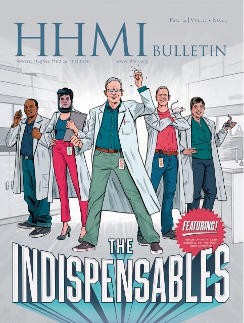
THE INDISPENSABLES<br />
12 The backbone of most labs, these quiet<br />
heroes do it all and then some.<br />
avant garde scientist<br />
18 With an outsider’s perspective,<br />
Leslie Vosshall has changed thinking<br />
about the meaning of olfaction—<br />
for humans and insects.<br />
THE FAT YOU CAN’T SEE<br />
24 In growing numbers of people, the liver<br />
holds a hidden, dangerous store of fat.<br />
Finding the triggers is step one.<br />
the view from here<br />
30 Unbounded creativity—and powerful<br />
computers—make possible the latest<br />
devices designed to peer into the<br />
deepest recesses of organs and cells.<br />
Departments<br />
PRESIDENT’S LETTER<br />
03 Stabilizing Forces<br />
CENTRIFUGE<br />
04 Good Stewards<br />
05 Undercover Triathlete<br />
06 Stepping into Sustainability<br />
UPFRONT<br />
08 Push and Pull<br />
10 Cancer’s Dead End<br />
PERSPECTIVES AND OPINIONS<br />
34 Connecting Cultures<br />
36 Q&A—What part of your job would<br />
people find the most surprising?<br />
CHRONICLE<br />
Science Education<br />
38 Teaching Genomics, Plainly<br />
<strong>Institute</strong> News<br />
40 HHMI Awards $50 Million to Colleges<br />
40 Fifty International Students Get<br />
Support from HHMI<br />
41 <strong>Medical</strong> Fellows Get a Chance to<br />
Try Research<br />
41 2012 Holiday Lectures on Science—<br />
Changing Planet: Past, Present, Future<br />
Lab Book<br />
42 The Yin and Yang of Plant Defense<br />
43 Like a Chinese Finger Trap<br />
44 Wiring the Brain<br />
Ask a Scientist<br />
45 Why do we develop distastes for<br />
certain foods?<br />
Nota Bene<br />
46 News of recent awards and other<br />
notable achievements<br />
OBSERVATIONS<br />
The Training Pipeline<br />
VISIT THE BULLETIN ONLINE FOR ADDITIONAL CONTENT AND ADDED FEATURES: www.hhmi.org/bulletin COVER IMAGE: CHRIS KING
editor’s letter<br />
Where’s My HHMI Bulletin?<br />
Something’s different, all right. You didn’t receive<br />
your August HHMI Bulletin. Not to worry; it just<br />
means that we’re going to a new publication<br />
schedule. Beginning with this issue, the Bulletin<br />
will be published three times a year, rather than<br />
quarterly. The issues—called Fall, Winter, and<br />
Spring—will come out in September, January, and<br />
May, respectively.<br />
Publication of the web and iPad editions of<br />
the Bulletin will follow the same schedule, with<br />
all the vibrant multimedia, interactive elements,<br />
and extra content you’ve come to expect. During<br />
the summer months, when many of you are busy<br />
with family and travel, we plan to make regular<br />
updates to our website with fresh content.<br />
We hope the new publication schedule will<br />
suit you, our readers, as we move forward in an<br />
increasingly digital age. And we hope that with<br />
this change we are lessening the impact on the<br />
planet, in at least a small way. While many readers<br />
tell us they prefer the look and feel of the<br />
print Bulletin, others have wholly embraced the<br />
web or iPad editions. Whatever your preference,<br />
we are committed to continue to bring you stories<br />
of scientific discovery.<br />
We hope, too, that you enjoy this issue, particularly<br />
our cover story on “the indispensables”—<br />
those research specialists who are critical to the<br />
success of myriad laboratory research programs.<br />
Having spent many years working at the bench,<br />
both in university medical centers and industry,<br />
I know just how essential these individuals can<br />
be. Though some shy away from the limelight, we<br />
take a moment to shine a light, however briefly,<br />
on the talents and contributions of a few of<br />
HHMI’s lab heroes—in this print issue and on our<br />
website. We hope their stories will inspire future<br />
generations to follow in their footsteps.<br />
HHMI TRUSTEES<br />
James A. Baker, III, Esq.<br />
Senior Partner / Baker Botts LLP<br />
Ambassador Charlene Barshefsky<br />
Senior International Partner<br />
WilmerHale<br />
Joseph L. Goldstein, M.D.<br />
Regental Professor & Chairman / Department of Molecular Genetics<br />
University of Texas Southwestern <strong>Medical</strong> Center at Dallas<br />
Hanna H. Gray, Ph.D.<br />
President Emeritus & Harry Pratt Judson<br />
Distinguished Service Professor of History<br />
The University of Chicago<br />
Garnett L. Keith<br />
Chairman / SeaBridge Investment Advisors, LLC<br />
Former Vice Chairman & Chief Investment Officer<br />
The Prudential Insurance Company of America<br />
Fred R. Lummis<br />
Chairman & CEO<br />
Platform Partners LLC<br />
Sir Paul Nurse<br />
President / The Royal Society<br />
Dame Alison Richard, Ph.D.<br />
Professor / Yale University<br />
Clayton S. Rose, Ph.D.<br />
Professor of Management Practice<br />
Harvard University<br />
Kurt L. Schmoke, Esq., Chairman<br />
Vice President & General Counsel / <strong>Howard</strong> University<br />
Anne M. Tatlock<br />
Director, Retired Chairman & CEO<br />
Fiduciary Trust Company International<br />
HHMI OFFICERS<br />
Robert Tjian, Ph.D. / President<br />
Cheryl A. Moore / Executive V.P. & Chief Operating Officer<br />
Craig A. Alexander / V.P. & General Counsel<br />
Sean B. Carroll, Ph.D. / V.P. for Science Education<br />
Jack E. Dixon, Ph.D. / V.P. & Chief Scientific Officer<br />
Mohamoud Jibrell / V.P. for Information Technology<br />
Nitin Kotak / V.P. & Chief Financial Officer<br />
Gerald M. Rubin, Ph.D. / V.P. & Executive Director,<br />
Janelia Farm Research Campus<br />
Kathy Wyszynski / V.P. for Human Resources<br />
Landis Zimmerman / V.P. & Chief Investment Officer<br />
HHMI BULLETIN STAFF<br />
Mary Beth Gardiner / Editor<br />
Cori Vanchieri / Story Editor<br />
Jim Keeley / Science Editor<br />
Nicole Kresge / Assistant Editor<br />
Maya Pines / Contributing Editor<br />
Additional ContrIButors<br />
Michelle Cissell, Mark Farrell, Lyn Garrity,<br />
Heather McDonald, Anne Sommer<br />
VSA Partners, NYC / Concept & Design<br />
Finlay Printing / Printing & Binding<br />
Telephone (301) 215.8855 • Fax (301) 215.8863 • www.hhmi.org<br />
©2012 <strong>Howard</strong> <strong>Hughes</strong> <strong>Medical</strong> <strong>Institute</strong><br />
The opinions, beliefs, and viewpoints expressed by authors in the HHMI Bulletin<br />
do not necessarily reflect the opinions, beliefs, viewpoints, or official policies of<br />
the <strong>Howard</strong> <strong>Hughes</strong> <strong>Medical</strong> <strong>Institute</strong>.<br />
Paul Fetters<br />
2 h h m i b u l l e t i n | Fall 2o12
president’s letter<br />
Stabilizing Forces<br />
James Kegley<br />
Walk into any high-level research lab today and you will<br />
find a few special individuals who are critical to that lab’s success.<br />
Chances are they’ve been a fixture in the lab for a while. Some of the<br />
very best research associates, research specialists, and lab managers<br />
are in permanent jobs. When you land a really good one—we refer<br />
to them as “the indispensables” in this issue of the Bulletin—you feel<br />
very lucky because they are hard to find. And they are often the glue<br />
that holds the lab together.<br />
In research labs, graduate students and postdocs are transient. It’s<br />
the nature of the system. They float in an out at a pretty rapid clip,<br />
moving on after about four to six years. On one hand, having that<br />
ebb and flow of trainees is good—essential even—as it’s a driving<br />
force for innovation and creativity. But at the same time, you need<br />
a core group to serve as the institutional memory of the lab, people<br />
who understand how we got where we are and why we are doing<br />
things a certain way—the progression, if you will.<br />
If a postdoc who develops a new methodology leaves, the lab<br />
head can feel like he or she has to start over again. That information<br />
can get lost if there is a gap between postdocs. With a few key<br />
long-term people in place, however, the postdoc can transfer that<br />
knowledge and expertise to them. And these staff scientists are very<br />
high level, many with a master’s, a Ph.D., and often even postdoctoral<br />
experience themselves. So they are extremely important for<br />
the continuity of the lab.<br />
In my own lab at the University of California, Berkeley, there<br />
are a handful of people without whom the lab would not function.<br />
One is my lab manager, Mallory Haggart. Mallory has kept the lab<br />
running for over 12 years. She coordinates all lab activities and takes<br />
care of all the supplies, equipment, and chemicals, as well as repairs.<br />
A little older than the students and postdocs, she’s the “mother hen”<br />
of the lab who helps establish the lab’s culture and atmosphere.<br />
Then there are our experts in specific technical areas. Carla Inouye,<br />
a biochemist, practices what is becoming a dying art: when a postdoc<br />
or student comes in and has to purify a protein, she’s the one<br />
who shows them how to do it. She’s been with me since 1998 and is<br />
fantastically valuable as the lab’s memory. Same for Shuang Zheng,<br />
who is in charge of cell culture—everyone in the lab relies on his<br />
skills in that arena. And Gina Dailey is our expert in the complicated<br />
molecular genetics, all the cloning and sequencing.<br />
These extremely skilled specialists are the stabilizing forces of<br />
the lab. Because graduate students and postdocs are trying to climb<br />
the academic ladder, they can be competitive, with sometimessharp<br />
elbows. You really need a small group of stalwarts to manage<br />
the whole thing.<br />
Relative to 25 or 30 years ago, the composition of a high-performing<br />
research lab has shifted. There are more of these backbone<br />
“Recognizing the role of research<br />
professionals in today’s laboratory<br />
organizations is important not<br />
only to the individuals who contribute<br />
their services but also to the<br />
research enterprise as a whole.<br />
”<br />
Robert Tjian<br />
employees on staff these days. At HHMI’s Janelia Farm Research<br />
Campus, the ratio of permanent staff is somewhat higher than in<br />
most organizations. Janelia has certain project teams where a scientist<br />
is not, classically speaking, a principal investigator. Instead, that<br />
person is there to provide very high-level service, such as creating<br />
transgenic animal models or performing cryo-electron microscopy.<br />
It’s a different model. And those scientists don’t feel at all that they<br />
are not appreciated. They know the work they are doing is highly<br />
regarded and valued.<br />
Recognizing the role of research professionals in today’s laboratory<br />
organizations is important not only to the individuals who contribute<br />
their services but also to the research enterprise as a whole. To that<br />
end, it is notable that the National <strong>Institute</strong>s of Health (NIH) has<br />
just released its “Biomedical Research Workforce Working Group<br />
Draft Report,” which makes recommendations for actions that NIH<br />
should take to support a future sustainable biomedical research infrastructure<br />
(see Observations, inside back cover). The report draws<br />
attention to the increasingly important and beneficial effect that staff<br />
scientists have on the system as a whole.<br />
The message is clear: these professional scientists are remarkably<br />
valuable to the progress of research. They are not looking for a fancy<br />
title or a bookshelf of awards—that’s not their motivation. They are<br />
no less effective as scientists; they’re just a different kind of scientist.<br />
They have our support, and they have our respect and appreciation.<br />
Fall 2o12 | h h m i b u l l e t i n<br />
3
centrifuge<br />
Good Stewards<br />
Spring is lively in the woods surrounding<br />
HHMI’s Janelia Farm Research Campus<br />
in Ashburn, Virginia. White-tailed deer<br />
rustle fallen leaves, pileated woodpeckers<br />
drum persistently overhead, and<br />
gray tree frogs trill in the treetops.<br />
But on one cloudy May afternoon, the<br />
sounds of nature are muffled by the<br />
cries of discovery. “Hey, Larry,” comes<br />
the call, “I found something!”<br />
A team of more than 20 explorers—<br />
Janelia Farm employees and their natureloving<br />
recruits—are combing the terrain:<br />
rolling aside logs, prodding leaf piles,<br />
and gently displacing stones. The reptiles<br />
and amphibians they seek out do<br />
little to announce themselves, but this<br />
is an observant group, and the finds<br />
come quickly.<br />
“Hey, Larry, is this another worm<br />
snake?” someone asks as an energetic<br />
wriggle of pink and gray catches his<br />
eye. “It’s amazing what you find if you<br />
just flip things over.” Another participant<br />
announces he’s found a frog—<br />
a pickerel, he guesses, judging by the<br />
spotty skin. Moments later, a salamander<br />
sighting evokes a flurry of activity<br />
as a group peers into the stream waiting<br />
for its yellow tail to reappear.<br />
Larry Mendoza, Janelia Farm’s biosafety<br />
and laser safety officer, scurries<br />
to each find. Mendoza is president of<br />
the Virginia Herpetological Society<br />
and a co-founder of Janelia’s Nature<br />
Club, through which he has gathered<br />
this crew for the second in a series of<br />
herpetological surveys of the Janelia<br />
Farm grounds.<br />
Reptiles and amphibians are particularly<br />
sensitive to environmental<br />
pressures, so monitoring the snakes,<br />
lizards, frogs, salamanders, and turtles<br />
that inhabit this land helps reveal the<br />
health of the local ecosystem, Mendoza<br />
explains. As future surveyors begin to<br />
track population changes over time,<br />
the data will become even more valuable.<br />
Just as important, adds Mendoza,<br />
is ensuring the event is educational<br />
and fun.<br />
Many of the scientists on the excursion<br />
have come recently to Janelia<br />
Farm and want to learn more about<br />
the local ecology. “We had very few<br />
reptiles of any kind in Seattle,” observes<br />
Matthew Iadanza, a member of<br />
Tamir Gonen’s structural biology lab,<br />
which relocated from the University<br />
of Washington last year. Others are<br />
here hoping to foster their children’s<br />
curiosity, and some of the day’s best<br />
finds—a slender water snake, an<br />
unusually vibrant box turtle—come<br />
from the group’s youngest naturalists.<br />
“I walked right by that turtle,” Mendoza<br />
says. “That’s why you’ve always got to<br />
bring kids!”<br />
After three and a half hours, the<br />
team has located 33 animals representing<br />
11 different species, and Mendoza<br />
declares the survey a success. He is<br />
particularly excited by the 15 eastern<br />
box turtles. The species may be threatened<br />
in Virginia because of loss of<br />
habitat, and Mendoza says their prevalence<br />
indicates good stewardship of<br />
this land.<br />
Mendoza has recorded the location,<br />
time, and discoverer of each find for<br />
inclusion in the Herpetological Society’s<br />
biannual publication, Catesbeiana. All<br />
participants will be asked to review<br />
the manuscript, he tells the group. As<br />
they disband, he reminds the team of<br />
upcoming Nature Club events—more<br />
herpetological surveys, bird walks, and<br />
perhaps a tour of insect life led by a<br />
resident entomologist. “We have 689<br />
acres here at Janelia,” he says. “That’s<br />
a lot of land—a lot of opportunity.”<br />
—Jennifer Michalowski<br />
web extra: For a sampling of what the naturalists<br />
found during their outing, see the audio slideshow at<br />
www.hhmi.org/bulletin/fall2012.<br />
Photo of Larry Mendoza by Adam Auel<br />
4 h h m i b u l l e t i n | Fall 2o12
Undercover<br />
Triathlete<br />
Emma Kelly<br />
When Erika Erickson’s California lab<br />
mates heard that she was just back<br />
from a triathlon competition in Beijing—<br />
where she earned gold in her age<br />
group—most were shocked. They knew<br />
she worked unusual hours for a graduate<br />
student, with lots of very early<br />
mornings and late nights, but they had<br />
no idea how she was spending her<br />
daytime hours.<br />
Her graduate adviser, HHMI-GBMF<br />
investigator Krishna K. Niyogi, knew<br />
what she was up to but can see how it<br />
might have been a surprise to others.<br />
“She’s pretty low-key about it,” he says.<br />
“She’ll really only tell you how it went<br />
if you ask her.”<br />
Erickson ran track and swam competitively<br />
as an undergraduate at the<br />
Massachusetts <strong>Institute</strong> of Technology<br />
but hadn’t tried triathlons until she moved<br />
to the University of California, Berkeley,<br />
for graduate study in plant biology.<br />
Two and a half years later, she has<br />
earned professional ranking in the sport,<br />
which means she can compete for<br />
prizes of $5,000 or more in events sanctioned<br />
by the sport’s main association,<br />
USA Triathlon. She trains 15 to 25 hours<br />
per week and fits in her research around<br />
workouts. “I don’t keep standard hours,”<br />
she laughs, saying it’s understandable<br />
that her lab mates were mystified by<br />
her trek to China. “And I don’t talk<br />
about my personal life too often in lab;<br />
I don’t want to give the impression<br />
that science is my second priority.”<br />
Along with training and travel to<br />
races, Erickson makes sure that her<br />
research on the structure and function<br />
of proteins involved in photosynthesis<br />
progresses at a reasonable pace. She<br />
says the dual focus gives her a bit of<br />
stability. “If I have a bad week in lab, my<br />
training can balance that out. Or if I<br />
have a bad week racing, a good week<br />
in lab can make up for it.”<br />
In her first professional race, the<br />
June 2 Pan American Cup in Dallas, her<br />
focus paid off. She placed 8th on the<br />
1,500-meter swim, 40-kilometer bike,<br />
10-kilometer run. Her time of 2:19:03<br />
was just 8 minutes, 41 seconds behind<br />
that of the leader, Laura Bennett, who<br />
represented the U.S. at the Olympics<br />
this summer.<br />
“The race went well overall, but the<br />
conditions were incredibly challenging—<br />
95 degrees and windy,” she says. And<br />
her competitors were very different<br />
from her usual running mates.<br />
“People weren’t at the Dallas race<br />
just to see if they could finish or just to<br />
have fun.” They were there for national<br />
pride, to win money and points in the<br />
ranking system. Those goals, she says,<br />
“bring an entirely different set of emotional<br />
responses to a race. I was really<br />
intimidated by my competitors. But I<br />
had fun and learned a lot.”<br />
As for her lab mates, their only firsthand<br />
experience with Erickson’s racing<br />
talents came from a 2-mile run around<br />
the Lawrence Berkeley National Laboratory.<br />
Niyogi offered to buy lunch for<br />
anyone in the lab who would participate<br />
with him or watch the race. “Several of<br />
us went, and Erika was just way, way<br />
out in front of all of us,” he says laughing.<br />
“She won the women’s division. I<br />
don’t think anybody was too surprised.”<br />
—Rabiya Tuma<br />
Fall 2o12 | h h m i b u l l e t i n<br />
5
centrifuge<br />
Stepping into Sustainability<br />
Peter Baumann will not go hungry if the<br />
local supermarket shuts down tomorrow.<br />
In fact, if all food production in the<br />
United States ground to a halt, he would<br />
probably be just fine.<br />
Baumann is an HHMI early career scientist<br />
at the Stowers <strong>Institute</strong> for <strong>Medical</strong><br />
Research in Kansas City, Missouri. In his<br />
spare time he hunts, grows, raises, and<br />
gathers almost 80 percent of the food<br />
that he and his wife, Diana, and their<br />
two dogs consume.<br />
The couple lives on 5 acres of land<br />
scattered with fruit trees, berry bushes,<br />
a large vegetable garden, and a pond<br />
stocked with bass and catfish. They have<br />
three freezers crammed with venison and<br />
chicken; a cellar overflowing with squash,<br />
potatoes, jams, and canned vegetables;<br />
and a coop of egg-laying chickens.<br />
“It sort of happened gradually,” says<br />
Baumann of his family’s foray into sustainable<br />
living.<br />
Baumann was lured by the memory<br />
of the taste of fresh vegetables grown<br />
in his mother’s extensive garden at their<br />
home in Germany. Diana’s childhood gardening<br />
memories, on the other hand, are<br />
limited to the few radishes her brother<br />
managed to coax from the ground in<br />
England. When the couple moved to a<br />
house in the countryside six years ago,<br />
they planted a few tomato seeds in pots<br />
on the patio. Unfortunately, raccoons<br />
got to the harvest before they could.<br />
The next year the Baumanns planted<br />
a small garden in their yard and adopted<br />
a dog to keep the raccoons away. It<br />
worked, and a year later they expanded:<br />
the garden got bigger and a second<br />
dog joined the family. “It really took off,”<br />
Baumann says. Today, the garden occupies<br />
a little more than half an acre and<br />
is home to a variety of beans, tomatoes,<br />
peppers, greens, squash, and herbs.<br />
What Baumann doesn’t grow he<br />
gathers, which he learned to do during<br />
weeks-long hikes in Europe and America.<br />
He’s been known to supplement his<br />
diet with unconventional items, such<br />
as the inner bark of a birch tree, which<br />
makes a tasty pasta substitute. His<br />
current foraging, however, is limited to<br />
mushrooms, cattails, pawpaws, and<br />
berries. “The inner shoots of the cattails<br />
are very similar to bamboo shoots,<br />
so you can put them in stir-fries or<br />
salads,” he says.<br />
For meat, Baumann kills about<br />
five deer each fall, which he and Diana<br />
butcher together. They use all the<br />
meat, keeping the good cuts for roasts<br />
and stews and grinding up the rest<br />
for the dogs, who also get the hoofs<br />
for chew toys.<br />
Chickens were a late addition to<br />
Baumann’s small-scale farm. It took some<br />
time for Diana to agree, but she came<br />
around and they mail-ordered 27 chicks<br />
in February 2011. This year, they added<br />
47 more chicks and 16 ducks, which<br />
roam around a half-acre enclosure. Some<br />
of the chickens end up on the dinner<br />
table and the rest produce about 40 to<br />
50 eggs per week.<br />
Baumann is ready to add goat meat<br />
and milk to the family’s diet. “We have<br />
the space and the ideal habitat for<br />
them,” he explains. Since Diana is now<br />
a pro at butchering deer and plucking<br />
chickens, he expects that something<br />
as innocuous as a few goats wandering<br />
the property won’t faze her at all.<br />
—Nicole Kresge<br />
web extra: View a slideshow of Peter Baumann’s smallscale<br />
farm at www.hhmi.org/bulletin/Fall2012.<br />
Emma Kelly<br />
6 h h m i b u l l e t i n | Fall 2o12
upfront<br />
08 PUsh and pull<br />
What appears chaotic is actually a well-orchestrated process<br />
for embryonic head-to-tail elongation.<br />
10 cancer’s dead end<br />
There is a way to restore p53’s tumor-suppressing prowess.<br />
web only content<br />
SAME BUT NOT EQUAL<br />
A code within the genetic code explains why identical proteins<br />
are produced at varying speeds.<br />
NERVE TONIC<br />
Axon degeneration—an active process after injury—can be delayed.<br />
Find these stories at www.hhmi.org/bulletin/fall2012.<br />
To work on broad and basic problems that touch on<br />
many different fields has been Jonathan Weissman’s<br />
goal. He made a recent leap to understand the<br />
nuances of protein production with encouragement<br />
from two stellar scientists. It’s a gift when<br />
one generation of researcher can lean on the one<br />
that came before—perhaps through their papers<br />
and lab notes or, even better, by chatting over<br />
a meal. Go to the online Bulletin to learn about<br />
Weissman’s inspirations.<br />
Fall 2o12 | h h m i b u l l e t i n<br />
7
upfront<br />
Push and Pull<br />
What appears chaotic is actually a well-orchestrated<br />
process for embryonic head-to-tail elongation.<br />
Few sights are as captivating as the movements of a developing<br />
embryo. Shapeless tissue remodels itself as cells migrate en masse to<br />
form elongated structures that, depending on the species, will become<br />
a frog spinal cord, a human gut, or the main body of a fly. Jennifer<br />
Zallen, an HHMI early career scientist at Memorial Sloan-Kettering<br />
Cancer Center, studies how embryonic tissues stretch along an anterior-posterior<br />
axis using the fruit fly Drosophila melanogaster as a<br />
model system. Shortly after formation of<br />
the embryo, the mass of fruit fly cells dramatically<br />
elongates over about a two-hour<br />
period and establishes a head at one end<br />
and a tail at the other, a process known as<br />
convergent extension.<br />
Over the past seven years, Zallen’s lab<br />
has used high-resolution, time-lapse imaging<br />
to track embryonic cell migration during<br />
that two-hour window. She has found that<br />
what looks like chaotic pushing and shoving<br />
between neighboring cells in elongating<br />
tissue is actually a highly cooperative and<br />
orderly process.<br />
Zallen’s group established the ground<br />
rules of the game in a 2006 Developmental<br />
Cell paper. Tracking single cells in a living<br />
embryo, they showed that cells consistently<br />
join and then exit pinwheel-like structures<br />
known as rosettes as a fly embryo becomes<br />
longer and thinner. Computational analysis<br />
indicated that most cells repeatedly move<br />
in and out of multiple 5-8 cell clusters as<br />
the embryo stretches outward. Each group<br />
of cells reorganizes to become longer and<br />
narrower, suggesting that rosette formation<br />
could drive morphological change.<br />
Since then, her laboratory has focused<br />
on understanding the multiple signals that<br />
encourage cells to move in and out of transient<br />
groups. “It is forces generated at the<br />
contacts between cells that cause the cells<br />
to rearrange and the tissue to elongate,”<br />
says Zallen. “These forces place special<br />
demands on the cell junctions, which must<br />
be dynamic enough to dismantle individual<br />
contacts but strong enough to prevent the<br />
group from ripping apart under tension.”<br />
Zallen’s lab group recently combined<br />
molecular techniques with Drosophila genetics<br />
to characterize one facet of that junctional<br />
regulation. In work published February 14,<br />
2012, in Developmental Cell, they identified<br />
a biochemical cue that allows cells to disengage<br />
from some neighbors in a rosette so they<br />
can find new ones. They found that a signaling<br />
factor, the tyrosine kinase protein Abl,<br />
makes the contact points (known as adherens<br />
junctions) between cells in a rosette more<br />
dynamic or fluid—in other words, it allows<br />
cells to glide more smoothly against one<br />
another. Fruit fly embryos genetically engineered<br />
to lack Abl show poor rosette formation<br />
and impaired elongation. Surprisingly, the<br />
more dynamic junctions in normal embryos<br />
are also stronger, resisting apparent breaks or<br />
tears seen in embryos lacking Abl.<br />
Equally essential for axis formation are<br />
mechanical signals that rope cells into a<br />
rosette. In work published in Developmental<br />
Cell in 2009, Zallen reported that cells<br />
perceive physical tension exerted by their<br />
8 h h m i b u l l e t i n | Fall 2o12
Brendan Monroe<br />
neighbors. These mechanical signals promote<br />
rosette cohesion by prompting the<br />
motor protein myosin to join long cables<br />
that extend across multiple cells and<br />
contract like a drawstring, pulling cells<br />
together to form a rosette.<br />
One way her group showed this experimentally<br />
was by literally tugging on a<br />
Drosophila embryo with a glass needle and<br />
then using live imaging to watch as fluorescently<br />
labeled myosin was recruited to the<br />
needle. This experiment helped explain<br />
one purpose that mechanical forces serve,<br />
namely, to bring cells together into multicellular<br />
gatherings.<br />
Whether the same mechanisms that<br />
push and pull cells in a simple organism<br />
like Drosophila drive convergent extension<br />
in vertebrate embryos remains unknown.<br />
“Right now, people have seen snapshots of<br />
something that looks like rosettes in other<br />
animals,” she says, noting that the vertebrate<br />
neural tube (the embryonic structure<br />
that gives rise to the central nervous system)<br />
exhibits pinwheel-like swirls of<br />
cells. “But rosette formation is a dynamic<br />
behavior that is difficult to assess in a static<br />
picture. Live imaging of vertebrate cells is<br />
needed to see if these cells move in a way<br />
that leads to elongation.”<br />
Even if she and others discover that<br />
vertebrates evolved a different way to form<br />
tubular organs, Zallen has an ambitious<br />
long-term goal: to figure out how large<br />
populations of cells act collectively. “Over<br />
the past 20 years, people have learned a lot<br />
about factors that determine cell identity,”<br />
she says. “But we know much less about<br />
how cells get to the right place to build<br />
a three-dimensional animal. This is the<br />
big unsolved question in developmental<br />
biology.” W – elise lamar<br />
web extra: See a video of rosette formation in<br />
Drosophila at www.hhmi.org/bulletin/Fall2012.<br />
Fall 2o12 | h h m i b u l l e t i n<br />
9
upfront<br />
Cancer’s Dead End<br />
There is a way to restore p53’s tumor-suppressing prowess.<br />
At first glance, the tumor suppressor gene p53 would seem<br />
like an ideal weapon against cancer. It puts the brakes on cancer<br />
progression and is impaired in about half of human tumors. <br />
In mouse models of cancer, HHMI investigators Tyler Jacks and Scott<br />
Lowe restored p53 activity in tumors and the tumors regressed.<br />
However, p53 activation kills some cancer<br />
cells, but not others, and no one knows<br />
why. HHMI early career scientist Joaquín<br />
Espinosa has set his sights on finding an<br />
answer, and with it, a strategy for making<br />
p53-based therapies effective. It’s a goal he’s<br />
pursued with relentless passion.<br />
“He’s an extremely creative thinker,”<br />
says former HHMI President Thomas Cech,<br />
who is an HHMI investigator and colleague<br />
of Espinosa’s at the University of Colorado<br />
at Boulder. Where a traditional approach<br />
might focus entirely on the p53 gene itself,<br />
Espinosa has adopted a broader and far<br />
more ambitious target—he aims to inventory<br />
all the genes and pathways that govern<br />
how cells react to p53 activation.<br />
Espinosa’s approach grew from his realization<br />
that if you want to harness p53’s<br />
tumor-squelching effects, you also need to<br />
block the myriad pathways that tumors use<br />
to circumvent p53’s killing orders. Espinosa<br />
sought to identify all of the p53-disarming<br />
pathways and the genes that control them<br />
with a technique called functional genomics.<br />
“This is a very new technology which<br />
allows us to interrogate the function of<br />
every gene in the human genome as it<br />
relates to p53,” Espinosa says. “With functional<br />
genomics, we can ask—which genes<br />
influence the programmed cell death<br />
mediated by p53?”<br />
To do functional genomics requires<br />
expensive equipment and huge libraries of<br />
costly reagents, and only a few such facilities<br />
exist in the world. Espinosa wanted one<br />
at the University of Colorado at Boulder.<br />
He had a vision for a functional genomics<br />
facility in Colorado that would not only<br />
advance his own research, but also put his<br />
colleagues at the forefront of biomedical<br />
research. “Joaquín enjoys doing things for<br />
the benefit of the scientific community,”<br />
Cech says. “He’s more than a team player,<br />
he’s an instigator of teamwork.”<br />
Espinosa’s first proposals to fund his<br />
functional genomics experiments were<br />
rejected by the National <strong>Institute</strong>s of<br />
Health and private foundations. But he<br />
persisted, eventually securing support from<br />
HHMI, the BioFrontiers <strong>Institute</strong>, and the<br />
University of Colorado Cancer Center.<br />
The Functional Genomics Facility at the<br />
University of Colorado at Boulder began<br />
operations in May 2010 with Espinosa at<br />
its helm. “We could finally do our dream<br />
experiment,” Espinosa says.<br />
In a series of investigations published<br />
online June 3, 2012, in Nature Chemical<br />
Biology, Espinosa’s group identified genes<br />
that, when inhibited, allow p53 to kill<br />
cancer cell types that wouldn’t otherwise<br />
respond. They accomplished this using an<br />
experimental drug called Nutlin-3, which<br />
activates the p53 protein, yet fails to induce<br />
cell death in most tumors. Using functional<br />
genomics, Espinosa’s team screened thousands<br />
of genes and identified a few hundred<br />
whose inhibition made Nutlin-3 effective at<br />
killing cancer cells. To their delight, they<br />
discovered that compounds to inhibit the<br />
protein products of two of these genes, ATM<br />
and MET, were already available.<br />
When they combined the ATM or<br />
MET inhibitors with Nutlin-3, the treatment<br />
destroyed cancer cells that didn’t<br />
respond to any of the drugs individually. “If<br />
you treat cancer cells with one of the drugs,<br />
nothing happens, but if you treat them with<br />
both drugs, the drugs kill them,” Espinosa<br />
says. The drug combination delivered a<br />
one-two punch—Nutlin-3 turned on p53,<br />
then the ATM or MET inhibitor blocked<br />
VSA Partners<br />
10 h h m i b u l l e t i n | Fall 2o12
the proteins that would allow the cancer<br />
cell to thrive despite p53 activation.<br />
“With one genetic screen, we identified a<br />
combination of drugs that will kill tumors<br />
that otherwise resist them,” Espinosa says.<br />
His team is collaborating with clinical<br />
investigators to test these drug combinations<br />
in animal models and human<br />
tumors grown in mice.<br />
By hitting multiple genetic targets,<br />
Espinosa hopes to drive cancer cells into a<br />
dead end. “You can block resistance genes<br />
before they ever have a chance,” he says.<br />
With this strategy, new treatments can be<br />
designed to overcome a tumor’s natural<br />
work arounds. “The future is not in isolated<br />
drugs, but rather in combinations of<br />
drugs,” Espinosa says.<br />
Clinical trials are costly and time-consuming,<br />
and Espinosa hopes his studies<br />
can help drug researchers predict and<br />
anticipate how their p53-related treatments<br />
will work in the clinic, long before they’ve<br />
spent millions on trials. “These are very<br />
basic experiments we can do in the lab to<br />
get ahead of problems we’ll see in clinic.”<br />
W –Christie Aschwanden<br />
Fall 2o12 | h h m i b u l l e t i n<br />
11
written by<br />
madeline drexler<br />
the backbone of most labs, these quiet<br />
heroes do it all and then some.<br />
ILLUSTRATED by<br />
CHRIS KING
oblem solvers, shelf stockers, bench<br />
scientists, record keepers, machine fixers,<br />
weekend warriors, den mothers, old hands,<br />
fresh eyes, mentors, managers. Every research<br />
lab has behind-the-scenes specialists without<br />
whom modern science could not get done.<br />
Their bland bureaucratic titles—research<br />
technician, resource manager—belie their vital<br />
contributions. These women and men are not wannabe<br />
principal investigators (PIs) or discouraged academics.<br />
Some have specialized expertise; others are jacks-of-all-trades.<br />
They are ambitious, not for fame, but for personal excellence<br />
and the chance to make a lasting contribution, however unheralded,<br />
to science.<br />
“There are very few laboratories that do not have at least one<br />
technician who often plays the role of lab manager, making sure<br />
the trains run on time, the supplies are ordered, the equipment<br />
gets repaired. It would be very hard to run a lab without such a<br />
person,” says Shirley Tilghman, a molecular geneticist and president<br />
of Princeton University.<br />
It’s a career option that needs serious consideration, according<br />
to Tilghman, who cochaired the National <strong>Institute</strong>s of Health<br />
Biomedical Research Workforce Working Group. The group’s<br />
June report calls for professionalizing the crucial research-affiliated<br />
roles of lab specialists and paying them accordingly. “Make<br />
them a professional category that academic medical centers and<br />
research institutes and universities recognize as valuable—in<br />
fact, invaluable—parts of the biomedical workforce.”<br />
A little recognition wouldn’t hurt, either. “As in most areas,<br />
credit flows up the hierarchy,” explains Gerry Rubin, director of<br />
HHMI’s Janelia Farm Research Campus, in Ashburn, Virginia.<br />
“The people at the top get way too much credit. And the technicians<br />
and managers don’t get enough. Academia very much<br />
rewards individual achievement—less so, team achievement.”<br />
Adds Rubin, “Labs are full of students and postdocs who come<br />
and go every three to four years. It’s important to have somebody<br />
with a sense of continuity and context.”<br />
Here are the stories of five indispensable lab team members,<br />
among many acknowledged by grateful HHMI investigators.<br />
my phone is<br />
never off.<br />
ROB edwards<br />
Research Technician III<br />
The Pulse of the Lab<br />
Seventeen years ago, Rob Edwards noticed an ad for a radio<br />
frequency engineer in the HIV-focused laboratory of HHMI<br />
investigator Michael Summers. On paper, Edwards may have<br />
14 h h m i b u l l e t i n | Fall 2o12
i’m with jeremy<br />
for the long haul.<br />
seemed an odd fit for the lab: no academic experience, just lots of<br />
work for defense contractors. “I don’t want to say this, but my old<br />
job depended on war—and sometimes, on destroying people’s<br />
lives,” he says. Today, after nearly two decades working alongside<br />
Summers, Edwards’ career has been recast. “What we do here<br />
saves people’s lives. It has a purpose.”<br />
At the University of Maryland, Baltimore County, Summers<br />
and his team are studying the architecture of HIV to understand<br />
how it and other retroviruses assemble, and how the viruses package<br />
their genetic material to infect other cells. This scientific<br />
pursuit hinges on nuclear magnetic resonance (NMR) spectroscopy—which<br />
is where Edwards enters the picture. He is<br />
responsible for two 600-megahertz NMR machines and a mammoth<br />
800-megahertz instrument with a cryogenic probe. “There<br />
are so many things that can go wrong,” he says. “The magnet, the<br />
probe, the platform, the software, the console, the amplifiers.”<br />
But NMR machines are just part of Edwards’ job. He designs<br />
the electrical and mechanical components of laboratory upgrades<br />
and ensures the continuous operation of centrifuges, water purification<br />
systems, electrical generators, and other critical research tools.<br />
“I keep everything up and running, because you never know when<br />
a grad student or a postdoc will have that sample—the protein<br />
they’ve been working on for a year,” Edwards says. “When they’re<br />
ready to roll, there can’t be any obstacles on the instrument side.”<br />
Edwards also mentors young scientists in Summers’ lab—<br />
including a large number of minority grad students and postdocs,<br />
who may not have received ample encouragement in the past. “He<br />
has served as a positive force for about 50 high-achieving minority<br />
students who have worked in my laboratory,” says Summers,<br />
“often privately enforcing his own interventions to challenge the<br />
students and support them during times of academic struggles.”<br />
As Edwards explains, “I’m a minority also; I know you can get<br />
down on yourself. So I always say, ‘I believe in you.’ That’s what<br />
Mike said to me when I first started here: ‘You can do it, I hired<br />
you for a reason, just be confident.’ Now that I have that confidence,<br />
when I see someone without it, I try to instill it.”<br />
Edwards brings this sense of purpose to every facet of his<br />
job. “My phone is never off. My vacation time is maxed,” he says<br />
with a laugh.<br />
phil smallwood<br />
Research Technician III<br />
maestro at the bench<br />
“Embryonic stem cells have to be tended and fed every day. They<br />
don’t know about weekends or holidays,” says Phil Smallwood,<br />
a research technician for 18 years in the laboratory of Jeremy<br />
Nathans, an HHMI investigator at the Johns Hopkins University<br />
School of Medicine who studies the mammalian visual system.<br />
“When I’m actively working with the cell cultures, I’m here<br />
seven days a week. I have been here Christmas Day, New Year’s<br />
Eve, New Year’s Day,” says Smallwood. “This is why Jeremy trusts<br />
me: he knows that it will get done and it will get done correctly.<br />
These cells are going to be turned into a mouse. I won’t cut corners,<br />
because Jeremy is basing his reputation on the conclusions<br />
that he will draw from these mice.”<br />
“Extraordinary experimentalists” is how Nathans describes<br />
Smallwood and research specialist Yanshu Wang, another essential<br />
lab team member (see Web Extra, Lab Heroes). “They have<br />
the same relationship to doing experiments that Itzhak Perlman<br />
has to playing the violin.”<br />
For Smallwood, who has created 40–50 mouse lines in Nathan’s<br />
lab—transgenics, knock-ins, knockouts—the musical analogy is<br />
apt: away from work, he is an accomplished flutist. “You always<br />
have to practice—you can’t rest on your laurels. It’s the same<br />
in science. You always have to keep learning new techniques,<br />
advancing with technology, or you’ll get left behind.”<br />
Smallwood also draws on another analogy to explain the craft<br />
of science: cooking. “The first time you make a cake, you follow<br />
the recipe. It’s the same with an experiment: you can’t change a<br />
variable right away. But after doing it for many years, you get a feel<br />
for how the cell behaves, for what works and what doesn’t. Take<br />
something as simple as pipetting an enzyme back and forth, such<br />
as in trypsinization, a process to chemically separate cells. I was<br />
originally taught that you add the trypsin once and place the colonies<br />
in the incubator. But the colonies don’t completely break up.<br />
I found that if you pipette the trypsin back and forth a few times<br />
and then put the colonies in the incubator, it’s like night and day.<br />
It’s a little trick that goes a long way.”<br />
Fall 2o12 | h h m i b u l l e t i n<br />
15
The co-inventor of 10 patents—beneficiary of Nathan’s uncommon<br />
largesse in sharing professional recognition—Smallwood<br />
doesn’t feel like an unsung hero. Yet while he appreciates the recognition,<br />
he doesn’t crave it. “The students, they come in, find<br />
a project, get published, go on for a postdoc, look for a job. I’m<br />
content to be in the background. Jeremy arrives in the morning<br />
and there I am at my bench. I’m a constant. I’m with Jeremy for<br />
the long haul.”<br />
we do a lot<br />
of quality<br />
control.<br />
peggy kroll-conner<br />
Research Technician III<br />
a star in the worm world<br />
Among the many niche communities in the giant enterprise of<br />
science, worm experts are a special cadre. Caenorhabditis elegans—the<br />
slender and sinuous nematode—may be a model<br />
organism, but it also requires model experimental techniques.<br />
And in the small guild of C. elegans specialists, Peggy Kroll-<br />
Conner is a star.<br />
Judith Kimble, an HHMI investigator at the University of<br />
Wisconsin–Madison who studies the fundamental controls of animal<br />
development, has for 16 years relied on Kroll-Conner’s finesse.<br />
“Peggy is the heart and soul of all our genetics,” says Kimble. “She<br />
has become the person who generates some of the most finicky<br />
strains in the lab. In the worm community, she is known as being<br />
exceptionally fastidious—her strains always have the correct genotype<br />
and, just as important, are not contaminated.”<br />
According to Kroll-Conner, the key to ensuring that frozen<br />
worm stocks are free of mold and bacteria is good sterile technique.<br />
She vigilantly labels items to prevent strain mix-ups. Once<br />
the worms are frozen, she thaws a portion to make sure the animals<br />
are viable and the stock pristine. She returns test thaws to the<br />
scientists who created the worms, so they can validate the strains.<br />
She also revalidates her own strains. “In other words,” says Kroll-<br />
Conner, “we do a lot of quality control.”<br />
Alongside the ever-present threat of contamination is the<br />
challenge of fashioning strains with multiple mutations. “Mother<br />
Nature doesn’t give up her secrets easily,” says Kroll-Conner. “I<br />
try to figure out a way to balance the mutations in a user-friendly<br />
way, to minimize the work needed to maintain the worms.”<br />
Certain mutations are more challenging than others, she says.<br />
In one experiment, she had to set up a mating with a strain of<br />
worms whose reproductive systems quickly developed tumors. “I<br />
set up numerous mating plates with large numbers of worms and<br />
ended up with only three cross-progeny. Only one of those had<br />
the genotype that I was looking for.”<br />
Such perseverance and attention to detail is essential in her<br />
role. It’s also about mutual support, she says, a concept that seems<br />
to have been bred into the C. elegans world by Janelia Farm senior<br />
fellow Sydney Brenner, a molecular biologist who shared the 2002<br />
Nobel Prize in Physiology or Medicine. “When Brenner started this<br />
science in the 1970s, he said that the worm community should<br />
work together in a spirit of cooperation. He felt that science would<br />
advance much faster that way,” she says. “I like that philosophy.”<br />
it’s those little<br />
steps i worry about.<br />
todd laverty<br />
shared resource manager<br />
working on the fly<br />
“A lot of things go on behind the scenes to make science happen,”<br />
says Todd Laverty, who for 29 years has supported the<br />
work of Gerry Rubin, director of HHMI’s Janelia Farm Research<br />
Campus. For 23 of those years, Laverty managed Rubin’s lab at<br />
the University of California, Berkeley. For the last six, he has overseen<br />
the Drosophila and Media Prep shared resources at Janelia.<br />
His specialty is feeding, sheltering, moving, breeding, and otherwise<br />
nurturing fruit flies, as well as preparing meals for other<br />
laboratory animals. “It was obvious, right from the beginning, that<br />
I had found my niche.”<br />
At Janelia, Laverty manages some 18,000 fly stocks—that<br />
is, 36,000 vials of flies, 50 to 100 flies per vial, each a unique<br />
genetic line. He shepherds the stocks through a 28-day lifecycle:<br />
egg, three larval stages, pupa, and hatched fly. Each morning,<br />
peering through a microscope at flies dozing on a pad suffused<br />
with carbon dioxide, he selects virgin females for genetic crosses.<br />
Each week, he starts new generations in fresh vials, using a duck<br />
feather to gently transfer the prospective parents from one plastic<br />
abode to the next. And each month, he manages the production<br />
of 130,000 vials of fly chow: a mixture of cornmeal, molasses,<br />
agar, and yeast—and gets four-star reviews from lab managers for<br />
quality and consistency.<br />
“I can remember telling people, when I started out, ‘What<br />
I’m going to be doing 20 or 30 years from now, I don’t know.<br />
But I’m happy now doing what I’m doing,’” Laverty says. After<br />
he’d worked for Gerry four or five years, he remembers riding<br />
in an elevator with a gray-haired co-worker. “I teased him: ‘Who<br />
gets gray hair working in a lab?’ Funny thing: now I’m that grayhaired<br />
guy!”<br />
16 h h m i b u l l e t i n | Fall 2o12
Laverty is the only person Gerry Rubin brought from his<br />
Berkeley lab to Janelia. “There was no one better than Todd to<br />
set up a facility to support all the Drosophila labs here. He is<br />
very calm, and he’s very good at managing people and building<br />
a team,” says Rubin. “In my Berkeley lab, at times he had to deal<br />
with as many as 15 postdoctoral fellows, all with different needs<br />
and demands. He does a remarkable job of keeping everybody<br />
happy.” In a high-pressure laboratory setting, Rubin adds, “That’s<br />
a very unusual ability.”<br />
Laverty describes himself as “a pleaser by nature. I get satisfaction<br />
out of starting a task and following through. PIs get the big<br />
ideas: ‘This is really cool, let’s figure this out. I’m not going to<br />
worry about the little steps right now.’ It’s those little steps that I<br />
worry about.”<br />
In Rubin’s lab at Berkeley, Laverty played a supporting role<br />
in more than 100 different research projects, including sequencing<br />
the Drosophila genome. Published in 2000, the sequence<br />
marked a scientific milestone because of the fruit fly’s pivotal<br />
role as a model organism in research, in areas ranging from<br />
aging and cancer to learning and memory. “There were hundreds<br />
of people in the field who could do the same thing that<br />
I was doing. But I had the opportunity to do it every day in<br />
the Rubin lab—and it was the Rubin lab that was sequencing<br />
the fly genome.”<br />
i think of cultures<br />
as my sheep.<br />
alicja trocha<br />
research specialist III<br />
making the impossible<br />
possible<br />
In 1989, Bruce Walker, an infectious disease specialist at Massa<br />
chusetts General Hospital, sifted through a tall stack of<br />
applications for a technician’s job in his lab. He immediately<br />
noticed the application from Alicja Trocha, a recent immigrant<br />
from Poland who had joined the anti-Communist Solidarity<br />
movement as a student in the 1980s. Trocha had worked in a lab<br />
on the rabies virus—like HIV, a deadly pathogen. She had earned<br />
a veterinary degree. And she had done field work with farmers,<br />
suggesting a level of real-world maturity not always seen in hothoused<br />
science majors.<br />
Walker interviewed Trocha in German, which she had picked<br />
up in an internment camp in Munich after fleeing Poland<br />
in 1986. Immediately impressed, he offered her the job.<br />
But Trocha, overwhelmed by Walker’s laboratory operation<br />
and by the technical terms she would need to master, initially<br />
“Labs are full of students<br />
and postdocs who come<br />
and go every three to four<br />
years. It’s important to have<br />
somebody with a sense<br />
of continuity and context.”<br />
gerry rubin<br />
declined. He persisted, and the result has been a professional<br />
match made in heaven.<br />
“Alicja has been invaluable to my career,” says Walker. “There<br />
is nobody in the world who can clone T cells better, and as my<br />
lab manager she has not only set the highest standards for performance<br />
and integrity but has done it in the most collegial<br />
way.” Today, Walker leads an international research effort to<br />
understand how some rare individuals, known as long-term nonprogressors—infected<br />
with HIV but never treated—can fight off<br />
the virus with their own immune systems. Insights into their biology<br />
could lead to a vaccine or new treatments for the disease.<br />
Trocha supports this effort at the lab bench. “Tissue cultures<br />
are like animals or kids,” she says. “You have to tend to them, not<br />
when you want, but all the time. My colleagues tease me that,<br />
since I was a veterinarian, I think of the cultures as my sheep. You<br />
may feed them twice a week, but some of them like to eat more,<br />
some less. I look at each flask and each culture individually,<br />
because they differ in their ability to proliferate. That variability<br />
in proliferation might be a clue to why some T cells are effective<br />
in inhibiting HIV in the body—and some are not.”<br />
As a lab manager, Trocha also deals with the minutiae of<br />
administration, regulatory requirements, and the exhaustive<br />
task of tracking a large repository of biological samples with bar<br />
codes. She documents growth and maintenance of the T cell<br />
clones (T cells derived from one “mother” cell that are 100 percent<br />
genetically identical), and she teaches fellows and postdocs<br />
how to do the same. She also catalogs the clones and the details<br />
of how they were established, so that when clinical questions arise<br />
years after, she has the answers.<br />
“I believe in myself because Bruce gave me that chance. I<br />
would have never known what I could do if I hadn’t come here,”<br />
she says. That brimming confidence extends to the science. “It<br />
is bigger than life to be with a group of people who have such<br />
bold plans and high aspirations. But this is what I like: trying to<br />
make the impossible possible. An AIDS vaccine would change<br />
the world.”<br />
W –madeline drexler<br />
web extra: Read about other indispensable lab heroes from HHMI labs by visiting<br />
www.hhmi.org/bulletin/Fall2012.<br />
Fall 2o12 | h h m i b u l l e t i n<br />
17
With an outsider’s perspective, Leslie Vosshall<br />
has changed thinking about the meaning of<br />
olfaction—for humans and insects.<br />
by Robin Marantz Henig<br />
photography by Jon Moe
hen Leslie Vosshall opens the door to her sprawling<br />
Manhattan apartment, I notice a child’s drawing on<br />
the front door asking visitors to take off their shoes. Vosshall would<br />
never ask me outright, but this crayoned request from her 10-yearold<br />
daughter Ophelia is too charming to ignore. I do as directed,<br />
and we conduct the interview in our socks.<br />
As it happens, Vosshall is wearing socks all over her body—thin<br />
nylon socks, one pair under her regular socks, and another pair<br />
with the feet cut off on her arms. The extra layer of clothing is all<br />
in the interest of science, specifically Vosshall’s decades-long fascination<br />
with the mechanics of the olfactory system. To generate<br />
enough samples of human scent for their insect experiments, the<br />
people in Vosshall’s lab occasionally wear socks on their arms and<br />
legs, while working up a sweat and foregoing deodorant or showers.<br />
After 24 hours the socks are removed, rolled up, and thrown<br />
into a freezer until needed. Her team uses the aromatic power<br />
balls to investigate how human scent attracts female mosquitoes.<br />
Other scientists might have left such vaguely unpleasant<br />
tasks to their assistants. But Vosshall, an HHMI investigator at<br />
Rockefeller University, engages in the stinky socks enterprise the<br />
way she engages in so many things—with a spirited and contagious<br />
enthusiasm. After our chat in her apartment, which includes<br />
a tour of the artwork she collects from contemporary painters and<br />
photographers (as well as a quick hello to Ophelia’s pet tarantula),<br />
we head crosstown to her lab. There she opens the freezer to show<br />
me where the socks are stored, and laughs at how many of the<br />
labeled specimens are hers.<br />
The science of smell has intrigued Vosshall for years, and<br />
wearing odiferous nylons is just a small piece of it. In her office at<br />
Rockefeller, where she is head of the Laboratory of Neurogenetics<br />
and Behavior, several small refrigerators house rows of another<br />
kind of art she collects, high-end perfume (her personal favorites<br />
are two scents from the Parisian house of Frédéric Malle:<br />
Lys Méditerranée for summer, Dans tes Bras for winter). She says<br />
refrigeration keeps the scents fresher. Another fridge contains a<br />
strictly scientific collection of small vials of smell samples, part of<br />
Vosshall’s attempt to categorize scents according to their chemical<br />
structure, a kind of periodic table of smell. Eventually, she<br />
hopes—but this is a long way off—scientists will be able to do for<br />
smell what is possible for vision and sound: provide an objective<br />
measurement (like a wavelength for a particular color or musical<br />
note) from which to infer a particular smell.<br />
Her work on the mechanics of mosquito olfaction carries<br />
implications for global public health. Because mosquitoes detect<br />
their human targets by smelling them, her research might one<br />
day reduce the incidence of common and deadly mosquito-borne<br />
infections, such as malaria and yellow fever.<br />
An Insects-Only Smell System<br />
Vosshall has been working in olfactory science since 1993, when<br />
she became a postdoc in the laboratory of neuroscientist and<br />
HHMI investigator Richard Axel of Columbia University. Her<br />
first discovery was controversial, since it described an olfactory<br />
receptor system that was unique to insects. This went against the<br />
conventional wisdom that most receptor systems are conserved<br />
from worms to humans. Her work on the insect olfactory receptor,<br />
called Orco, was originally done in the fruit fly Drosophila melanogaster.<br />
Later, Vosshall and her colleagues decoded an olfactory<br />
sensory map in the fruit fly brain, through which the activation<br />
of a particular fly odorant receptor leads to a particular behavior.<br />
In 2008, Vosshall’s lab group, which consists of six to nine postdocs<br />
and about five graduate students, began to shift its attention<br />
to mosquitoes—which explains the socks in the freezer. In 2008<br />
and in subsequent studies in 2011, her group elucidated the first<br />
modern scientific explanation for how the popular anti-mosquito<br />
compound DEET works—it confuses the insects by jamming<br />
their odor receptors. Ultimately, Vosshall says, she hopes to help<br />
develop more targeted, more efficient insect repellants, a crucial<br />
weapon against mosquito-borne diseases like malaria.<br />
Enticing aromas<br />
Reaching into her office fridge, Vosshall opens a vial and smells<br />
it, then passes it to me. “This is violet,” she says, and I sniff. The<br />
smell is luscious and a little heady, almost more food than flower.<br />
Reluctantly, I give it back to her. (I probably couldn’t have continued<br />
smelling with such pleasure even if I had held on to it; even<br />
a strong scent will pretty much disappear when you try to inhale<br />
it a second time.)<br />
She opens another vial, sniffs, and visibly shudders. “Gaaaah,”<br />
she says. “I didn’t realize what this was. Here, smell.” Foolishly, I<br />
sniff the vial that had thoroughly disgusted her. But I can’t smell<br />
anything at all. Vosshall seems delighted.<br />
“That’s androstenone,” she tells me. “It’s a component of male<br />
sweat, and a small percentage of people are unable to smell it.”<br />
Most people are totally grossed out by the scent, she says. Another<br />
15 percent or so find it less disgusting—some even like it, describing<br />
it as smelling sort of like vanilla. And within that group is an<br />
unknown minority of people who, like me, can’t smell androstenone<br />
at all. The key is the gene for the olfactory receptor OR7D4.<br />
Vosshall probably has two good copies. I probably have two faulty<br />
20 h h m i b u l l e t i n | Fall 2o12
copies. She’s doing some studies now to test the sensitivity of<br />
young women to these odors at the most fertile point in the menstrual<br />
cycle. The women sniff from two vials of androstenone in<br />
different concentrations, as well as two control vials, while their<br />
sweat response and cortisol response are measured. Her “longterm<br />
dream,” she says, is to study the effect of OR7D4 and other<br />
receptors on general sociability or sexual responsiveness, but<br />
these studies are especially difficult to conduct.<br />
“The only available experiments are very indirect,” she says.<br />
Asking a person to sit in a laboratory and smell a sex-related odor<br />
and give a rating of how pleasant or unpleasant the scent is, she<br />
says, “is very far abstracted from human sexuality and does not<br />
really capture the biology well.”<br />
Vosshall discovered OR7D4 with her<br />
Rockefeller collaborator Andreas Keller<br />
and a team led by Hiroaki Matsunami at<br />
Duke University. When they announced<br />
their finding of the androstenone-sensing<br />
receptor in 2007, there was a little ripple<br />
of amazement in the olfactory science<br />
world. A Newsweek report referred to<br />
some good-natured jealousy among other<br />
smell investigators.<br />
Working at the fringes<br />
“There are some people you meet and<br />
you know that they will do something very<br />
meaningful in science,” Vosshall’s postdoc<br />
advisor Axel told me in a phone conversation<br />
laden with superlatives. “She’s<br />
really strikingly dynamic, and that’s coupled<br />
with experimental fearlessness and<br />
thoughtfulness.”<br />
Axel said he is particularly impressed<br />
by Vosshall’s perseverance, no matter how<br />
many wrong turns she takes along the way.<br />
“Leslie is able to enter into new and difficult<br />
experimental arenas with the<br />
knowledge that it’s going to take a depth of<br />
understanding and time to make a meaningful<br />
contribution.”<br />
Vosshall’s discovery of a unique olfactory<br />
system for insects was a bonanza, she<br />
says. “The fact that the target proteins are<br />
only present in insects is a huge convenience.”<br />
It means scientists will be able to<br />
prevent and reduce mosquito biting in a<br />
way that should have no effect on humans.<br />
To move in that direction, she is collaborating<br />
with Bayer CropScience in Germany<br />
to devise an insect repellant that blocks the insect’s ability to smell.<br />
The goal is to find a product as effective as DEET that is longer<br />
lasting, less oily, less toxic, and safe enough to use on infants.<br />
Vosshall says she has always preferred working at the fringes<br />
of science, where the questions are most interesting and least<br />
explored. Her rule of thumb for whether to pursue a specific scientific<br />
inquiry: unless precisely three laboratories are already working<br />
on it, forget it. More than three and the topic is already too popular<br />
for her. Fewer and there aren’t enough colleagues who are knowledgeable<br />
about the subject with whom she can exchange ideas.<br />
When she studied the function of insect odorant receptors,<br />
for instance, the only other people she says were working on the<br />
problem were Kazushige Touhara in Tokyo and Bill Hansson in<br />
With an eye for the uncommon, Leslie Vosshall’s life is imbued with art, enterprising<br />
research, and community engagement—which feeds back into her science.<br />
Fall 2o12 | h h m i b u l l e t i n<br />
21
Germany, both of whom she eventually collaborated with. “When<br />
the field gets very large,” she says, “collaborations become harder<br />
to arrange, and secrecy is more of a barrier.”<br />
Vosshall’s outsider status goes back to childhood. She was born<br />
in Lausanne, Switzerland, the eldest of three children of nomadic,<br />
adventurous parents. Before she was eight years old, she had<br />
moved six times to four countries, going back to Switzerland every<br />
summer for family vacations crammed into a bare-bones shack<br />
high in the Swiss Alps. On the first day of third grade in suburban<br />
New Jersey, where the Vosshalls finally settled, young Leslie didn’t<br />
speak a word of English. She picked it up quickly—she’s pure<br />
New Yorker now—but she always held on to that feeling of being<br />
just outside the normal flow of things.<br />
“Even in high school she was fairly counterculture,” says her<br />
sister, Nicki Dugan, a public relations executive in San Francisco.<br />
“We spent a lot of time poring through the Salvation Army men’s<br />
department looking for ties and David Byrne–style big jackets. She<br />
tended to want to stand out.”<br />
Stylistically, Dugan says, Vosshall was into angular haircuts,<br />
multiple ear piercings, and hair dyed colors that nature never<br />
intended. As a young teenager she spent weekend afternoons<br />
exploring Manhattan on her own, and took solo summer trips, first<br />
to Greece and then to Cape Cod. It was during two summers at<br />
the Marine Biological Laboratory in Woods Hole, Massachusetts,<br />
where she worked for her uncle, Philip Dunham of Syracuse<br />
University, that she fell in love with science.<br />
“Mainstream was just taboo for Leslie,” says her sister. But<br />
even though Vosshall favored the punk scene and avant-garde<br />
music, she was an attentive student who graduated in 1983 as<br />
valedictorian at Kinnelon High School. She chose Columbia<br />
over the other Ivy League schools she could have attended, partly<br />
because she liked being in its pioneering first coed class, and<br />
partly because it was in New York, a city she always wanted to live<br />
in and now can’t imagine leaving.<br />
From Columbia, where she majored in biochemistry, Vosshall<br />
went to Rockefeller University for her doctorate, where she<br />
studied circadian rhythms in the fruit fly. “Even back then she<br />
was fearless,” says Marina Picciotto, a close friend from graduate<br />
school who is now a professor of psychiatry, neurobiology, and<br />
pharmacology at Yale School of Medicine. “In every interaction<br />
you ever have with Leslie, she’s always herself, always speaks her<br />
mind.” Picciotto and Vosshall have remained close friends, both<br />
married to neuroscientists and with children about the same age.<br />
Sometimes the families attend scientific conferences together, so<br />
one of the parents can take the kids to the zoo while the other<br />
three attend the scientific sessions.<br />
Vosshall’s arm offers a pleasing scent to this mosquito, which detects its prey through smell. She aims to block<br />
that olfactory system to stop diseases like malaria, which kills up to 1 million people each year.<br />
Leslie Vosshall lab<br />
22 h h m i b u l l e t i n | Fall 2o12
In grad school, Vosshall held on to a separate, bohemian life<br />
in addition to her workaday scientific world. She tried her hand<br />
at experimental filmmaking. “I made lots of really bad Super 8<br />
nonnarrative films,” she says. “I lived downtown at the time—I was<br />
married to a musician and I had a lot of friends who were filmmakers.<br />
I wanted to get into the action, too.”<br />
Picciotto remembers being cast in one of Vosshall’s films as<br />
Julia Child in the laboratory, whipping up a preparation of brain<br />
specimen in a Sunbeam blender. That artsy phase was shortlived,<br />
Vosshall says. “You have to know your limits. Now I just<br />
buy a lot of art.”<br />
The Woman’s Role<br />
Vosshall takes her role as a woman in science seriously, but she<br />
doesn’t put it at the forefront of her activities as some woman scientists<br />
do. “She’s not afraid of taking risks or of being perceived<br />
as exactly who she is,” Picciotto says, “which is an incredibly brilliant<br />
scientist with an extremely strong aesthetic sense—and also a<br />
woman in a world dominated by men.”<br />
At age 47, Vosshall says she is lucky to be part of the bridge<br />
generation, when it became much easier to be a woman in science.<br />
“I felt like, with a few exceptions, people expected me to<br />
be a scientist and encouraged me.” She always had lots of female<br />
peers and colleagues, she says—that is, until recently. After their<br />
postdoctoral work, a “disappointing” number of women drop out<br />
of the tenure-track academic career path that she was on—meaning<br />
that now, as a full professor, she says she is “constantly the only<br />
woman in the room.”<br />
It’s often the career-versus-family quandary that forces women<br />
to leave science, but Vosshall says she managed to have a child<br />
without breaking stride largely because Rockefeller made it easy.<br />
“There is terrific day care right here on campus,” she says. And<br />
her husband, Kevin Lee, executive director of the Ellison <strong>Medical</strong><br />
Foundation, is a hands-on father, taking Ophelia to fencing, art,<br />
and rock guitar lessons so Vosshall can spend time in the lab, or at<br />
home at her desk in a corner of the expansive living room.<br />
Public Talks and Schnozmopolitans<br />
Vosshall takes her public role seriously and is committed to<br />
explaining her work to the public. She was featured last year at the<br />
World Science Festival, a three-day annual event in New York<br />
that brings together smart lay people and marquee-name scientists.<br />
At the session called “The Science of Smell,” Vosshall entertained<br />
her audience with tales of “strip club science” (studies showing<br />
that female strippers get more tips when they’re ovulating, possibly<br />
because they’re sending off unconscious scent signals) and, as she<br />
put it, the “empirical” and “artistic” nature of perfumery.<br />
She was also an early participant in the popular Secret Science<br />
Club, which meets monthly at a Brooklyn bar where people go<br />
to talk science, ask questions, and drink thematic cocktails. The<br />
Mainstream<br />
WAs just taboo<br />
for Leslie.<br />
FOR MORE INFORMATION: To see Vosshall speak at the 2011World Science Festival, visit<br />
www.worldsciencefestival.com/videos/how_do_we_smell.<br />
–Nicki Dugan<br />
one cooked up for Vosshall’s lecture about smell was called the<br />
“Schnozmopolitan.”<br />
Through these public appearances, Vosshall is not only disseminating<br />
scientific information to the public, she’s also indirectly<br />
finding hundreds of volunteers for her smell studies. Her work on<br />
mosquitoes was detailed in The New Yorker, which brought forth<br />
hundreds of people willing to tell their mosquito-biting stories.<br />
Some people seem never to get bitten, others are mosquito magnets,<br />
and everyone has a pet theory about what distinguishes the<br />
two in terms of what they eat, what color clothing they wear, what<br />
their blood type is, or what cologne they use.<br />
Vosshall is aiming at a more scientific analysis, bringing those<br />
volunteers to Rockefeller for a formal study involving blood analysis,<br />
detailed medical histories, and follow-up to see if she can<br />
find any genetic or other traits that make some people absolutely<br />
irresistible to mosquitoes.<br />
She is also conducting a study of more than 1,000 volunteers<br />
who are brought in to give blood samples (for genetic analysis) and<br />
to describe a battery of smells according to 146 standard descriptors<br />
(such as smoke, soil, excrement, urine, gasoline, or rose). Her<br />
goal, she says, is to “try to correlate their olfactory idiosyncrasies<br />
to their DNA.”<br />
With so many different projects grabbing her interest at once,<br />
Vosshall is demonstrating the style and pacing that she’s maintained<br />
her whole career. As her friend Picciotto says, “Leslie is<br />
always thinking of next steps rather than last steps.” W<br />
Fall 2o12 | h h m i b u l l e t i n<br />
23
THE FAT<br />
YOU CAN’T SEE<br />
in growing numbers of people, the liver<br />
holds a hidden, dangerous store of fat.<br />
finding the triggers is step one.<br />
written by Sarah C.P. Williams & illustrated by Graham Roumieu
a shiny, pinkish-brown triangle tucked under the<br />
right rib cage, a healthy liver is a marvel. nutrientrich<br />
blood from the intestines pulses into one<br />
side, and the liver goes to work removing toxins,<br />
converting digested food to energy, storing vitamins<br />
and minerals, and controlling how much fat and<br />
sugar is sent back out to the rest of the body.<br />
Without the liver acting as a filter and energy producer, a person<br />
can’t survive, and no artificial organ can perform all of its duties.<br />
But in one in three Americans—and similar numbers in other<br />
developed nations—the liver has lost its luster. It is swollen, yellowish,<br />
congested with fat, and doesn’t function up to par. Over<br />
time, this condition, called fatty liver disease, can lead to inflammation,<br />
scarring, and hardening of the organ, and eventually,<br />
liver failure. In some people, it causes liver cancer.<br />
“Fatty liver disease is the number one liver disease in this<br />
country in both adults and children,” says HHMI investigator<br />
Gerald Shulman of the Yale School of Medicine. Rates of fatty<br />
liver disease have risen dramatically over the past two decades.<br />
“Furthermore, it is strongly linked to hepatic insulin resistance<br />
and type 2 diabetes,” he says. Understanding these triggers and<br />
how the disease progresses may stop the uptick in its occurrence.<br />
Scientists know that fat buildup in the liver is more common<br />
in people who are overweight, have type 2 diabetes, or drink<br />
excessive amounts of alcohol. But beyond that, not much about<br />
fatty liver disease is well understood. Does the diabetes or fatty<br />
liver come first? Do certain genes predispose people to fatty liver<br />
disease? How can the disease be detected early and treated? Do<br />
other diseases contribute to fat in the liver? These are only some<br />
of the questions that Shulman and others are pondering as they<br />
study who gets fatty liver disease and why.<br />
“This is a slowly progressing disease,” says hepatologist Rohit<br />
Loomba of the University of California, San Diego, School of<br />
Medicine. “If we want to prevent liver disease down the road, we<br />
need to act now.” According to a 2008 estimate, fatty liver disease<br />
will be the leading reason for liver transplants by 2020, overtaking<br />
hepatitis C.<br />
With a sense of urgency, scientists are pushing forward with<br />
studies on the basic biochemistry behind fatty liver. They are<br />
coming at the problem from different disciplines—genetics, endocrinology,<br />
immunology, and biochemistry—and their findings<br />
show a complex, sometimes contradictory story of what pollutes<br />
the liver with fat.<br />
from food and genes to fat<br />
Fatty liver disease is rarely detected because of symptoms. Most<br />
often, a patient gets a routine blood test and the doctor notices<br />
altered levels of proteins made by the liver. Even then, there’s no<br />
one test to give a definitive diagnosis of fatty liver disease. By ruling<br />
out other causes of abnormal liver proteins in the blood—such as<br />
a hepatitis infection or tumor—a doctor may conclude that the<br />
patient has fatty liver disease. If the patient is not a heavy drinker,<br />
then the diagnosis is nonalcoholic fatty liver disease (NAFLD).<br />
Today, more than three-quarters of fatty liver cases are NAFLD.<br />
“It’s an incredibly broad diagnosis,” says HHMI investigator<br />
Helen Hobbs of the University of Texas Southwestern <strong>Medical</strong><br />
Center. “It can mean anything from mild fatty liver to severe<br />
inflammation.”<br />
As recently as a decade ago, not much more was known about<br />
the prevalence of NAFLD, says Hobbs. Clinicians had a poor<br />
grasp of the overall incidence, how it progressed, or how to identify<br />
patients at risk for developing it in the first place. So when<br />
Hobbs launched the Dallas Heart Study, tracking the health<br />
of 6,000 individuals in the Dallas area, she and her colleagues<br />
included questions about all aspects of cardiovascular and metabolic<br />
health. Among topics such as cholesterol and diabetes, they<br />
also homed in on NAFLD.<br />
“One thing we were very curious about was whether nonalcoholic<br />
fatty liver disease has a genetic underpinning,” says Hobbs.<br />
As part of the study, Hobbs and her colleagues used a special<br />
type of magnetic resonance imaging (MRI) scan to quantify liver<br />
26 h h m i b u l l e t i n | Fall 2o12
fat in 2,349 participants. If the liver fat content was greater than<br />
5.5 percent, the participant was classified as having NAFLD;<br />
about one-third of the total population fit the diagnosis.<br />
“The biggest surprise for us wasn’t the incidence—we had<br />
suspected it would be high,” says Hobbs. “It was the differences<br />
among races.”<br />
Among Hispanics, her team found, 45 percent had NAFLD.<br />
But only 33 percent of European Americans met the diagnosis<br />
criteria, and only 24 percent of African Americans had NAFLD.<br />
Even when they factored in obesity and diabetes, there were still<br />
major differences in the rate of NAFLD among ethnic groups.<br />
Hobbs and her colleagues delved into genetic data from the<br />
patients and tested whether any genetic mutations could explain<br />
the different frequencies of NAFLD. They discovered that one<br />
variant of a gene called PNPLA3 seemed to predispose people<br />
to fatty liver. Moreover, the gene variant was most common in<br />
Hispanics, and least common in African Americans. It explained<br />
more than 70 percent of the differences in NAFLD incidence<br />
between races.<br />
“Within all populations studied to date, the variation has<br />
been associated with fatty liver,” Hobbs says. “If you have the risk<br />
allele of the gene, you tend to have higher triglyceride content<br />
in the liver.”<br />
In the liver, the PNPLA3 protein is responsible for breaking<br />
down triglycerides, the main building block of fats. So Hobbs<br />
suspected that the variant of PNPLA3 stopped the protein from<br />
working, or from being expressed at all in the liver, which would<br />
lead to an accumulation of fats. In mouse studies, however,<br />
her team has shown that an excess of the variant PNPLA3 protein<br />
causes fatty liver. Hobbs’ team is working to flesh out the<br />
mechanism involved. Solving the mystery will help uncover the<br />
biochemical pathways involved in fatty liver disease progression,<br />
but it’s unlikely to explain the full story of fatty liver.<br />
“This is a gene-environment interaction,” says Hobbs. “If you<br />
have this gene and you’re thin, you won’t have fatty liver disease.<br />
But if you have this gene and you’re obese, it is very likely you will.”<br />
the diabetes link<br />
Even if there’s a genetic component to NAFLD, that doesn’t<br />
explain the increase in rates. The change in incidence can most<br />
likely be tracked back to changes in people’s diets—particularly<br />
an increase in sugar consumption—over the past few decades,<br />
says Loomba, whose San Diego clinic sees hundreds of NAFLD<br />
patients a year.<br />
“If you take any normal, healthy person and do an MRI of<br />
their liver, and then start giving them three cans of soda a day,<br />
you can scan their liver again two weeks later and see liver fat<br />
already going up.”<br />
Soda doesn’t have fat in it, but a diet high in sugar changes<br />
the way the body deals with nutrients, including fat. Normally,<br />
the hormone insulin produced by the pancreas after eating a<br />
meal causes the liver to store sugars—taken up from the blood<br />
Shulman: Brian Park Hobbs: David Gresham / UT Southwestern <strong>Medical</strong> Center Flavell: Yale University<br />
HHMI investigators Gerald Shulman, Helen Hobbs, and Richard Flavell are each studying fatty liver disease<br />
from different perspectives and finding unique roads into the complex disease and its causes.<br />
Fall 2o12 | h h m i b u l l e t i n<br />
27
Normal Energy Storage & Use<br />
when you eat, insulin produced by the pancreas puts your body into storage mode to<br />
save all the energy you’ve ingested (left side of diagram). in the liver, glucose is<br />
converted to glycogen for long-term storage. in fat deposits around the body, molecules<br />
come together to form fats. and in muscles, the building blocks of proteins assemble.<br />
once you’ve digested the food, glucose and insulin levels drop and the molecules that have<br />
been assembled for energy storage start being broken down as your body needs them.<br />
in the form of glucose and fructose—for later (see diagram). And<br />
some of the glucose is repackaged into fat molecules. But a diet<br />
high in sugar can lead to insulin insensitivity, or insulin resistance.<br />
The body, including the liver, becomes less efficient at<br />
responding to insulin’s signals. Eventually, it stops responding at<br />
all. And rather than send the fat molecules into the blood, the<br />
liver retains the fat it produces.<br />
“The liver is clearly central to normal glucose homeostasis,”<br />
says Shulman, who is determined to sort out the intricate<br />
interplay between sugar and fat metabolism in the liver. The critical<br />
questions, he says, are what factors lead to the development<br />
of NAFLD and how do alterations in the liver’s metabolism of fat<br />
contribute to insulin resistance in the liver.<br />
Shulman’s lab group has been studying the development of<br />
insulin resistance in muscle and the liver for nearly three decades.<br />
They want to know the precise sequence of events that occur<br />
when patients develop muscle insulin resistance, liver insulin<br />
resistance, NAFLD, and type 2 diabetes. So they have developed<br />
28 h h m i b u l l e t i n | Fall 2o12
novel magnetic-resonance scanning techniques to noninvasively<br />
measure the concentrations of metabolites within liver and skeletal<br />
muscle in patients who are prone to develop type 2 diabetes<br />
and in those with well-established type 2 diabetes. Using this<br />
approach, they have found that healthy, young, lean, insulinresistant<br />
offspring of parents with type 2 diabetes, who have a high<br />
likelihood of developing type 2 diabetes, have insulin resistance<br />
only in skeletal muscle and not in the liver.<br />
“We have shown that selective insulin resistance in skeletal<br />
muscle—the earliest defect we can observe in these otherwise<br />
young, healthy, lean individuals—can predispose them to hyperlipidemia,<br />
NAFLD, and liver insulin resistance by diverting<br />
ingested carbohydrate away from muscle, where it is normally<br />
stored as glycogen, to the liver, where it is converted to fat,” says<br />
Shulman. “By screening patients for muscle insulin resistance,<br />
and understanding the progression to fatty liver and diabetes,<br />
researchers may be able to uncover new ways to stop these diseases<br />
in their early stages,” he says.<br />
In a separate study, Shulman and his colleagues studied<br />
healthy, young, lean individuals in the New Haven, Connecticut,<br />
community from five different ethnic groups and found a striking<br />
increase in the prevalence of NAFLD associated with insulin<br />
resistance in the lean Asian-Indian male volunteers. “Just about<br />
everyone will develop NAFLD if they become obese, independent<br />
of ethnicity,” says Shulman. “But there appears to be<br />
something going on in the Asian-Indian men that predisposes<br />
them to develop NAFLD and hepatic insulin resistance at a<br />
much-lower body mass index.”<br />
To further investigate this question, Shulman’s group teamed<br />
up with HHMI investigator Richard Lifton at Yale and found<br />
a different mutation than Hobbs’ team had uncovered. They<br />
pinpointed mutations in a gene called APOC3. While Hobbs’<br />
PNPLA3 is involved in triglyceride breakdown within the liver,<br />
the APOC3 protein regulates the breakdown of triglycerides in<br />
the blood for storage in fat cells. Increased plasma concentrations<br />
of APOC3, which these gene variants have been shown to cause,<br />
will predispose these individuals to both NAFLD and increased<br />
triglyceride concentrations in the blood.<br />
In transgenic mouse studies, Shulman’s team found that mice<br />
overexpressing human APOC3 developed both fatty liver and<br />
hepatic insulin resistance when fed a high-fat diet, supporting his<br />
theory that increased plasma concentrations of APOC3 predisposes<br />
an individual to the development of NAFLD, and that fatty<br />
liver can cause hepatic insulin resistance and contribute to the<br />
development of type 2 diabetes.<br />
“I think the APOC3 variants that we describe predispose lean<br />
individuals to NAFLD and hepatic insulin resistance reflecting a<br />
gene-environment interaction. Just having high plasma concentrations<br />
of APOC3 will not do anything in itself, as reflected by<br />
the lack of fatty liver and hepatic insulin resistance in the APOC3<br />
transgenic mice fed a regular chow diet,” says Shulman. “But if you<br />
add a little bit of fat to their diet, they get fatty liver and hepatic<br />
insulin resistance.” When the young, lean, Asian-Indian men<br />
with NAFLD lose a relatively small amount of weight, Shulman’s<br />
team has found, their fatty liver and hepatic insulin resistance can<br />
be reversed.<br />
Hobbs’ and Shulman’s evidence of two different genes with<br />
profound effects on NAFLD hints at the complexity of this metabolic<br />
disease. More research will be needed to sort out how<br />
these genes, and likely others, affect the development of this<br />
devastating syndrome.<br />
bacteria’s role<br />
Doctors in the clinic need a way to identify patients who are at<br />
risk of developing worse forms of the disease—inflammation and<br />
scarring of the liver. After all, some people with fatty liver do<br />
just fine without treatment (although if Shulman is right, their<br />
fatty liver could be contributing to diabetes). In other patients,<br />
however, the liver becomes inflamed, clogged with immune molecules,<br />
and eventually begins to harden and stop working.<br />
“We need to find out who is at risk for developing full-on liver<br />
disease and who isn’t,” says Loomba. “And then we need to focus<br />
our energy on stopping liver disease in those at risk.”<br />
Patients with more risk factors—diabetes, smoking, poor diet,<br />
and alcohol consumption—are more likely to develop worse<br />
stages of liver disease. But even without those risk factors, some<br />
patients end up worse off than others.<br />
Now, a theory is emerging out of left field on why some<br />
patients may develop more severe inflammation in the liver.<br />
HHMI investigator Richard Flavell at Yale studies bacteria that<br />
inhabit people’s guts. He recently discovered problems in the lining<br />
of the gut that lead to the body’s inability to control these<br />
types of bacteria in mice with susceptibility to inflammatory<br />
bowel disease (IBD).<br />
“Once we had found this problem in the gut, it occurred to<br />
us that it was unlikely to be limited to just the intestines,” says<br />
Flavell. Researchers knew that the blood vessels between the<br />
intestines and liver of people with IBD often had leaks, letting<br />
unwanted material through.<br />
“The function of these blood vessels is to carry food to the<br />
liver,” says Flavell. “But we hypothesized that there could be a<br />
way in which the bad components of bacteria—or even whole<br />
bacteria—were traveling to the liver.”<br />
So he took mice with mutations that predispose them to IBD<br />
and put them on diets that usually lead to NAFLD. “We immediately<br />
saw that if the mice don’t have this pathway in the intestines<br />
working, they get much worse fatty liver,” he says.<br />
The experiment, which included help from Shulman, was<br />
the first to suggest that NAFLD isn’t just a metabolic disease, but<br />
could have a link to bacteria. The gut bacteria, while likely not<br />
causing fatty liver in the first place, could explain the inflammation<br />
that can lead—in a fraction of NAFLD patients—to more<br />
severe liver disease. Flavell plans to study how the bacterial populations<br />
in the intestines of people with NAFLD vary from those in<br />
healthy people. It’s an angle of the disease that may someday lead<br />
(continued on page 48)<br />
Fall 2o12 | h h m i b u l l e t i n<br />
29
The<br />
View<br />
From<br />
Here<br />
Unbounded creativity—and powerful<br />
computers—make possible the latest<br />
devices designed to peer into the<br />
deepest recesses of organs and cells.<br />
Written by Ivan Amato<br />
Illustrated by mitch blunt
Photography has come a long way in<br />
the two decades since digital cameras alL<br />
but relegated picture taking on film to a chapter in<br />
the history book of technology. Image-processing tools like<br />
Photoshop and mobile apps like Instagram have turned anyone with<br />
aspirations into a postproduction touch-up artist. Revolutionary,<br />
yes. But it’s nothing compared to the evolutionary explosion<br />
in imaging techniques that the digital crossover has unleashed<br />
in laboratories.<br />
Every major advance in imaging technology precipitates a new<br />
round of breakthroughs in cell biology,” says structural biologist<br />
Grant Jensen, an HHMI investigator at the California <strong>Institute</strong> of<br />
Technology. Seeing is the quickest route to understanding, says<br />
Jensen, who has been using a technique similar to a computed<br />
tomography (CT) scan to render visible the molecular machinery<br />
inside cells in three-dimensional splendor.<br />
By combining advanced microscopes, new-generation cameras,<br />
innovative methods of acquiring raw data, and computational<br />
processing of the data, HHMI scientists and others are creating<br />
unprecedented depictions of biology’s magnificent marriage of<br />
form and function. Think of the leaps from still photography to<br />
movies, from black-and-white to color, and from silent to sound,<br />
and you begin to get a sense of how much more of life these<br />
pumped-up imaging tools are allowing researchers to see.<br />
Neuroscientist Mark Schnitzer, an HHMI investigator at<br />
Stanford University, has been working with colleagues to extend<br />
the reach of their microscopes to watch ensembles of cells deep<br />
inside the brains of live, mobile animals. Schnitzer has been<br />
thinking hard about the evolution of imaging in biology. As he<br />
sees it, there has been a three-phase progression since the 17th<br />
century when the likes of Robert Hooke in England and Antonie<br />
van Leeuwenhoek in Holland first ushered microscopy into scientific<br />
investigations.<br />
That first phase, based on what scientists could see in real time<br />
with their eyes peering through a microscope, lasted for centuries.<br />
First they recorded what they saw with hand drawings and then<br />
later with photographs. Their recorded images, says Schnitzer,<br />
were essentially data in pictorial form.<br />
With phase 2 and the advent of digital cameras and laser-scanning<br />
techniques over the past few decades, “the image becomes<br />
a set of measurements,” says Schnitzer. Each pixel has associated<br />
with it digital values of light intensity, color, or some other parameter.<br />
More than just a pictorial rendition of what is visible, he<br />
notes, “the image becomes a numerical representation of reality.”<br />
The camera’s automatic digital accounting of the images’<br />
characteristics opened up a world of computer-assisted analyses<br />
of what once were only pictures. It also opened options for novel<br />
data syntheses, among them visual reconstructions of threedimensional<br />
biological structures from two-dimensional images<br />
of consecutive “slices” of a biological sample.<br />
“The third phase of biological imaging is where things have<br />
gotten really interesting,” Schnitzer says, adding that the ready<br />
availability of powerful computers is as much the driver here as<br />
are the microscopes and cameras. Here, he notes, the meaning of<br />
the raw data is hard to interpret by eye until a computer processes<br />
and reconstructs it into, say, a two- or three-dimensional view with<br />
discernible cellular or molecular features. This is akin to the way<br />
today’s high-end sonography equipment can reconstruct images<br />
of a fetus from sound echoing from it, or the way magnetic resonance<br />
imaging machines convert electromagnetic signals from<br />
molecules in the body into portraits of internal anatomy. It takes<br />
a lot of computing power to turn those raw signals into medically<br />
useful pictures.<br />
The Third Phase<br />
Jensen has been pushing this third phase of imaging into the<br />
molecular domain. He combines a freeze-prep step—designed<br />
to keep water inside cells from evaporating when inside an electron<br />
microscope’s vacuum sample chamber—with a multiple-view<br />
reconstruction process. The process yields enough 3D clarity, he<br />
says, that the resulting images alone can provide sufficient evidence<br />
32 h h m i b u l l e t i n | Fall 2o12
Jill Connelly / AP<br />
to choose between competing theories about roles played by otherwise<br />
mysterious molecular complexes. One example he likes to<br />
talk about is the syringe-like mechanism of a molecular defense<br />
complex in bacteria whose action had been a matter of speculation.<br />
(See Web Extra sidebar, “A CT Scan for Protein Complexes.”)<br />
At HHMI’s Janelia Farm Research Campus, developmental<br />
biologist Philipp Keller has been pushing a different multiview,<br />
computer-intensive technique into new biological territory.<br />
Called simultaneous multiview light-sheet microscopy, the<br />
technique has allowed Keller, a Janelia Farm fellow, to acquire<br />
breathtaking movies—they literally evoke gasps—of the daylong<br />
embryonic development of fruit flies. He combines computerized<br />
tracking of cell lineage, proliferation, and migration<br />
with color-coding techniques to make movies of some of biology’s<br />
most spectacular performances. (See Web Extra sidebar,<br />
“Embryogenesis in Motion.”)<br />
Meanwhile, Anthony Leonardo, a group leader at Janelia Farm,<br />
has been applying a variety of sometimes Hollywood-esque techniques<br />
to help answer questions about whole organisms. Here’s<br />
one of his questions: How do dragonflies deploy brain-encoded<br />
“guidance rules” to execute the acrobatic, high-speed task of capturing<br />
prey during flight? In time, he will<br />
map the “microstructure of behavior”—<br />
among them, details of how the hunting<br />
dragonfly orients its head and body—captured<br />
in the data from video recordings of<br />
the insects’ less-visible neurophysiological<br />
and muscular control system. (See Web<br />
Extra sidebar, “Eye on the Fly.”)<br />
In a nearby lab, Janelia Farm fellow<br />
Kristin Branson has been ramping up camera-<br />
and computer-based observations of<br />
fly behavior to an industrial scale. Putting<br />
her computer science background to work,<br />
she transformed what amounts to a surveillance<br />
camera system into an automatic<br />
behavior detector that she and co-workers<br />
have used to spy on 300,000 genetically<br />
diverse fruit flies. The scientists teach the<br />
computers to associate specific fly behaviors<br />
with particular patterns of data, which<br />
are then embedded within the image files<br />
recorded by the cameras. Rather than having<br />
someone perform the time-consuming<br />
task of making direct observations of flies,<br />
one by one, this automatic machine-based<br />
approach sums high-throughput observations<br />
on aggression and courtship, among<br />
other behaviors. The hope is that the<br />
quantitative leap in data gathering and<br />
analysis will lead to a qualitative leap in<br />
understanding how genes orchestrate nervous systems and related<br />
behaviors. (See Web Extra sidebar, “Capturing Behavior.”)<br />
Fly Me Through the Cell<br />
Schnitzer is interested in virtual tours of inaccessible places. His<br />
group has been developing techniques for simultaneously imaging<br />
hundreds of brain cells in live mice to tease out the neural circuitry<br />
in the cerebellar cortex that underlies learning and memory. He<br />
and co-workers also have developed a microendoscopy technique<br />
that enables them to apply fluorescence microscopy to deep-brain<br />
structures that had been inaccessible to conventional microscopy,<br />
at least in living and behaving mice. Schnitzer’s team mounts tiny<br />
microscopes that weigh a fraction of an ounce onto the heads of<br />
mice. Central to these devices are micro-optical needles with a<br />
lens that can magnify like a typical microscope’s much larger,<br />
curved lens—needles that can penetrate deep into the brain and<br />
image living cells and tissue there.<br />
And in a project that complements Branson’s, Schnitzer aims<br />
to develop instrumentation for massively parallel brain imaging<br />
of up to 100 fruit flies at once—both normal flies and those with<br />
(continued on page 48)<br />
Grant Jensen flash freezes specimens to lock water in place then gets 3D images<br />
of cells in near-native state. He likens the result to a CT scan for protein complexes.<br />
Fall 2o12 | h h m i b u l l e t i n<br />
33
perspectives & opinions<br />
Dianne Newman<br />
connecting<br />
cultures<br />
medical researchers should<br />
seize the tools of geochemistry.<br />
Darcy Hemley<br />
34 h h m i b u l l e t i n | Fall 2o12
<strong>Medical</strong> microbiologists, whether studying tuberculosis or cystic fibrosis,<br />
often ignore the chemical environment of the tissues in which microbes<br />
naturally thrive. Dianne Newman argues that geobiologists like herself—<br />
who study how microbes have coevolved with the earth—have tools that<br />
could be applied to the medical sciences.<br />
During a sabbatical at the University of Oxford last fall,<br />
I spent hours in a beautiful 16th-century library. The shelves<br />
included headings in Latin denoting the subject of the volumes<br />
in each row. One day, I did a double take at a heading<br />
that paired two fields rarely discussed together: MEDICINA<br />
& HIST. NAT., or medicine and natural history.<br />
This pairing makes perfect sense. Both Earth and the<br />
human body are complex ecosystems that depend on coevolution,<br />
where life impacts the chemical and physical properties<br />
of the environment, and the environment, in turn, feeds back<br />
into life’s evolutionary adaptations. And for both, it is impossible<br />
to do controlled experiments on the whole. Instead,<br />
we must perform creative but imperfect analog experiments<br />
or make careful measurements of the natural system.<br />
Experimental Earth scientists, unable to reproduce the<br />
complexity of the natural world in a lab, have focused their<br />
efforts on developing analytically dazzling ways to measure<br />
dynamic environments. For example, geochemists have<br />
dated Earth’s age by measuring radioactive isotopes of lead<br />
in meteorites and tracked the rise of carbon dioxide in the<br />
atmosphere by analyzing trapped bubbles in ancient ice<br />
cores. In my lab, we are collaborating with organic geochemists<br />
to make sense of molecular fossils from bacteria in<br />
billion-year-old sedimentary rocks.<br />
Although they share an interest in microorganisms,<br />
medical and environmental microbiologists have separate<br />
scientific cultures: they don’t typically attend the same conferences<br />
or publish in the same journals. As a consequence,<br />
we know shockingly little about the chemistry of the places<br />
in the body where microorganisms reside.<br />
Yet, the same methods geochemists apply to sediments<br />
and ice cores can be tweaked for cells, tissues, and organs.<br />
Take the area of infectious disease. <strong>Medical</strong> researchers<br />
typically culture bacteria after isolating them from their natural<br />
microenvironments. While these scientists are adept<br />
at identifying genes, proteins, and pathways involved in lab<br />
models of disease, whether their findings extend beyond<br />
their specific model system is not always clear. What’s more,<br />
most pathogen studies have focused on the microbes’ early<br />
rapid-growth stages, even though that represents only a<br />
fraction of their life in the human host. Partly due to these<br />
artificial experiments, antibiotics that work on organisms in<br />
the lab often fail in the clinic.<br />
So, what can geobiology offer the study of infectious disease?<br />
To give just one example, my colleague Victoria Orphan<br />
combines fluorescence in situ hybridization and secondary<br />
ion mass spectrometry to study slow-growing bacterial<br />
populations in methane seeps on the ocean’s floor. These<br />
populations cannot yet be cultured in the lab, so she learns<br />
about their metabolic activities by measuring their RNA and<br />
isotopic composition. The same approach could be used to<br />
study the metabolism of slow-growing pathogens inhabiting<br />
the mucus-filled lungs of patients with cystic fibrosis (CF).<br />
By understanding the chemical composition of the mucus<br />
in space and time, and how the organisms are coevolving<br />
with their environment, we could potentially devise better<br />
ways to disrupt the microbes in their natural niches.<br />
Geobiologists have much to learn from the mechanistic<br />
approaches taken by medical microbiologists, as well. For<br />
example, a major question in Earth history is, when did<br />
oxygenic photosynthesis arise? Although geochemists have<br />
uncovered a wealth of molecular fossils spanning vast time<br />
periods, they are limited in their ability to interpret them.<br />
By studying the functions of their lipid counterparts in living<br />
cells using the tools of molecular and cellular biology,<br />
geobiologists could gain insight into the fossils’ evolutionary<br />
significance.<br />
Building bridges across these two scientific cultures will<br />
take more than the buy-in of a few research labs. These<br />
ideas should germinate in college and graduate school,<br />
when budding researchers are most open-minded. One of<br />
my graduate students, HHMI international student research<br />
fellow Sebastian Kopf, is using the isotope-measuring techniques<br />
he learned as a geology student in Germany and at<br />
Massachusetts <strong>Institute</strong> of Technology to measure bacterial<br />
growth rate in sputum samples from cystic fibrosis patients.<br />
While I encouraged Sebastian to pick the CF lung as his<br />
“field site,” his strategy to study the microbial community<br />
in this environment is a product of his own creativity and<br />
training in the geosciences.<br />
My ultimate hope is that future generations of scientists<br />
will feel comfortable pairing medicine and Earth science—<br />
just like the builders of that library in the 16th century.<br />
Interview by Virginia <strong>Hughes</strong>. Dianne Newman is an<br />
HHMI investigator at the California <strong>Institute</strong> of Technology.<br />
Fall 2o12 | h h m i b u l l e t i n<br />
35
Q & A<br />
What part of your job would people<br />
find the most surprising?<br />
They do everything from making fly food to fixing centrifuges.<br />
Here, a few lab managers and research specialists<br />
describe some of the more unusual aspects of their jobs.<br />
—Edited by Halleh B. Balch<br />
Heike Pelka<br />
R esearch SpeCIaLIst III<br />
Matthew Adams<br />
Research TechnICIan II<br />
Sarah Sarsfield<br />
Lab Manager<br />
Frank Wilson<br />
Lab Manager II<br />
Danny R einberg lab<br />
New Yor k Universit y<br />
“I think what people will<br />
find most surprising is that,<br />
as a lab manager who has<br />
worked at the bench for<br />
33 years, I do a lot of architectural<br />
work. I even have<br />
my own architectural ruler<br />
now. The biochemistry<br />
wing at NYU was recently<br />
renovated, and I designed<br />
a section of the labs. Of<br />
course, people sometimes<br />
say, “That’s not in your<br />
job description.” Well,<br />
I don’t believe in job<br />
descriptions. You do what<br />
you are capable of.”<br />
Chr istopher Plowe lab<br />
Universit y of MarYLand<br />
sChool of MeDICIne<br />
“The part of my job that I<br />
find most surprising is our<br />
challenge study. We give<br />
patients malaria to study the<br />
disease process and its<br />
symptoms, and then we<br />
cure them. It’s humbling<br />
and interesting to me that<br />
people come to us to get<br />
malaria. They’re sacrificing<br />
a little bit of their health so<br />
that we can learn something<br />
new to create a vaccine or<br />
to learn about the parasite.<br />
Our duty to them is to<br />
make sure they aren’t sick<br />
for too long.”<br />
DavID Gint y lab<br />
Johns Hopkins University<br />
“Some of the procedures<br />
with mice—like checking<br />
plugs—would probably be<br />
among the weirdest stuff<br />
I’ve done. We do developmental<br />
neuroscience,<br />
which means that we study<br />
embryonic development. So<br />
we need to determine when<br />
mice have mated and then<br />
track the pregnant mice.<br />
The morning after you put a<br />
male and a female together,<br />
you check for a plug. If the<br />
mice mated, the female<br />
will have a sperm plug. As a<br />
scientist, I don’t really think<br />
much of it, but when people<br />
ask what I do, I don’t usually<br />
answer, ‘Oh, I spent yesterday<br />
morning checking<br />
80 female mice to see if they<br />
had sex the night before.’”<br />
Pietro De CaMILLI lab<br />
Yale University<br />
“I’m responsible for maintaining<br />
the lab’s infrastructure<br />
along with other operationsrelated<br />
duties. People are<br />
surprised to learn that,<br />
rather than call in a specialist,<br />
I often repair the lab’s<br />
instrumentation myself. My<br />
father was adept at repairing<br />
and constructing electronic<br />
devices, and in my early<br />
years I picked up a lot by<br />
watching him at work in the<br />
basement. I’m not afraid<br />
to open up an instrument,<br />
even if I don’t normally<br />
operate it, and I can usually<br />
diagnose the point of failure<br />
if a system is faulty. I find it<br />
a welcome change from<br />
tedious administrative tasks.”<br />
Heike Pelka, Matthew Adams, Sarah Sarsfield, Frank Wilson<br />
36 h h m i b u l l e t i n | Fall 2o12
chronicle<br />
Vikas Gupta / Poss Lab<br />
38 scIENCE EDUCATION<br />
Teaching Genomics, Plainly<br />
40 INSTITUTE NEWS<br />
HHMI Awards $50 Million to Colleges / Fifty<br />
International Students Get Support from HHMI /<br />
<strong>Medical</strong> Fellows Get a Chance to Try Research /<br />
2012 Holiday Lectures on Science—Changing<br />
Planet: Past, Present, Future<br />
42 LAB BOOK<br />
The Yin and Yang of Plant Defense / Like a Chinese<br />
Finger Trap / Wiring the Brain<br />
45 ASK A SCIENTIST<br />
Why do we develop distastes for certain foods?<br />
46 NOTA BENE<br />
Fourteen Elected to National Academy of Sciences /<br />
Quake Receives Lemelson-MIT Prize / Horwich Awarded<br />
Shaw Prize<br />
A tiny zebrafish has just hatched from its egg. Under a microscope,<br />
the slim, translucent fish lies motionless on its side, too<br />
young even to swim. The only movement is its beating heart.<br />
At this early stage of life, the single ventricle of the fish’s heart<br />
is a hollow tube composed of only about 120 muscle cells.<br />
Within three months, those cells, called cardiomyocytes, will<br />
replicate, morph, and spread to form a full-sized adult heart.<br />
HHMI early career scientist Kenneth Poss recently visualized<br />
this dramatic transformation using a colorful new technique<br />
to label heart cells like the ones seen here. To read more about<br />
this research, visit www.hhmi.org/bulletin/fall2012.<br />
Fall 2o12 | h h m i b u l l e t i n<br />
37
science education<br />
Teaching Genomics, Plainly<br />
Day one of neurochemistry class left junior Nathan Achilly<br />
dumbfounded. He had sauntered in, confident that this course would<br />
follow the same path as his previous lab experiences at Franklin &<br />
Marshall College (F&M), in Lancaster, Pennsylvania: a predetermined<br />
project, a known outcome, a 100-point lab report.<br />
But on that first day in 2010, professor Rob Jinks stood waving a<br />
list of seven mysterious genes. Researchers at the nearby Clinic for<br />
Special Children had recently linked mutations in those genes with<br />
diseases in Amish and Mennonite patients. The students’ assignment<br />
was to find out if the genes were the culprits.<br />
The clinic team, two physicians and a molecular geneticist, did<br />
not have the resources to determine whether the mutations actually<br />
caused the slew of neurological and other symptoms. That required<br />
“functional data,” observations in cells and animals confirming that<br />
the mutations incited problems that could explain the diseases.<br />
Jinks sent his class after that functional data. “It was like, OK, here<br />
are some genes and a list of symptoms. Pick one and go,” says Achilly.<br />
Aided by an HHMI grant and Jinks’ steady encouragement, the<br />
students boldly advanced. Through a semester—followed by a year<br />
of independent study for some—pairs of students searched databases<br />
of gene sequences, engineered bacteria to shuttle mutated<br />
genes into cells, and captured images of how those cells reacted to<br />
the alterations. In December 2010, over shared pizza, the students<br />
handed their evidence to the clinicians.<br />
“We just sat there with our mouths open,” recalls clinic physician<br />
Kevin Strauss. Jinks, his 13 undergraduate students, and the<br />
clinic group published their results on January 17, 2012, in PLoS<br />
ONE, revealing the mechanisms behind seven perplexing genetic<br />
conditions plaguing the local community.<br />
The audacious and controversial experiment was a far cry from<br />
the typical college “canned lab,” in which students might count<br />
wrinkled peas or extract caffeine from coffee. With real-life gene<br />
sleuthing as the focus, students would need to find time to learn the<br />
neuroscience content necessary to pass a standardized exam. And<br />
their teacher would have to manage seven diverging projects.<br />
Jinks wasn’t worried. The students taking this upper-level course<br />
had already taken a prerequisite neurobiology course; they would<br />
simply be learning content to solve problems rather than to pass a<br />
Naomi Wilkinson<br />
38 h h m i b u l l e t i n | Fall 2o12
test. And he had experience managing several students doing independent<br />
study projects at once.<br />
Jinks knew he could do it—with HHMI support. Awarded in<br />
2008, the $1.3 million HHMI grant was structured broadly to bolster<br />
genetic analysis and bioinformatics at F&M, allowing for the recruitment<br />
of instructors, the career development of current faculty, and<br />
outreach to high school teachers to help boost their understanding<br />
of bioinformatics. Jinks, who had long studied vision in horseshoe<br />
crabs, first tapped those HHMI funds in 2009, to embark on a<br />
sabbatical at the University of Pennsylvania. There he performed<br />
protein experiments, learned DNA sequencing, and analyzed genetics<br />
to better understand vision-related diseases in children.<br />
When he returned to F&M, he approached the Lancaster clinic’s<br />
research team to collaborate. Physicians Strauss and Holmes Morton,<br />
alongside molecular geneticist Erik Puffenberger, had already been<br />
teaching an F&M course, describing in colorful case studies their<br />
work with patients from the Amish and Mennonite communities.<br />
These so-called Plain People are named for their eschewing<br />
of cars, televisions, and other modern conveniences. Because the<br />
50,000 Amish in Lancaster County descended from only 50 to 200<br />
original settlers, and because they tended to intermarry, they—<br />
along with the Mennonites—have a high rate of certain genetic<br />
disorders. The smaller gene pools and clear family histories make it<br />
easier for researchers to trace possible genetic causes. Since 2009,<br />
the clinic team has been discovering new genetic illnesses at a rate<br />
of 5 to 10 each year.<br />
Their success fueled Jinks’ bold and risky idea:<br />
to restructure his neurochemistry course to support<br />
the clinic team’s genetic discoveries. “It was<br />
probably ambition coming off a really exciting sabbatical,”<br />
he says. “But it is a very unique population<br />
of patients and a great group of students.”<br />
Mutations and Behavior Changes<br />
Jinks went forward with the restructured course, teaching undergraduates<br />
how to make strong connections between the bedside and<br />
bench. Some students did so literally, by going to the clinic to meet<br />
patients and taking that experience back to the lab. Independent study<br />
student Rebecca Willert, for example, studied a Mennonite family in<br />
which five members displayed a form of intellectual disability characterized<br />
by learning delays. Through gene mapping and sequencing of<br />
family members’ genomes, the clinic team had come up with a single<br />
gene as the possible culprit. Puffenberger handed over that information<br />
to Willert, in addition to—with the patients’ consent—medical<br />
records data detailing symptoms and onset.<br />
By searching bioinformatic databases, Willert hypothesized that<br />
the normal gene, called CRADD, regulates how brain cells sprout<br />
and maintain proper connections. Her cellular studies showed<br />
that the mutated version of CRADD found in the Mennonite family<br />
alters the ability of the CRADD protein to interact with other<br />
proteins necessary to initiate programmed cell death. Thus, her<br />
findings offered a potential reason for the patients’ cognitive and<br />
learning deficits.<br />
With Strauss and team, Willert shared her scientific sleuthing<br />
story with the family and at the annual meeting of the Society for<br />
Neuroscience in Washington, D.C., in November 2011.<br />
“We were looking for answers to whatever would cause this,” says<br />
family member Geneva Martin, whose three sons, brother-in-law,<br />
and sister-in-law are affected. “It helps just to know.”<br />
Willert says that meeting the family helped her gain a better<br />
focus in the lab. “I could put a name and face on what I had been<br />
studying for so long,” she says, “and actually see the kids and how<br />
they interacted.”<br />
Now she can imagine how a mutation in a nerve cell might<br />
translate into the behavioral changes that she saw. It also helped her<br />
better design the experiments that she conducted this past summer<br />
when she was hired by the clinic team to continue her work.<br />
Shaking off those first-day jitters, Achilly took to the project as<br />
well. After two years uncovering how a mutation in an enzyme<br />
involved in protein synthesis might cause Usher syndrome type<br />
“The critical thinking involved,<br />
thinking of these things as puzzles,<br />
has been captivating.<br />
”<br />
nathan achilly<br />
IIIB, characterized by hearing loss and progressive vision loss,<br />
Achilly, who graduated in May, has expanded his sights beyond a<br />
medical degree. He now plans to pursue an M.D., Ph.D. in translational<br />
neuroscience.<br />
“The two years of experience that I gained from Rob’s lab have<br />
changed my life in many ways,” he says. “The critical thinking<br />
involved, thinking of these things as puzzles, has been captivating.”<br />
W –trisha gura<br />
FOR MORE INFORMATION: Among other colleges and universities, Franklin & Marshall<br />
College recently won another HHMI grant to advance its work with the Clinic for Special Children<br />
(see <strong>Institute</strong> News).<br />
Fall 2o12 | h h m i b u l l e t i n<br />
39
institute news<br />
HHMI Awards $50 Million<br />
to Colleges<br />
Forty-seven small colleges and<br />
universities have accepted a challenge: to<br />
create more engaging science classes, bring<br />
real-world research experiences to students,<br />
and increase the diversity of students who<br />
study science.<br />
With HHMI grants totaling more than<br />
$50 million, these U.S. institutions will discuss<br />
strategies and share know-how with an<br />
eye toward eventually disseminating effective<br />
models more broadly.<br />
The expectation is that the four-year<br />
grants, ranging from $800,000 to $1.5 million,<br />
will have a big impact on these small<br />
schools, which should be more nimble than<br />
larger research universities and better able<br />
to quickly develop and test new ideas.<br />
Hope College in Holland, Michigan,<br />
plans to use some of its grant money to<br />
fund a research program for incoming<br />
freshmen the summer before they enroll.<br />
The grant will also provide continued funding<br />
for Hope’s Fostering a Community of<br />
Excellence in Science program, which<br />
pairs freshman and upperclassman minority<br />
students to encourage them to stay<br />
in the sciences. The program has already<br />
supported nearly 40 students and will<br />
expand to help meet the needs of transfer<br />
students in the coming years.<br />
“HHMI is investing in these schools<br />
because they have shown they are superb<br />
incubators of new ideas and models that<br />
might be replicated by other institutions to<br />
improve how science is taught in college,”<br />
says Sean B. Carroll, vice president of science<br />
education at HHMI.<br />
The <strong>Institute</strong> invited 215 schools to apply<br />
for the competition. Of those, 187 schools<br />
submitted proposals. After two rounds of<br />
peer review, a panel of 23 leading scientists<br />
recommended that 43 awards be made to<br />
47 schools (a joint award went to five undergraduate<br />
schools in southern California<br />
known as the Claremont Colleges).<br />
Each school chose to work on one<br />
of six overarching objectives (see box).<br />
“The strategic theme-based approach is<br />
a new opportunity that enables the grantees<br />
to organize into smaller groups so that<br />
faculty from schools can come together<br />
throughout the next four years to share<br />
ideas, challenges, solutions,” said David J.<br />
Asai, director of HHMI’s precollege and<br />
undergraduate program. “We anticipate<br />
that the theme-based programs will provide<br />
useful models that will inform other institutions,<br />
including larger research universities,<br />
about strategies that might be replicated.” W<br />
FOR MORE INFORMATION: To see a list of the 2012<br />
awardees, visit www.hhmi.org/news/hhmicolleges20120524b.html.<br />
College Initiative<br />
STRATEGIC Themes<br />
• Preparing undergraduates to become<br />
K-12 teachers who understand inquirybased<br />
learning.<br />
• Creating curricula that emphasize<br />
learning competencies.<br />
• Defining and assessing what it means for<br />
a student to be scientifically literate.<br />
• Developing effective strategies that promote<br />
the persistence of all students in science.<br />
• Creating course-based research experiences<br />
that will help students learn science by<br />
doing authentic research.<br />
• Encouraging students to engage in<br />
research through one-on-one apprenticebased<br />
experiences.<br />
Fifty International Students Get Support from HHMI<br />
Elisa Araldi is studying in the United<br />
States to advance science in Italy. “The<br />
reason I came to the U.S. for my graduate<br />
education was to train in the best scientific<br />
environment in the world and bring my<br />
knowledge and expertise back to Italy,” she<br />
says. Thanks to an HHMI fellowship, she’s<br />
one step closer to her goal.<br />
Araldi is one of 50 graduate students<br />
from 19 countries who were awarded<br />
HHMI International Student Research<br />
Fellowships. The program provides $43,000<br />
a year to international science students<br />
studying in the United States. The fellowships<br />
fund the students during their third,<br />
fourth, and fifth years of graduate school—<br />
a pivotal point in their studies when they<br />
must delve into intense laboratory research<br />
for their doctoral dissertations.<br />
“Now in its second year, HHMI’s International<br />
Student Research Fellowships<br />
Program is supporting nearly 100 outstanding<br />
future scientists from 28 different<br />
countries,” says William R. Galey, director<br />
of HHMI’s graduate and medical education<br />
programs. “We anticipate that the program<br />
will eventually support 150 outstanding biomedical<br />
science graduate students in the<br />
crucial years of their Ph.D. work.” Funding<br />
for the program this year is $2.15 million,<br />
up from $2 million last year.<br />
Sixty-three Ph.D.-granting institutions<br />
were eligible to nominate their graduate<br />
students for the fellowships. Of the<br />
19 countries represented in this year’s slate<br />
of awardees, six—Australia, Bulgaria, Chile,<br />
Italy, Latvia, and Switzerland—are new to<br />
the program.<br />
HHMI started the program because<br />
it recognized that international students<br />
in the U.S. often have difficulty securing<br />
funding to support their graduate studies.<br />
“Being an international student, it is<br />
virtually impossible to find fellowships<br />
that fully support my stipend,” says Araldi,<br />
who is studying macrophage biology at<br />
New York University’s Sackler <strong>Institute</strong>.<br />
“Part of my project involves very sophisticated<br />
and expensive experiments, and<br />
this fellowship will allow me to perform<br />
more experiments.”<br />
And more experiments for Araldi means<br />
more scientific expertise for Italy. W<br />
FOR MORE INFORMATION: The names of the<br />
International Student Research fellows can be found at<br />
www.hhmi.org/news/popups/isrf2012.html.<br />
40 h h m i b u l l e t i n | Fall 2o12
<strong>Medical</strong> Fellows Get a Chance<br />
to Try Research<br />
This past summer, 70 medical, dental,<br />
and veterinary students put their courses<br />
and rotations on hold to focus on laboratory<br />
research. The students, who came<br />
from 27 schools across the country, are<br />
part of HHMI’s <strong>Medical</strong> Research Fellows<br />
Program, a $2.5 million annual initiative<br />
to increase the training of future<br />
physician-scientists.<br />
The program gives the students the<br />
opportunity to spend a year doing biomedical<br />
research at laboratories across the<br />
United States. Started in 1989, the fellowship<br />
has enabled more than 1,400 students<br />
to participate in research.<br />
When applying for the program, each<br />
student submits a research plan outlining<br />
the work they will do with a mentor of their<br />
choice. Dylan Wolman, a fellow from Tufts<br />
University School of Medicine, applied to<br />
work at HHMI’s Janelia Farm Research<br />
Campus with JFRC fellow Davi Bock<br />
and group leader Karel Svoboda. Wolman<br />
will be spending his year using light and<br />
electron microscopy to study the neural<br />
connectivity between the mouse motor and<br />
barrel cortices.<br />
“A year of research provides an avenue to<br />
practice what should be an essential skill in<br />
any scientific field: questioning ‘why,’” says<br />
Wolman. “It is a thought exercise [that will<br />
teach] you to question why an unexplained<br />
symptom in a particular disease constellation<br />
occurs, and perhaps even help you<br />
develop the spark necessary to pursue that<br />
question to its answer.”<br />
In a first for the program, one of the medical<br />
fellows will be spending part of his year<br />
in Durban, South Africa, at the KwaZulu-<br />
Natal Research <strong>Institute</strong> for Tuberculosis<br />
and HIV (K-RITH). Eric Kalivoda, a<br />
student from the University of Vermont<br />
College of Medicine, will spend several<br />
months in Durban, assisting HHMI investigator<br />
William Jacobs with his research on<br />
tuberculosis. He will then spend the rest of<br />
the year in Jacobs’ lab at the Albert Einstein<br />
College of Medicine in New York.<br />
“K-RITH offers the unique perspective<br />
to study and address [the co-infection of<br />
HIV and drug-resistant TB] at its epicenter,<br />
and I look forward to working with<br />
my mentor and the K-RITH team of scientists<br />
to develop improved diagnostics<br />
for rapid TB drug-susceptibility testing,”<br />
says Kalivoda.<br />
This year HHMI also launched a<br />
Summer <strong>Medical</strong> Fellows Program, providing<br />
opportunities for 27 medical students<br />
to do research for 8 to 10 weeks in the labs<br />
of HHMI or Janelia Farm scientists.<br />
The yearlong <strong>Medical</strong> Research<br />
Fellows Program has an important impact<br />
on the awardees. Seven of the 71 medical<br />
fellows involved in the 2011–2012<br />
program are continuing for a second<br />
year, and ten students have decided to<br />
pursue a Ph.D. in addition to their medical<br />
degree. W<br />
FOR MORE INFORMATION: For more about the <strong>Medical</strong><br />
Research Fellows Program and a list of the 2012–2013 fellows,<br />
go to hhmi.org/medfellowships.<br />
web extra: Go to www.hhmi.org/bulletin/fall2012 to<br />
hear from a medical research fellow alumnus.<br />
2012 Holiday Lectures on Science—<br />
Changing Planet: Past, Present, Future<br />
Just over 4 and a half billion years<br />
ago, a cloud of gas and dust came together<br />
to form our planet. Since then, it has undergone<br />
massive change. In HHMI’s 2012<br />
Holiday Lectures on Science, three leading<br />
scientists will explore the history of life<br />
HOLIDAY LECTURES<br />
on Earth and the forces that have shaped,<br />
and will continue to shape, our ever-changing<br />
planet.<br />
Andrew H. Knoll of Harvard University<br />
will use the fossil-record geochemistry<br />
to explain the history of life on Earth, as<br />
Changing Planet: Past, Present, Future<br />
well as discuss his exploration of the geology<br />
and possibility of life on Mars. Naomi<br />
Oreskes of the University of California, San<br />
Diego, will present past scientific models of<br />
the forces responsible for shaping the physical<br />
features of our planet and describe the<br />
current understanding of plate tectonics.<br />
Daniel P. Schrag of Harvard University will<br />
discuss climate change over geologic time<br />
and explain how understanding the past<br />
can help us comprehend the impact of our<br />
activities on current and future climate.<br />
The Holiday Lectures will take place<br />
November 15–16, 2012, at HHMI headquarters<br />
in Chevy Chase, Maryland. You<br />
can sign up to view a live webcast at www.<br />
holidaylectures.org. W<br />
Fall 2o12 | h h m i b u l l e t i n<br />
41
lab book<br />
The Yin and Yang of Plant Defense<br />
A plant’s response to infection is controlled<br />
by a concentration gradient of salicyclic acid.<br />
When a plant suffers a microbial infection, it produces salicylic<br />
acid. But the role this hormone plays in protecting plants has been<br />
somewhat of a mystery. A team of researchers led by HHMI-GBMF<br />
investigator Xinnian Dong has proposed a model to explain how<br />
salicylic acid controls both cell death at the site of infection and cell<br />
survival and immune activation in noninfected tissue.<br />
When exposed to infection, plant cells respond either by dying,<br />
to protect the rest of the plant, or by activating resistance measures<br />
to survive. Salicylic acid and its signal transducer, NPR1, both influence<br />
the outcome, but they do not interact directly. Dong and her<br />
research team at Duke University studied how two other proteins—<br />
NPR3 and NPR4—interact with salicylic acid and NPR1 before and<br />
after infection to modulate levels of plant defense.<br />
Observing mutants of the model plant Arabidopsis thaliana that<br />
lacked NPR3, NPR4, or both, Dong’s team concluded that NPR3 and<br />
NPR4 both promote degradation of NPR1, whose buildup prevents<br />
cells from dying. Next, the team found that salicylic acid had opposite<br />
effects on these two protein-protein interactions. It stimulates NPR3’s<br />
degradation of NPR1—but only with high levels of salicylic acid—and<br />
prevents NPR4’s degradation of NPR1 in the presence of salicylic acid.<br />
Dong’s group proposes that without infection, NPR1 levels are<br />
kept low by NPR4-mediated degradation. Upon infection, salicylic<br />
acid forms a concentration gradient with higher levels at the infection<br />
site and lower levels outward. A high level of salicylic acid in<br />
infected cells facilitates NPR3-based degradation of NPR1, and the<br />
infected cells die. In cells distant from the damage, the lower salicylic<br />
acid level limits the binding of salicylic acid to NPR3 and NPR4,<br />
blocking degradation of NPR1, and those distant cells survive.<br />
The team’s findings, published June 14, 2012, in Nature, may<br />
be applied to both agriculture and medicine. “But our dynamic<br />
model has to be tested first,” Dong says. “That is not going to be easy.”<br />
W –Nora Taranto<br />
A gradient of the protein NPR1 (green)<br />
causes cell death near an infection site on<br />
an Arabidopsis leaf.<br />
IN BRIEF<br />
Memory Plays Muse<br />
An overactive enzyme called HDAC2 may<br />
be at the core of memory decline, according<br />
to HHMI investigator Li-Huei Tsai,<br />
whose lab team showed that blocking<br />
the enzyme restores memory in mice with<br />
Alzheimer’s disease.<br />
HDAC2 switches off genes implicated<br />
in learning and memory. When the enzyme<br />
was overexpressed in mice, memory function<br />
dropped and there was a significant<br />
loss of synapses—the information transfer<br />
stations between brain neurons. When<br />
the researchers reduced HDAC2 in neurons<br />
of mice exhibiting Alzheimer’s disease,<br />
they found a dramatic increase in the<br />
number of active synapses. After only four<br />
weeks, the HDAC2-blocked mice were<br />
indistinguishable from healthy mice in<br />
memory tests.<br />
The researchers, whose work was published<br />
February 29, 2012, in Nature, also<br />
discovered what might put HDAC2 into<br />
overdrive. Two known Alzheimer’s indicators,<br />
the protein amyloid- and oxidative<br />
stress—an excess of a reactive form of<br />
oxygen—can activate the protein glucocorticoid<br />
receptor 1, which in turn switches<br />
on rampant production of HDAC2.<br />
Tsai, who is at the Massachusetts <strong>Institute</strong><br />
of Technology, is eager to see whether<br />
blocking HDAC2 is effective in humans.<br />
“We need to test this hypothesis in humans<br />
to see whether this concept holds up,” she<br />
says. “While all the data look very promising<br />
in animal models, human studies are a<br />
completely different ball game.”<br />
Following a Fish’s Evolution<br />
After the last ice age, when glaciers melted<br />
and fresh water streamed across land<br />
masses, many animals adapted to new<br />
habitats. HHMI investigator David Kingsley<br />
and a team of international scientists have<br />
followed a fish—the three-spine stickleback—to<br />
determine what genetic changes<br />
allowed it to thrive in new environments in<br />
North America, Europe, and Asia.<br />
Kingsley’s team at Stanford University<br />
School of Medicine and scientists from the<br />
Broad <strong>Institute</strong> of MIT and Harvard collaborated<br />
with international scientists to<br />
uncover the complete genome sequences<br />
of 21 members of the stickleback family<br />
collected from around the world. As<br />
described in Nature, on April 5, 2012, the<br />
scientists looked for DNA sequences that<br />
had changed as the fish adapted.<br />
Amazingly, 147 regions in the genome<br />
were consistently altered across the 21 sticklebacks.<br />
The genetic regions are associated<br />
with metabolism, developmental processes,<br />
behavior, and body armor formation, identifying<br />
a suite of changes that enabled the<br />
formerly saltwater fish to survive in fresh<br />
water. More than 80 percent of the changes<br />
were found in regulatory sequences rather<br />
than protein-coding regions, helping to<br />
answer a long-standing debate about the<br />
relative importance of regulatory and coding<br />
changes during adaptive evolution. By<br />
studying the diverse stickleback genome,<br />
Kingsley says, “we can find the key genes<br />
that control evolutionary change.”<br />
The Autism Network<br />
Scientists estimate that about one quarter<br />
of autism spectrum disorders (ASDs)<br />
may be caused by mutations in the eggs<br />
or sperm of parents who have no history of<br />
ASD. Tracking the origins of some of these<br />
so-called sporadic mutations, HHMI investigator<br />
Evan Eichler and Jay Shendure, at<br />
the University of Washington, found that<br />
many of the mutations affect a large protein<br />
network that functions in cell cycle<br />
regulation and neuronal development.<br />
Nodoka Oka and Yasuomi Tada / Kagawa University, Japan, and Xinnian Dong / Duke University<br />
42 h h m i b u l l e t i n | Fall 2o12
Like a Chinese Finger Trap<br />
Scientists zero in on disease-causing<br />
amyloid structure.<br />
In neurodegenerative diseases like Alzheimer’s, the needle-like<br />
fibers that accumulate in the brain are not the real damage-doers.<br />
The culprits are intermediate protein structures, called small amyloid<br />
oligomers, made of a few proteins that misfold and aggregate.<br />
But the oligomer’s fleeting existence—sometimes lasting only minutes<br />
before forming the longer fibers—make them nearly impossible<br />
to study. HHMI investigator David Eisenberg has at last pinned<br />
down the structure of an amyloid oligomer.<br />
“We wanted to find the toxic agent,” Eisenberg, at the University<br />
of California, Los Angeles, says about his research published March<br />
9, 2012, in Science. “You can’t design drugs if you don’t know the<br />
structure of the toxic agent.”<br />
Though the oligomers in major amyloid diseases such as<br />
Alzheimer’s, Parkinson’s, and even type 2 diabetes are short-lived,<br />
Eisenberg found one that lasts longer. The needle-like amyloid fibers<br />
in some cataracts take decades to form, so the oligomer state of these<br />
misfolded proteins can be easily trapped. Eisenberg and graduate<br />
student Arthur Laganowsky took advantage of these unhurried aggregates<br />
to study the cataract-forming protein, αß crystalline (ABC).<br />
Lagonowsky used a computational algorithm to find the segments<br />
of the ABC protein responsible for forming the fibers. He<br />
then confirmed that the ABC oligomer had antibody affinities and<br />
toxicity patterns similar to those<br />
in the major amyloid diseases.<br />
Finally, using x-ray diffraction,<br />
Lagonowsky saw that the small<br />
oligomer consists of a cylinder of<br />
six protein chains. They dubbed<br />
the structure “cylindrin.”<br />
“This cylinder looks sort of<br />
like those toys you get in Chinatown,<br />
where you put your fingers<br />
in and realize they’re stuck,”<br />
says Eisenberg. “It has a structure<br />
unlike any of the 70,000<br />
structures catalogued in the opensource<br />
Protein Data Bank.”<br />
Eisenberg hopes that understanding<br />
the cylindrin structure<br />
may lead to new approaches to<br />
The toxic agents in many<br />
neurodegenerative diseases are<br />
intermediate structures that<br />
form small cylindrical oligomers.<br />
studying the structures of the more elusive oligomers associated<br />
with major diseases. “The fundamentals are absolutely critical to<br />
understanding medicine,” he says. “Work in structure and computation<br />
of amyloid diseases is just starting.” W –Halleh b. Balch<br />
IN BRIEF<br />
David Eisenberg / University of California, Los Angeles<br />
Comparing autistic children’s genes<br />
to those of their parents, the scientists<br />
found more than 250 mutations unique<br />
to the children. Some mutations had been<br />
linked to autism in past studies; others<br />
were strong new candidates.<br />
Analyzing the data further, the researchers<br />
found that the number of mutations<br />
increased with the father’s age, and that<br />
39 percent of the mutations affected proteins<br />
within the same network.<br />
“I was surprised to see so many of them<br />
as part of a highly interconnected set of<br />
proteins,” Eichler says. Skeptical at first,<br />
the scientists sequenced the exomes (the<br />
protein-encoding genes) of 50 nonautistic<br />
siblings; none carried the mutations found<br />
in the network. The research was published<br />
April 4, 2012, in Nature.<br />
“Understanding how the mutations<br />
affect cells will require further experiments,”<br />
Eichler says, “but converging on<br />
a network is a step forward in the field of<br />
autism genetics.”<br />
It Does a Body Good<br />
Milk may contain the calcium that helps<br />
build strong bones, but we rely on subtle<br />
biological interactions to absorb and make<br />
use of the mineral. In one of these interactions,<br />
called phosphorylation, a kinase<br />
enzyme affixes phosphates to proteins.<br />
With correct phosphorylation, proteins<br />
are able to bind calcium to form bones<br />
and teeth.<br />
For over a century, scientists have known<br />
that the milk protein casein contains phosphate.<br />
In a study published June 1, 2012, in<br />
Science, researchers led by Jack E. Dixon,<br />
HHMI vice president and chief scientific<br />
officer, found that Fam20C—a member<br />
of the seldom-studied Fam20 family of<br />
proteins—is responsible for the phosphorylation<br />
of casein.<br />
Scouring old scientific reports, the<br />
researchers also discovered something<br />
unexpected: mutations in Fam20C had<br />
been reported in patients with Raine<br />
syndrome—a fatal disease in which bones<br />
become too dense. Dixon’s team developed<br />
a cell line with the Raine syndrome<br />
mutations. “Every single one of these<br />
mutations inactivated the Fam20C kinase<br />
and prevented it from being secreted,”<br />
says Dixon. The mutations affected phosphorylation<br />
of casein as well as several<br />
other proteins involved in enamel and<br />
bone formation.<br />
“What we’ve discovered isn’t just the<br />
casein kinase,” Dixon says. “It’s a whole<br />
new branch on the kinase tree that seems<br />
to play an important role in bone and<br />
teeth formation.”<br />
Following CHI to Its Perfect Form<br />
Joe Noel calls chalcone isomerase, or<br />
CHI, the “perfect enzyme” because of its<br />
extreme speed.<br />
Noel, an HHMI investigator at the Salk<br />
<strong>Institute</strong>, wanted to know how CHI evolved<br />
to catalytic perfection. He discovered that<br />
CHI likely evolved from a noncatalytic<br />
protein that played a completely different<br />
role in plants, and that CHI and its noncatalytic<br />
relatives coexist today.<br />
CHI is crucial to production of flavonoids,<br />
which customize plants to varied<br />
terrestrial environments. Flavonoids provide<br />
UV sunscreens, for example, and colors<br />
that lure pollinators.<br />
Noel and colleagues combed through<br />
the genome of Arabidopsis and found that<br />
CHI shares genetic similarities with three<br />
members of the FAP protein family.<br />
To learn how the two protein families<br />
were related, the scientists introduced FAP<br />
genes into E. coli bacteria, and then studied<br />
Fall 2o12 | h h m i b u l l e t i n<br />
43
lab book<br />
Wiring the Brain<br />
Development in some parts of the brain is controlled<br />
BY neural activity rather than experience.<br />
A newborn’s brain develops by forming and strengthening new<br />
synapses—connections between neurons—in response to sights,<br />
sounds, and experiences. But one part of the brain, the basal ganglia,<br />
doesn’t receive inputs from the external world, so how does it build<br />
its wiring shortly after birth?<br />
Research by HHMI investigator Bernardo Sabatini suggests that<br />
self-reinforcing loops of neural activity may drive the development<br />
of synapses in the basal ganglia, a region of the brain that uses sensory<br />
and social context to direct movement.<br />
The basal ganglia’s main input center—the striatum—orchestrates<br />
movement through two pathways: a direct pathway that<br />
stimulates movement and an indirect pathway that inhibits movement.<br />
To learn how striatal activity affects neuronal development,<br />
Sabatini and his colleagues at Harvard <strong>Medical</strong> School bred mice<br />
whose indirect or direct pathways were turned off because they were<br />
incapable of releasing the chemical messenger GABA.<br />
The scientists expected that silencing these neurons would<br />
stop them from forming connections between the striatum and<br />
receiving neurons. Instead, even with the striatum’s output signal<br />
silenced, the synapses grew normally. However, silencing the direct<br />
pathway prevented the formation of connections delivering input<br />
to the striatum, and silencing the indirect pathway increased the<br />
growth of these input synapses.<br />
The circuit was basically wiring<br />
itself—output controlled the<br />
development of input.<br />
In a follow-up experiment,<br />
the group reduced the activity in<br />
neurons providing input to the<br />
striatum during development.<br />
When these mice reached adulthood,<br />
their brains had fewer<br />
neuronal connections in their<br />
striatum than normal mice, suggesting<br />
that wiring changes in<br />
An electrode fills an indirect<br />
pathway neuron with a red fluorophore<br />
to help measure its growth.<br />
the basal ganglia during early development can have lasting effects.<br />
This research, says Sabatini, reveals the existence in basal ganglia<br />
development “of a very important positive feedback loop where something<br />
can establish itself by driving its own maturation into the right<br />
state.” Sabatini’s team published its results May 31, 2012, in Nature.<br />
“We went into the study with hypotheses that were completely<br />
disproven by the study,” says Sabatini. Ready for more surprises, he will<br />
continue to engineer mice, activating and inactivating specific neurons<br />
to reveal how perturbations affect wiring later in life. W –Nora taranto<br />
IN BRIEF<br />
the expressed FAP proteins with x-ray crystallography.<br />
Unexpectedly, the researchers<br />
saw pockets in FAPs that held tightly to<br />
fatty-acid molecules from the bacteria. In<br />
a similar way, CHI grabs chalcone to turn it<br />
into plant flavonoid.<br />
Tracking FAP proteins in Arabidopsis,<br />
the team found that they play a role in<br />
fatty acid biosynthesis and regulation in<br />
chloroplasts. Even algae, the modern-day<br />
example of early plant ancestors, rely on<br />
FAPs. Based on these findings, Noel posits<br />
that FAPs are the immediate precursor to<br />
CHI. The work was published May 13, 2012,<br />
in Nature.<br />
What Makes Your Clock Tick?<br />
Two proteins, CLOCK and BMAL1, are responsible<br />
for the circadian rhythms that<br />
regulate our sleep patterns, blood pressure,<br />
and metabolism on a 24-hour schedule.<br />
Now, HHMI investigator Joseph S. Takahashi<br />
has solved the three-dimensional structure<br />
of these proteins.<br />
CLOCK and BMAL1 are “the batteries<br />
of the biological clock,” says Takahashi,<br />
at the University of Texas Southwestern<br />
<strong>Medical</strong> Center.<br />
Despite the proteins’ important role,<br />
scientists had been unable to resolve their<br />
structures. Since the discovery of the Clock<br />
gene in 1997, researchers had tried in vain<br />
to isolate the CLOCK and BMAL1 proteins<br />
in the crystalline form required for x-ray<br />
crystallography. Takahashi’s group succeeded<br />
by coexpressing the two proteins<br />
and then paring them down to isolate<br />
the areas that interact with each other.<br />
They discovered that CLOCK is entwined<br />
around BMAL1 and that tight interactions<br />
occur in three different regions. In one<br />
region, the proteins are interlocked, and in<br />
a second region, a single amino acid from<br />
BMAL1 fits snugly into a groove in CLOCK.<br />
With the structure in hand, published<br />
May 31, 2012, in Science, scientists can<br />
delve into how other proteins vie for the<br />
binding spot on CLOCK, and how gene<br />
mutations of the protein-binding site<br />
alter circadian rhythms. Takahashi next<br />
hopes to solve the proteins’ full-length<br />
structures and their complexes with other<br />
CLOCK proteins.<br />
Not Your Parents’ Home Video<br />
Suspended in liquid and illuminated by<br />
beams of light, thousands of cells divide<br />
and differentiate into the neurons, muscle,<br />
and skin of a fruit fly embryo. These are the<br />
kinds of movies scientists can capture with<br />
a new microscope designed by biophysicist<br />
Philipp Keller.<br />
Named SiMView, for simultaneous<br />
multiview light sheet microscopy, the<br />
microscope captures millions of images,<br />
allowing scientists to watch developmental<br />
biology unfold with unprecedented<br />
detail. The images rely on two sheets of<br />
laser light that scan back and forth over<br />
the embryo for hours on end. The fluorescently<br />
labeled cells emit light in response<br />
to the laser illumination, allowing Keller<br />
and his colleagues at HHMI’s Janelia Farm<br />
Research Campus to watch each cell<br />
move and divide throughout the embryo’s<br />
development.<br />
On a large table filled with optical lenses<br />
and mirrors angled to precision, the lasers<br />
are flawlessly aligned, and the two cameras<br />
straddling the sample chamber must be<br />
in perfect calibration to achieve the highresolution<br />
videos. “Instead of looking at the<br />
sample from one direction” like most microscopes<br />
do, Keller says, “we look at it from<br />
four directions at the same time.” The result<br />
is a view of the developmental process as<br />
it occurs—from every side of the embryo,<br />
start to finish.<br />
The results were published June 3, 2012,<br />
in Nature Methods.<br />
Bernardo Sabatini / Harvard <strong>Medical</strong> School<br />
44 h h m i b u l l e t i n | Fall 2o12
q<br />
We all have<br />
foods we like<br />
and dislike,<br />
but from an<br />
evolutionary<br />
standpoint<br />
it seems like we<br />
shouldn’t turn<br />
up our noses at<br />
anything that<br />
helps us survive,<br />
and we shouldn’t<br />
have different<br />
tastes from other<br />
people living<br />
in the same<br />
environ ment.<br />
Why do we<br />
develop distastes<br />
for certain<br />
foods?<br />
Asked by a college student from Maine<br />
FURTHER reading:<br />
Garcia Bailo B, Toguri C, Eny KM, El-Sohemy, A.<br />
Genetic variation in taste and its influence<br />
on food selection. OMICS 2009; 13:69–80.<br />
A<br />
Our genes and the environment influence<br />
our food preferences. Our taste buds<br />
distinguish between bitter, sour, sweet,<br />
umami (savory), and salty flavors, and we<br />
may even have a “fat” taste receptor. Scientists<br />
have identified some of the genes<br />
responsible for these different receptors<br />
and believe that variations within these<br />
genes may affect our individual food preferences.<br />
Environmental pressures that<br />
are no longer an issue probably caused<br />
our taste buds to evolve, leading to preferences<br />
that may not make sense in the<br />
modern world.<br />
For example, scientists believe that<br />
the ability to detect bitterness evolved<br />
to prevent consumption of poisonous—<br />
and often bitter—plants. Unfortunately,<br />
some healthy plant-based foods, such<br />
as broccoli, also trigger the same bitter<br />
taste perception and associated negative<br />
reaction. The ability to perceive the<br />
bitterness of a chemical called phenylthiocarbamide<br />
(PTC) reflects a particular<br />
genetic variation affecting the range of a<br />
person’s bitter taste recognition. Interestingly,<br />
different populations have different<br />
sensitivities to PTC. One study showed<br />
that only 3 percent of West Africans do<br />
not perceive PTC as bitter, while about<br />
30 percent of Caucasian North Americans<br />
do not (see Further Reading). One<br />
can imagine that populations in West<br />
Africa may have developed this advantage<br />
based on greater exposure to certain<br />
bitter-tasting poisonous plants.<br />
Sour taste may also serve as a protective<br />
mechanism. Most animals reject<br />
sour-tasting foods. This reaction probably<br />
evolved to protect against eating<br />
spoiled food or unripe fruit.<br />
Our preference for sweet foods may<br />
be due to the fact that sweet foods are<br />
usually high in energy, and our prehistoric<br />
ancestors would have needed<br />
high-energy meals to survive. The many<br />
generations of humans preceding us<br />
ask a scientist<br />
did not have the food luxuries we have<br />
today. Food was scarcer, and an active<br />
lifestyle kept them from developing<br />
obesity. While the genetic variations<br />
underlying the preference for sweetness<br />
are still being studied, one can imagine<br />
that these variations could have arisen<br />
based on the availability of food sources<br />
for a particular population. Overall,<br />
pref erence for sweetness is a complex<br />
trait—one influenced by genetics and<br />
the environment.<br />
Umami food—that is, food with a<br />
savory, meaty flavor—is another taste<br />
that appeals to humans. Since the body<br />
uses similar molecular processes to<br />
detect sweet and umami foods, our affinity<br />
for this high-energy food group is<br />
not surprising.<br />
Our preference for salt seems to be<br />
influenced most by environmental factors,<br />
mainly childhood exposure to salty<br />
foods. Studies suggest that people with<br />
lower sodium exposure as infants have<br />
less fondness for salty foods as adults,<br />
compared with those who had higher<br />
exposure to sodium in infancy.<br />
Recent research indicates that mice<br />
recognize and prefer fatty foods. A receptor<br />
on the tongue called CD36 appears to<br />
control how mice perceive fat. Though<br />
humans have a version of CD36, its<br />
role in human fatty-taste perception is<br />
unknown. As with sweet and umami<br />
taste preferences, an attraction to fatty<br />
foods would have maximized survival<br />
chances of our ancestors by directing<br />
them to eat high-calorie foods.<br />
We’d likely be better off if we were less<br />
quick to choose the sweet or meaty, highcalorie<br />
foods that supported our ancestors’<br />
survival and, instead, went for the bittertasting<br />
broccoli or mustard greens.<br />
Answered by Roxana Daneshjou,<br />
a 2011 HHMI medical fellow at<br />
Stanford University School of Medicine.<br />
Science is all about asking questions, exploring the problems that confound or intrigue us. But answers<br />
can’t always be found in a classroom or textbook. At HHMI’s Ask a Scientist website, working scientists<br />
tackle your tough questions about human biology, diseases, evolution, animals, and genetics.<br />
Visit www.hhmi.org/askascientist to browse an archive of questions and answers, find helpful Web links,<br />
or toss your question into the mix. What’s been puzzling you lately?<br />
Fall 2o12 | h h m i b u l l e t i n<br />
45
nota bene<br />
spotlight<br />
Fourteen Elected to National Academy of Sciences<br />
top row: NANCY M. BONINI, GIDEON DREYFUSS, EVAN E. EICHLER, K. CHRISTOPHER GARCIA, RACHEL GREEN, GREGORY J. HANNON, LIQUN LuO<br />
bottom row: roy parker, nikola p. pAVLETICH, louis J. ptÁČ ek, alexander y. rudensky, xiaowei zhuang, xinnian dong, karl deisseroth<br />
Twelve HHMI investigators, an HHMI-Gordon and Betty Moore Foundation investigator, and an HHMI early career<br />
scientist were elected to the National Academy of Sciences. The investigators are Nancy M. Bonini, University<br />
of Pennsylvania; Gideon Dreyfuss, University of Pennsylvania School of Medicine; Evan E. Eichler, University of<br />
Washington School of Medicine; K. Christopher Garcia, Stanford University School of Medicine; Rachel Green, Johns<br />
Hopkins School of Medicine; Gregory J. Hannon, Cold Spring Harbor Laboratory; Liqun Luo, Stanford University; Roy<br />
Parker, University of Arizona; Nikola P. Pavletich, Memorial Sloan-Kettering Cancer Center; Louis J. Ptáček, University<br />
of California, San Francisco; Alexander Y. Rudensky, Memorial Sloan-Kettering Cancer Center; and Xiaowei Zhuang,<br />
Harvard University. The HHMI-GMBF investigator is Xinnian Dong, Duke University, and the early career scientist is<br />
Karl Deisseroth, Stanford University.<br />
Nine HHMI investigators and one HHMI<br />
professor are among 220 new members and<br />
17 foreign honorary members elected to the<br />
American Academy of Arts and Sciences.<br />
HHMI’s newly elected members are James<br />
J. Collins, Boston University; Brian J.<br />
Druker, Oregon Health and Science<br />
University; Sarah C. R. Elgin, HHMI professor,<br />
Washington University in St. Louis;<br />
Susan Ferro-Novick, University of<br />
California, San Diego; Tyler E. Jacks,<br />
Massachusetts <strong>Institute</strong> of Technology;<br />
Richard P. Lifton, Yale School of Medicine;<br />
Liqun Luo, Stanford University<br />
School of Medicine; Danny F. Reinberg,<br />
New York University School of Medicine;<br />
BreNDA A. ScHULmAN, St. Jude Children’s<br />
Research Hospital; and Steven A.<br />
Siegelbaum, Columbia University. David<br />
W. Oxtoby, a member of the HHMI Science<br />
Education Advisory Board, was also<br />
elected to the academy.<br />
Foldit, an online game created by HHMI<br />
investigator David Baker of the University<br />
of Washington, took first place in the interactive<br />
game category in the National<br />
Science Foundation’s 2011 International<br />
Science and Engineering Visualization Challenge.<br />
Baker’s free game allows players to fold<br />
proteins for fun while contributing to scientific<br />
research.<br />
HHMI investigators Cornelia I. Bargmann<br />
of the Rockefeller University,<br />
Bonnie L. Bassler of Princeton University,<br />
and Jack Szostak of the Massachusetts<br />
General Hospital were elected to<br />
the American Philosophical Society. The<br />
APS is the oldest learned society in the<br />
United States; its members are selected from<br />
spotlight<br />
stephen quake<br />
a variety of disciplines ranging from the arts<br />
to the physical sciences.<br />
The Norwegian Academy of Sciences and<br />
Letters awarded its 2012 Kavli Prize in Neuroscience<br />
to Cornelia I. Bargmann, an<br />
HHMI investigator at the Rockefeller University;<br />
Winfried Denk, a senior fellow at<br />
Janelia Farm Research Campus; and Ann<br />
M. Graybiel of the Massachusetts <strong>Institute</strong><br />
of Technology. The three scientists, who<br />
are recognized for elucidating basic neuronal<br />
mechanisms underlying perception<br />
Quake Receives Lemelson-MIT Prize<br />
HHMI investigator Stephen Quake of Stanford Univer -<br />
sity was awarded the 2012 Lemelson-MIT Prize,<br />
which honors outstanding midcareer inventors dedicated<br />
to improving the world through technological<br />
invention. Quake is being recognized for his work in<br />
drug discovery, genome analysis, and personalized<br />
medicine. His group was the first to use microfluidic<br />
technology in the determination of protein structure<br />
through x-ray crystallography.<br />
Bonini: Paul Fetters Dreyfuss: Paul Fetters Eichler: Ron Wurzer / AP Garcia: Barbara Ries Green: Paul Fetters Hannon: Zack Seckler / AP Luo: Charlene Liao Parker: Margaret Hartshorn<br />
Pavletich: Paul Fetters Ptáček: Paul Fetters Rudensky: Memorial Sloan-Kettering Cancer Center Zhuang: Cheryl Senter / AP Dong: Jim Bounds A/P Deisseroth: Darcy Padilla Quake: George Nikitin / AP<br />
46 h h m i b u l l e t i n | Fall 2o12
Jerry Domian<br />
and decision, will share an award of one<br />
million dollars.<br />
HHMI investigator Bonnie L. Bassler of<br />
Princeton University and HHMI vice president<br />
and chief scientific officer Jack E.<br />
Dixon have been elected as foreign members<br />
of the Royal Society. Fellows and foreign<br />
members gain membership in the society for<br />
contributions to fundamental research and<br />
for leading scientific and technological progress<br />
in industry and research establishments.<br />
HHMI investigator JoANNe cHory of the<br />
Salk <strong>Institute</strong> for Biological Studies was<br />
awarded the 2012 Genetics Society of America<br />
Medal for outstanding contributions to<br />
genetics for the last 15 years. Chory studies the<br />
mechanisms plants use to alter their growth<br />
in response to changes in the environment.<br />
The American Phytopathological Society<br />
presented Jeffery L. Dangl, an HHMI-<br />
GBMF investigator at the University of North<br />
Carolina at Chapel Hill, with the 2012 Ruth<br />
Allen Award. Dangl was honored for his<br />
research on the molecular mechanisms<br />
underlying the plant immune system’s ability<br />
to discriminate between friend and foe.<br />
Karl Deisseroth, an HHMI early career<br />
scientist at Stanford University, was awarded the<br />
University of North Carolina’s 12th Perl-UNC<br />
Neuroscience Prize. Deisseroth shares the prize<br />
with Edward Boyden and Feng Zhang of the<br />
Massachusetts <strong>Institute</strong> of Technology for their<br />
development and application of optogenetics<br />
to study neural circuit function. Deisseroth<br />
also received the 2012 Klaus Joachim Zülch<br />
Neuroscience Prize from the Gertrud<br />
Reemtsma Foundation in Germany and the<br />
2012 Rush and Helen Record Neuroscience<br />
Award from the Baylor College of Medicine.<br />
The Educational Resources Group<br />
in the HHMI science education department<br />
recently won two Telly Awards for the short<br />
film The Making of the Fittest: Natural Selection<br />
and Adaptation. The awards are given to<br />
film and video productions, both broadcast<br />
and online.<br />
StePHen J. eLLedge, an HHMI investigator<br />
at Brigham and Women’s Hospital, has<br />
spotlight<br />
Horwich Awarded Shaw Prize<br />
arthur horwich<br />
won the American-Italian Cancer Foundation’s<br />
2012 Prize for Scientific Excellence in<br />
Medicine. Elledge studies the regulation of<br />
the cell cycle and is also interested in the<br />
development of genetic technologies to aid<br />
in gene and drug discovery.<br />
The National Hemophilia Foundation presented<br />
Katherine A. High, an HHMI<br />
investigator at the Children’s Hospital of<br />
Philadelphia, with its Leadership in Research<br />
Award. High studies the molecular basis of<br />
blood coagulation and the development of<br />
novel therapeutics for the treatment of bleeding<br />
disorders.<br />
HHMI investigator Helen H. Hobbs of the<br />
University of Texas Southwestern <strong>Medical</strong><br />
Center received the International Atherosclerosis<br />
Society’s inaugural Antonio M.<br />
Gotto Jr. Prize in Atherosclerosis Research.<br />
Hobbs’ research focuses on defining genetic<br />
factors that contribute to variations in the<br />
levels of cholesterol in the blood.<br />
The Gruber Foundation presented HHMI<br />
investigators Lily Y. Jan and Yuh Nung<br />
Jan, both of the University of California, San<br />
Francisco, with its 2012 Neuroscience Prize.<br />
The scientists were recognized for their work<br />
on how potassium channels control brain<br />
activity and how brain cells diversify and specialize<br />
during embryonic development.<br />
Eric R. Kandel, an HHMI investigator at<br />
the Columbia University College of Physicians<br />
and Surgeons, received the American<br />
Psychiatric Association’s 2012 Adolf Meyer<br />
Award. Kandel studies the basic molecular<br />
Arthur Horwich, an HHMI investigator at the Yale<br />
School of Medicine, received the 2012 Shaw Prize in<br />
Life Science and Medicine. He shares the $1 million<br />
award with Franz-Ulrich Hartl of the Max Planck<br />
<strong>Institute</strong> of Biochemistry for their contributions to<br />
understanding the molecular mechanism of protein<br />
folding. Horwich and Hartl determined that<br />
the protein chaperonin acts as a cage-like folding<br />
“machine” that provides a safe place for proteins to<br />
fold, away from outside interference.<br />
mechanisms underlying learning and memory<br />
in animals.<br />
HHMI investigator William G. Kaelin Jr.<br />
of the Dana-Farber Cancer <strong>Institute</strong> is a<br />
recipient of the 2012 Lefoulon-Delalande<br />
Foundation’s Scientific Grand Prize. He<br />
shares the prize with Peter J. Ratcliffe of the<br />
University of Oxford and Gregg L. Semenza of<br />
the Johns Hopkins University School of Medicine<br />
for research on how tissues and organs<br />
sense and adapt to changing oxygen levels.<br />
Jeremy Nathans, an HHMI investigator at<br />
the Johns Hopkins University School of Medicine,<br />
is the recipient of the 2012 Albert C.<br />
Muse Prize, given by the Eye and Ear Foundation<br />
of Pittsburgh. Nathans uses molecular<br />
genetic approaches to study the development<br />
of the mammalian retina and embryo.<br />
HHMI investigator Charles L. Sawyers<br />
of the Memorial Sloan-Kettering Cancer<br />
Center was elected president of the American<br />
Association for Cancer Research. In this<br />
role, Sawyers will work with the AACR membership<br />
and board of directors to further the<br />
association’s mission to accelerate progress in<br />
the prevention and cure of cancer.<br />
The 11th annual Wiley Prize in Biomedical<br />
Sciences was awarded to Ronald D. Vale,<br />
an HHMI investigator at the University of<br />
California, San Francisco. Vale shares the<br />
prize with Michael Sheetz at Columbia University<br />
and James Spudich at Stanford<br />
University for explaining how cargo is moved<br />
by molecular motors inside cells.<br />
Fall 2o12 | h h m i b u l l e t i n<br />
47
continued from page 29<br />
(THE FAT YOU CAN’T SEE)<br />
to ways to identify patients at risk for severe liver disease, or to distinct<br />
treatment methods from those targeting obesity and blood sugar.<br />
For now, though, scientists agree on the best way to treat—and<br />
reverse—NAFLD: weight loss, exercise, and a balanced diet.<br />
In the same way three cans of soda a day can quickly lead to fatty<br />
liver, Loomba says, weight loss and exercise can rapidly reverse it.<br />
He’s seen patients, within weeks of beginning an exercise regimen,<br />
show improvement in both liver fat levels and insulin resistance<br />
in liver cells.<br />
“If we get patients with type 2 diabetes to lose relatively small<br />
amounts of weight by diet alone, we can cure most cases of fatty<br />
liver, hepatic insulin resistance, and type 2 diabetes,” says Shulman.<br />
“But unfortunately getting patients to lose weight and keep it off is<br />
one of the most challenging clinical endeavors.”<br />
There are multiple drugs in clinical trials for treating later<br />
stages of liver disease—the inflammation and scarring—but no pill<br />
to cure early NAFLD. And because of the complex interconnections<br />
between diabetes, weight, and liver fat, a single cure-all drug<br />
targeting only the liver is unlikely to be discovered. But with each<br />
discovery of genes, molecular pathways, and bacteria that contribute<br />
to the disease, scientists feel they are getting closer to being<br />
able to identify patients most at risk and help lessen the severity<br />
of their disease.<br />
“I think we have probably just moved from the infancy to<br />
the adolescence of understanding this disease,” says Loomba.<br />
“The hope is that our understanding of fatty liver is really going to<br />
explode in the next five years. I think we are on the cusp of something<br />
big.” W<br />
continued from page 33<br />
(THE VIEW FROM HERE)<br />
genetically induced changes. He aims to learn how neural circuits<br />
associated with those genetic variations produce and control specific<br />
animal behaviors.<br />
Jensen is pushing computational reconstruction of reality in<br />
pursuit of scientific understanding in yet another direction. He and<br />
visualization experts in his laboratory have been transforming their<br />
electron cryotomography data into mechanism-revealing animations.<br />
Says Jensen, “If a picture is worth a thousand words, then an<br />
animation is worth a million.”<br />
“We have done movies where we fly into cells and look at things<br />
from different points of view,” says Jensen. “We have tried to illustrate<br />
mechanisms of how things work inside cells.” He believes<br />
animations can be powerful educational tools. “Some people still<br />
don’t believe that HIV is the causative agent of AIDS, for instance,<br />
so they decline treatments that could help them,” says Jensen. “Our<br />
animations of HIV’s molecular transformations will help them<br />
understand that we are not making this up.”<br />
As researchers move through the early part of Schnitzer’s third<br />
phase of biological imaging, Jensen is already imagining what might<br />
be a fourth phase. “The ultimate would be a microscope where you<br />
could see down to individual atoms while they were being arranged<br />
and rearranged inside a cell in its living state,” Jensen muses. “If you<br />
could develop an imaging tool like that, the study of cell biology<br />
would be all but over,” Jensen says, noting, however, that it would<br />
take a century or more to digest such a comprehensive portrait of<br />
life. An outlandish vision for revealing biology’s deepest structures<br />
and mechanisms, to be sure, but that is just the sort of thinking it<br />
takes to unveil molecular biology in all of its elaborate minutiae. W<br />
GO GREEN<br />
Prefer to receive your<br />
Bulletin digitally? Go to<br />
hhmi.org/bulletin/subscribe.<br />
This paper is certified by SmartWood<br />
for FSC standards, which promote<br />
environmentally appropriate, socially<br />
beneficial, and economically viable<br />
manage ment of the world’s forests.<br />
Cutting-edge science<br />
in the palm of your hands.<br />
Get interactive<br />
mini-lessons in<br />
biology with the<br />
Click and Learn<br />
app from HHMI's<br />
BioInteractive.<br />
View online or<br />
download free<br />
from the iTunes<br />
app store today!<br />
hhmi.org/<br />
biointeractive/click<br />
48 h h m i b u l l e t i n | Fall 2o12
30<br />
From small beginnings come great things. It’s hard to believe<br />
that this brightly studded balloon will within hours become<br />
a flight-ready fruit fly. By following color-coded cells in a<br />
Drosophila embryo, such as the one seen here, Philipp Keller<br />
tracks the origin and movement of cells over time using a<br />
microscopy technique he developed at Janelia Farm Research<br />
Campus. See his video of the embryo’s development from<br />
start to finish at www.hhmi.org/bulletin/fall2012.<br />
Keller lab / Janelia Farm Research Campus
4000 Jones Bridge Road<br />
Chevy Chase, Maryland 20815-6789<br />
www.hhmi.org<br />
Out of Africa<br />
This beauty is Aedes aegypti, a mosquito that can transmit<br />
dengue and yellow fever viruses, among others. Originally from<br />
Africa, it is now found in tropical and subtropical regions<br />
around the world. While this mosquito prefers to feast on animals<br />
over humans, it lives in close proximity to blood-hungry relatives<br />
that will choose a human meal every time. HHMI investigator<br />
Leslie Vosshall and her lab group are studying the evolutionary<br />
changes behind the insects’ diverse dietary habits. Their research may<br />
help reduce mosquito-borne illnesses. Read about Vosshall and<br />
the arc of her scientific career in “Avant Garde Scientist,” page 18.<br />
Leslie Vosshall