Download PDF - Howard Hughes Medical Institute
Download PDF - Howard Hughes Medical Institute
Download PDF - Howard Hughes Medical Institute

You also want an ePaper? Increase the reach of your titles
YUMPU automatically turns print PDFs into web optimized ePapers that Google loves.
Jill Connelly / AP<br />
to choose between competing theories about roles played by otherwise<br />
mysterious molecular complexes. One example he likes to<br />
talk about is the syringe-like mechanism of a molecular defense<br />
complex in bacteria whose action had been a matter of speculation.<br />
(See Web Extra sidebar, “A CT Scan for Protein Complexes.”)<br />
At HHMI’s Janelia Farm Research Campus, developmental<br />
biologist Philipp Keller has been pushing a different multiview,<br />
computer-intensive technique into new biological territory.<br />
Called simultaneous multiview light-sheet microscopy, the<br />
technique has allowed Keller, a Janelia Farm fellow, to acquire<br />
breathtaking movies—they literally evoke gasps—of the daylong<br />
embryonic development of fruit flies. He combines computerized<br />
tracking of cell lineage, proliferation, and migration<br />
with color-coding techniques to make movies of some of biology’s<br />
most spectacular performances. (See Web Extra sidebar,<br />
“Embryogenesis in Motion.”)<br />
Meanwhile, Anthony Leonardo, a group leader at Janelia Farm,<br />
has been applying a variety of sometimes Hollywood-esque techniques<br />
to help answer questions about whole organisms. Here’s<br />
one of his questions: How do dragonflies deploy brain-encoded<br />
“guidance rules” to execute the acrobatic, high-speed task of capturing<br />
prey during flight? In time, he will<br />
map the “microstructure of behavior”—<br />
among them, details of how the hunting<br />
dragonfly orients its head and body—captured<br />
in the data from video recordings of<br />
the insects’ less-visible neurophysiological<br />
and muscular control system. (See Web<br />
Extra sidebar, “Eye on the Fly.”)<br />
In a nearby lab, Janelia Farm fellow<br />
Kristin Branson has been ramping up camera-<br />
and computer-based observations of<br />
fly behavior to an industrial scale. Putting<br />
her computer science background to work,<br />
she transformed what amounts to a surveillance<br />
camera system into an automatic<br />
behavior detector that she and co-workers<br />
have used to spy on 300,000 genetically<br />
diverse fruit flies. The scientists teach the<br />
computers to associate specific fly behaviors<br />
with particular patterns of data, which<br />
are then embedded within the image files<br />
recorded by the cameras. Rather than having<br />
someone perform the time-consuming<br />
task of making direct observations of flies,<br />
one by one, this automatic machine-based<br />
approach sums high-throughput observations<br />
on aggression and courtship, among<br />
other behaviors. The hope is that the<br />
quantitative leap in data gathering and<br />
analysis will lead to a qualitative leap in<br />
understanding how genes orchestrate nervous systems and related<br />
behaviors. (See Web Extra sidebar, “Capturing Behavior.”)<br />
Fly Me Through the Cell<br />
Schnitzer is interested in virtual tours of inaccessible places. His<br />
group has been developing techniques for simultaneously imaging<br />
hundreds of brain cells in live mice to tease out the neural circuitry<br />
in the cerebellar cortex that underlies learning and memory. He<br />
and co-workers also have developed a microendoscopy technique<br />
that enables them to apply fluorescence microscopy to deep-brain<br />
structures that had been inaccessible to conventional microscopy,<br />
at least in living and behaving mice. Schnitzer’s team mounts tiny<br />
microscopes that weigh a fraction of an ounce onto the heads of<br />
mice. Central to these devices are micro-optical needles with a<br />
lens that can magnify like a typical microscope’s much larger,<br />
curved lens—needles that can penetrate deep into the brain and<br />
image living cells and tissue there.<br />
And in a project that complements Branson’s, Schnitzer aims<br />
to develop instrumentation for massively parallel brain imaging<br />
of up to 100 fruit flies at once—both normal flies and those with<br />
(continued on page 48)<br />
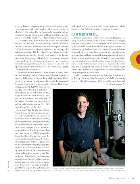
Grant Jensen flash freezes specimens to lock water in place then gets 3D images<br />
of cells in near-native state. He likens the result to a CT scan for protein complexes.<br />
Fall 2o12 | h h m i b u l l e t i n<br />
33