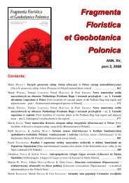
66 T. Majewski Aknowlogments. The author would like <strong>to</strong> thank Mr Roman K rólik and Mr Rafał Ruta as well as the collection cura<strong>to</strong>rs in Łomna and Kraków, Dr. Violetta Tomaszewska and Dr. Daniel K ubisz, for kindly providing insects for examination. The specimens listed in this study are in the author’s collection which will be handed over <strong>to</strong> the herbarium of the Institute of Botany, Polish Academy of Sciences. DESCRIPTIONS OF SPECIES Dimeromyces corynetis Thaxter Dimeromyces corynetis Thaxter, Proc. Amer. Acad. Arts 48: 157. 1912 (‘D. Corynitis’). Female thallus yellowish, 290 μm long. Receptacle 50 μm long (without the foot), consisting of four cells; the basal cell large, elongated, the second one flattened, bearing laterally a short, slender secondary appendage; the third cell isodiametric, bearing laterally the perithecium, and the fourth cell isodiametric, bearing laterally a long, slender, 6-celled secondary appendage. Primary appendage being a continuation of the receptacle axis, unicellular, elongated, as long as the basal cell of the receptacle. Perithecium fusiform, 175 x 45 μm, with indistinct neck, stalk cell 75 μm long. Male thallus not found (Fig. 1a). On Necrobia ruficollis (Fabricius) (Coleoptera, Cleridae): Wierzbica Górna, Kluczbork poviat, ex cadaver of Capreolus capreolus (L.), leg. Roman Królik, 28.8.1990, TM 10100 (host ex coll. R. Królik). Only one complete thallus was found on the host’s elytron. Its features are consistent with the descriptions and figures given by Thaxter (1924) and S antamari a (2003). Dimeromyces corynetis, an infrequent species, parasitizes representatives of the genera Corynetes and Necrobia. It is known from America (Argentina and Hawaii) and from Western Europe, namely from Spain, Great Britain, France, and Italy (Santamaria et al. 1991; Santamaria 2003). Euphoriomyces magnicellulatus Santamaria Euphoriomyces magnicellulatus Santamaria, Revista Iberoam. Micol. 8: 48. 1991. Thallus hyaline, 85-135 μm long. Receptacle consisting of 6-8 superposed cells, 40-63 μm long. The basal cell usually somewhat elongated, the remaining cells rather flattened in the lower part of the receptacle and isodiametric in its upper part. Except the basal one, the receptacle cells cut off bilaterally basal cells of short secondary antheridial blanchlets or stalk cells of perithecia. Primary appendage is a prolongation of the receptacle, not exceeding the perithecial apex. It consists of one or two cells bearing antheridia laterally and distally. Perithecia one or two per thallus, 50-75 x 18-25 μm, elongated, broadest near the middle, with indistinct neck. Fig. 1: b-e. On Hydnobius multistriatus (Gyllenhall) (Coleoptera, Leiodidae): Pieskowa Skała, Kraków poviat, 18.8.2001, leg. Rafał Ruta, TM 10512–10514 (host ex coll. R. Ru t a ). Many thalli were found on the elytra of one insect in an en<strong>to</strong>mological collection. They are mostly consistent with the description given by Santamaria (1991, 2003); however, thalli with two mature or maturing perithecia are frequent. Euphoriomyces magnicellulatus seems <strong>to</strong> be a very rare species. It has been reported only from Spain on Hydnobius spinipes (Gyllenhal) (type host), Leiodes sp. and Bap<strong>to</strong>linus affinis (Paykull), and from the Balearic Islands on Hydnobius sp.
<strong>Some</strong> <strong>additional</strong> <strong>Laboulbeniales</strong> (<strong>Ascomycetes</strong>) <strong>new</strong> <strong>to</strong> <strong>Poland</strong> 67 Fig. 1. Dimeromyces corynetis Thaxter. a – female thallus (drawn from TM. 10100). Euphoriomyces magnicellulatus Santamaria. b – paired thalli with one perithecium (TM 10513), c-e – thalli with two perithecia (TM 10514). Laboulbenia manubriolata Thaxter. f – mature thallus (TM 10161). Scale bar: 100 μm.