PDF - BioMed Central
PDF - BioMed Central
PDF - BioMed Central
Create successful ePaper yourself
Turn your PDF publications into a flip-book with our unique Google optimized e-Paper software.
Dhakal et al. BMC Cancer 2013, 13:506<br />
http://www.biomedcentral.com/1471-2407/13/506<br />
RESEARCH ARTICLE<br />
Open Access<br />
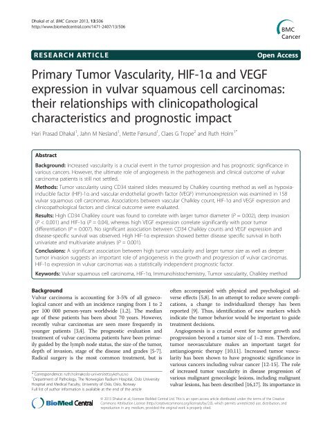
Primary Tumor Vascularity, HIF-1α and VEGF<br />
expression in vulvar squamous cell carcinomas:<br />
their relationships with clinicopathological<br />
characteristics and prognostic impact<br />
Hari Prasad Dhakal 1 , Jahn M Nesland 1 , Mette Førsund 1 , Claes G Trope 2 and Ruth Holm 1*<br />
Abstract<br />
Background: Increased vascularity is a crucial event in the tumor progression and has prognostic significance in<br />
various cancers. However, the ultimate role of angiogenesis in the pathogenesis and clinical outcome of vulvar<br />
carcinoma patients is still not settled.<br />
Methods: Tumor vascularity using CD34 stained slides measured by Chalkley counting method as well as hypoxiainducible<br />
factor (HIF)-1α and vascular endothelial growth factor (VEGF) immunoexpression was examined in 158<br />
vulvar squamous cell carcinomas. Associations between vascular Chalkley count, HIF-1α and VEGF expression and<br />
clinicopathological factors and clinical outcome were evaluated.<br />
Results: High CD34 Chalkley count was found to correlate with larger tumor diameter (P = 0.002), deep invasion<br />
(P < 0.001) and HIF-1α (P = 0.04), whereas high VEGF expression correlate significantly with poor tumor<br />
differentiation (P = 0.007). No significant association between CD34 Chalkley counts and VEGF expression and<br />
disease-specific survival was observed. High HIF-1α expression showed better disease specific survival in both<br />
univariate and multivariate analyses (P = 0.001).<br />
Conclusions: A significant association between high tumor vascularity and larger tumor size as well as deeper<br />
tumor invasion suggests an important role of angiogenesis in the growth and progression of vulvar carcinomas.<br />
HIF-1α expression in vulvar carcinomas was a statistically independent prognostic factor.<br />
Keywords: Vulvar squamous cell carcinoma, HIF-1α, Immunohistochemistry, Tumor vascularity, Chalkley method<br />
Background<br />
Vulvar carcinoma is accounting for 3-5% of all gynecological<br />
cancer and with an incidence ranging from 1 to 2<br />
per 100 000 person-years worldwide [1,2]. The median<br />
age of these patients has been about 70 years. However,<br />
recently vulvar carcinomas are seen more frequently in<br />
younger patients [3,4]. The prognostic evaluation and<br />
treatment of vulvar carcinoma patients have been primarily<br />
guided by the lymph node status, the size of the tumor,<br />
depth of invasion, stage of the disease and grades [5-7].<br />
Radical surgery is the most common treatment, but is<br />
* Correspondence: ruth.holm@oslo-universitetssykehus.no<br />
1 Department of Pathology, The Norwegian Radium Hospital, Oslo University<br />
Hospital and Medical Faculty, University of Oslo, Oslo, Norway<br />
Full list of author information is available at the end of the article<br />
often accompanied with physical and psychological adverse<br />
effects [5,8]. In an attempt to reduce severe complications,<br />
a change to individualized therapy has been<br />
reported [9]. Thus, identification of new markers which<br />
indicate the tumor behavior would be important to guide<br />
treatment decisions.<br />
Angiogenesis is a crucial event for tumor growth and<br />
progression beyond a tumor size of 1–2 mm. Therefore,<br />
tumor neovasculature makes an important target for<br />
antiangiogenic therapy [10,11]. Increased tumor vascularity<br />
has been shown to have prognostic significance in<br />
various cancers including vulvar cancer [12-15]. The role<br />
of increased tumor vascularity in disease progression of<br />
various malignant gynecologic lesions, including malignant<br />
vulvar lesions, has been described [16,17]. Its importance in<br />
© 2013 Dhakal et al.; licensee <strong>BioMed</strong> <strong>Central</strong> Ltd. This is an open access article distributed under the terms of the Creative<br />
Commons Attribution License (http://creativecommons.org/licenses/by/2.0), which permits unrestricted use, distribution, and<br />
reproduction in any medium, provided the original work is properly cited.
Dhakal et al. BMC Cancer 2013, 13:506 Page 2 of 9<br />
http://www.biomedcentral.com/1471-2407/13/506<br />
vulvar cancer has been emphasized by the increased<br />
vascularity in preinvasive lesions and invasive vulvar<br />
carcinomas [15-21]. Vulvar carcinoma patients with increased<br />
vascularity were reported to have poor prognosis<br />
in some studies [6,15,19], whereas other showed no<br />
significance [20].<br />
Hypoxia-inducible factor (HIF)-1α, a transcription factor,<br />
is a key regulator of angiogenesis when a growing<br />
tumor experiences hypoxic stress and acts through various<br />
intracellular signalling pathways. Such activation results<br />
in the secretion of vascular endothelial growth<br />
factor (VEGF) and other factors related to tumor metabolism<br />
necessary for hypoxia compensation and tumor<br />
cell survival [22]. It is known to be expressed in various<br />
solid tumors including vulvar squamous cell carcinomas<br />
[23-30]. The relation between primary tumor vascularity<br />
and HIF-1α expression in head and neck and oesophageal<br />
squamous cell carcinoma has been reported<br />
[24,25], and the prognostic impact of HIF-1α expression<br />
in cancer is varied [26,27,29-32]. HIF-1α expression investigated<br />
recently in normal epithelium, intraepithelial<br />
neoplasia and invasive carcinoma of vulva did not show<br />
significant differences [28]. To our knowledge, no study<br />
of HIF-1α expression and its connection with prognosis<br />
in vulvar carcinoma patients has been reported. VEGF, a<br />
potent angiogenic molecule over-expressed in a hypoxic<br />
state, is crucial to induce tumor angiogenesis and acts<br />
through the receptors VEGFR1 and VEGFR2 [22,33]. It<br />
is expressed in various human cancers including vulvar<br />
malignancy [21,28,34,35]. A significant variation in expression<br />
of VEGF in nonneoplastic epithelium, preneoplastic<br />
lesions and invasive squamous cell carcinoma of<br />
vulva has been described [21,28,35]. Its expression in<br />
vulvar cancer and relationship with vascularity has been<br />
reported [19]. The prognostic impact of VEGF expression<br />
in invasive vulvar carcinoma is still not settled<br />
[19,36].<br />
In the present study, we have evaluated a large series<br />
of primary vulvar squamous cell carcinomas for primary<br />
tumor vascularity and expression of HIF-1α and VEGF<br />
and elucidated their relationships with various clinicopathological<br />
parameters and clinical outcome.<br />
Methods<br />
Patient materials<br />
A retrospective study was performed on a cohort of 158<br />
patients with vulvar squamous cell carcinoma. All patients<br />
had undergone a resection at The Norwegian Radium<br />
Hospital between 1977 and 2006. The median age<br />
at diagnosis was 75 years (range, 41–92 years). In 108<br />
(68%) of these cases radical surgery (a total vulvectomy<br />
plus a bilateral inguinal lymphadenectomy) had been<br />
performed, whereas the remaining 50 (32%) patients had<br />
non-radical surgery. Postoperative therapy had been<br />
administered to 44 patients including irradiation in 40<br />
(25%) cases and irradiation/chemotherapy in four (3%)<br />
cases. Seventy-four (47%) of the patients died as a result<br />
of their vulvar cancer. All patients were followed up<br />
from the time of their confirmed diagnosis until death<br />
or 1. September, 2009. The median follow-up time for patients<br />
still alive was 108 months (range, 43 to 347 months).<br />
All tumors were staged based on the new International<br />
Federation of Gynecology and the Obstetrics (FIGO) classification<br />
from 2009 [37]. The Regional Committee for<br />
Medical Research Ethics South of Norway (S-06012), The<br />
Social and Health Directorate (04/2639 and 06/1478) and<br />
The Data Inspectorate (04/01043) approved the current<br />
study protocol. In this study we have used paraffin embedded<br />
tumor tissue from vulvar cancer patients diagnosed<br />
between 1977 and 2006. As many of these patients are<br />
dead or very old we did not have the opportunity to obtain<br />
patient consent. Permission to perform this study without<br />
patient consent was obtained from The Social and Health<br />
Directorate (04/2639).<br />
Histological specimens were reviewed by the coauthor<br />
J.M.N. without access to any clinical information<br />
on the patients. The tumors were classified according to<br />
the World Health Organization recommendations [38].<br />
All 158 tumors were classified as keratinizing/non-keratinizing<br />
squamous cell carcinomas.<br />
Immunohistochemistry<br />
Three micrometer sections were processed for immunohistochemistry<br />
using the Dako EnVision Flex+ System<br />
(K8012; Dako, Glostrup, Denmark) and the Dako Autostainer.<br />
Deparaffinization and the unmasking of epitopes<br />
were performed using PT-Link (Dako) and EnVision Flex<br />
target retrieval solution at a high pH. After treatment with<br />
0.03% hydrogen peroxide (H 2 O 2 ) for 5 min to block<br />
endogenous peroxidase activity, the sections were incubated<br />
with monoclonal antibodies raised against CD34<br />
(30 min at room temperature, clone QBEND-10, 1:1000,<br />
1μg IgG 1 /ml) purchased from Monosan (Uden, The<br />
Netherlands), HIF-1α (over night at 4°C, clone 54/HIF-1α,<br />
1:100, 2.5 μg IgG 1 /ml) purchased from BD Transduction<br />
Laboratories (San Jose, CA, USA) and VEGF (over night<br />
at 4°C, clone VG1, 1:100, 0.45 μg IgG 1 /ml) purchased<br />
from Dako. Then the slides were incubated with EnVision<br />
Flex+ mouse linker (15 min), EnVision Flex/HRP<br />
enzyme (30 min) and 3’3-diaminobenzidine tetrahydrochloride<br />
(DAB) (10 min). After counterstaining with<br />
hematoxylin the samples were dehydrated and mounted<br />
in Richard-Allan Scientific Cyto seal XYL (Thermo<br />
Scientific, Waltham, MA, USA). All of the sample series<br />
included positive controls known to be positive for CD34,<br />
HIF-1α and VEGF. As negative controls, the primary antibodies<br />
were replaced with mouse myeloma protein IgG 1<br />
at the equivalent concentration.
Dhakal et al. BMC Cancer 2013, 13:506 Page 3 of 9<br />
http://www.biomedcentral.com/1471-2407/13/506<br />
Quantification of tumor vascularity<br />
Chalkley method was used for quantification of tumor<br />
vascularity as recommended in a consensus meeting<br />
[39]. The method has been described in detail earlier<br />
[14]. Three most vascularized areas in the CD34 stained<br />
tumor section known as “hotspots” were identified<br />
under the low power magnification after scanning<br />
first at ×40 and then ×100 magnification following the<br />
Weidner’s method of selection of vascular hotspots [40].<br />
Then a 25 point Chalkley eyepiece graticule fixed in one<br />
of the eyepieces of the microscope was applied to each<br />
vascular hotspot at ×200 magnification [Chalkley grid<br />
area of 0.1886 mm 2 (Nikon microscope, Eclipse E400)]<br />
in such a way that maximum number of black dots in<br />
Chalkley graticule fell on or within immunostained<br />
microvessels. The number of these dots that have fallen<br />
on or within the immunostained microvessels were<br />
counted in each selected hotspot area and recorded as<br />
Chalkley count. Sclerotic and necrotic area was avoided<br />
and count was done in only invasive carcinoma including<br />
margin. The highest count among the 3 hotspots counts<br />
from each tumor was used for further analyses. Measurement<br />
of vascularity was performed without the knowledge<br />
of clinicopathological data or clinical outcome.<br />
Evaluation for HIF-1α and VEGF expression<br />
Expression of HIF-1α was evaluated on immunostained<br />
slides semiquantitatively into four classes and only nuclear<br />
immunoreactivity of the tumor cells was taken into account.<br />
Due to similar staining intensity of the HIF-1α<br />
positive cases we did not consider the intensity of immunostaining.<br />
Based on the number of HIF-1α positively<br />
stained tumor cells, tumors were grouped into: 0% of the<br />
cells; < 10% of the cells; 10-50% of the cells and > 50% of<br />
the cells. For further analyses, HIF-1α expression in nucleus<br />
in more than 50% of the tumor cells was considered<br />
as high. VEGF positive cases showed different staining intensity<br />
and both intensity and number of positive tumor<br />
cells were evaluated. Cytoplasmic expression of VEGF was<br />
categorized semiquantitatively on the basis of intensity of<br />
the signal (absent, 0; weak, 1; moderate, 2; strong, 3) and<br />
the percentage of positive tumor cells (absent, 0; < 10%, 1;<br />
10-50%, 2; > 50%, 3). The composite score was calculated<br />
as fraction of positive tumor cells score multiplied by intensity<br />
score, and range from 0 to 9. For further analyses,<br />
cytoplasmic VEGF immunostaining with a composite<br />
score ≥ 6 was classified as high expression. Examination of<br />
immunostaining was performed in a blinded fashion with<br />
no knowledge of the clinicopathological variables and patient<br />
outcomes.<br />
Statistical analyses<br />
The associations between the HIF-1α and VEGF expression<br />
and CD34 Chalkley counts of primary tumor vascularity<br />
and the clinicopathological variables were evaluated by<br />
the Pearson chi-square (χ 2 ), Fisher’s exact test and linearby-linear<br />
association as required. The disease-specific survival<br />
analysis, based on death from vulvar cancer only, was<br />
performed using the Kaplan Meier method and P value<br />
computed by log-rank test. A Cox proportional hazards<br />
regression model was used for both univariate and multivariate<br />
evaluation of survival rates. In the multivariate<br />
analysis, a backward regression was performed and variables<br />
with a P ≤ 0.05 in univariate survival analysis were<br />
included in the model. The vulvar carcinoma tissues in<br />
our cohort have been collected over an extensive period<br />
from 1977–2006. Due to the large variation in storage<br />
time and given that the fixation protocol for these tissues<br />
up to 1987 was acid formalin, whereas from 1987–2006<br />
was buffered formalin, Mann–Whitney U test was performed<br />
to evaluate whether this has any influence on the<br />
CD34, HIF-1α and VEGF immunostaining. The Mann–<br />
Whitney U test showed that the distribution of CD34,<br />
Figure 1 Representative images of CD34 staining of primary<br />
vulvar carcinoma vascularization. (A) Low vascularity (low Chalkley<br />
count) and (B) High vascularity (high Chalkley count). Images were<br />
taken by a Leica DFC 320 digital camera with a Plan-neofluar 10×<br />
objective lens in Axiophot microscope (Zeiss Germany).
Dhakal et al. BMC Cancer 2013, 13:506 Page 4 of 9<br />
http://www.biomedcentral.com/1471-2407/13/506<br />
HIF-1α and VEGF expression was the same between samples<br />
processed before and after 1987. All analyses were<br />
processed using the SPSS 18.0 statistical software package<br />
(SPSS, Chicago, IL). Statistical significance was considered<br />
for P 50% tumor cells) in the nucleus was observed in 57<br />
(36%) and low levels (≤ 50% tumor cells) in 101 (64%)<br />
cases (Figure 2A and B), whereas high VEGF expression<br />
(score ≥ 6) in the cytoplasm was identified in 63 (40%)<br />
and low low level (score < 6) in 95 (60%) cases (Figure 2C<br />
and D).<br />
CD34 Chalkley count, HIF-1α and VEGF expression in<br />
relation to clinicopathological parameters are shown in<br />
Table 1. High CD34 Chalkley count was found to correlate<br />
significantly with larger tumor diameter (P = 0.002)<br />
and deeper invasion (P < 0.001), whereas high VEGF<br />
expression correlate significantly with poor tumor differentiation<br />
(P = 0.007). High level of HIF-1α was significantly<br />
correlated to high CD34 Chalkley counts (P =0.04).<br />
VEGF expression did not show any association with<br />
CD34 Chalkley count and HIF-1α levels.<br />
In univariate survival analysis, high HIF-1α expression was<br />
associated with better disease-specific survival (P =0.001)<br />
(Figure 3), whereas no significant association between<br />
CD34 Chalkley counts and VEGF expression and diseasespecific<br />
survival (P =0.16andP = 0.45, respectively) was<br />
observed. In multivariate analysis, lymph node metastases,<br />
age and HIF-1α expression retained independent prognostic<br />
significance (Table 2).<br />
Discussion<br />
We observed that primary tumor vascularity, quantified<br />
by Chalkley method, had a significant association with<br />
tumor size and depth of invasion in invasive vulvar carcinomas.<br />
Tumor size has been reported to predict local<br />
lymph node metastasis [41] and is an important prognostic<br />
marker in vulvar cancer patients. Tumor size is at<br />
present used to stratify patients into different risk groups<br />
and acts as a determinant for surgical treatment [6,7].<br />
In vulvar carcinomas, depth of tumor invasion is also indicative<br />
of the aggressiveness of primary tumor and is<br />
reported to be associated with lymph node metastases<br />
[41] and reduced survival [6]. Inguinofemoral lymph<br />
node status is the most powerful indicator of poor prognosis<br />
in vulvar cancer [42-44] and a significantly reduced<br />
survival in the current study has been confirmed. In the<br />
present study, no prognostic significance of tumor vascularity<br />
was observed for patients with vulvar carcinoma.<br />
Figure 2 Representative images of HIF-1α and VEGF immunoexpression in primary vulvar carcinoma. (A) high HIF-α nuclear expression<br />
and (B) low HIF-α nuclear expression (C) high VEGF cytoplasmic staining and (D) low VEGF cytoplasmic staining. 40× objective lens.
Dhakal et al. BMC Cancer 2013, 13:506 Page 5 of 9<br />
http://www.biomedcentral.com/1471-2407/13/506<br />
Table 1 CD34 Chalkley count, HIF-1α and VEGF expression in relation to clinicopathological variables in vulvar<br />
carcinomas<br />
Variable Total CD34 Chalkley count HIF-1α VEGF<br />
N Low High (%) P value Low High (%) P value Low High (%) P value<br />
Age 0.25 1 0.28 1 0.68 1<br />
25–69 59 30 29 (49) 34 25 (42) 36 23 (39)<br />
70–84 81 29 52 (64) 55 26 (32) 46 35 (43)<br />
85+ 18 8 10 (56) 12 6 (33) 13 5 (28)<br />
FIGO 0.67 2 0.22 2 0.08 2<br />
Ia 0 0 0 (0) 0 0 (0) 0 0 (0)<br />
Ib 77 35 42 (55) 51 26 (34) 48 29 (38)<br />
II 7 2 5 (71) 4 3 (43) 7 0 (0)<br />
IIIa 30 14 16 (53) 13 17 (57) 20 10 (33)<br />
IIIb 26 8 18 (69) 18 8 (31) 11 15 (58)<br />
IIIc 7 2 5 (71) 5 2 (29) 4 3 (43)<br />
IVa 1 1 0 (0) 1 0 (0) 0 1 (100)<br />
IVb 7 3 4 (57) 6 1 (14) 4 3 (43)<br />
Not available 3<br />
Lymph node metastasis 0.21 3 0.54 3 0.11 3<br />
None 87 39 48 (55) 58 29 (33) 55 31 (37)<br />
Unilateral 44 19 25 (57) 25 19 (43) 29 15 (34)<br />
Bilateral 24 6 18 (75) 15 9 (38) 10 14 (58)<br />
Not available 3<br />
Tumor diameter (cm) 0.002 1 0.95 1 0.98 1<br />
0.3–2.5 32 19 13 (41) 19 13 (41) 20 12 (38)<br />
2.6–4.0 51 24 27 (53) 33 16 (31) 30 21 (41)<br />
4.1–20.0 72 21 51 (71) 44 28 (39) 44 28 (39)<br />
Not available 3<br />
Tumor differentiation 0.07 3 0.23 3 0.007 3<br />
Well 35 19 16 (46) 26 9 (26) 19 16 (46)<br />
Moderate 92 32 60 (65) 54 38 (41) 64 28 (30)<br />
Poor 31 16 15 (48) 21 10 (32) 12 19 (61)<br />
Depth of invasion (mm) 50% tumor cells.<br />
Low: VEGF score < 6, High: VEGF score ≥ 6.
Dhakal et al. BMC Cancer 2013, 13:506 Page 6 of 9<br />
http://www.biomedcentral.com/1471-2407/13/506<br />
Figure 3 Survival curves using the Kaplan-Meier method. The<br />
Kaplan-Meier curve of disease-specific survival in relation to the HIF-<br />
1α showed that patients whose tumors expressed low levels of HIF-<br />
1α had a worse prognosis than those with high levels.<br />
This is in accordance with an earlier study on vulvar<br />
cancer [20], but in contrast to others [6,15,19]. These<br />
conflicting reports on primary tumor vascularity and<br />
prognosis might be due to methodological differences,<br />
different study cohort or biological factors [6,19,20]. We<br />
used the Chalkley counting method for vascular quantification<br />
which measures the relative vascular area [45],<br />
as recommended in a consensus meeting for quantification<br />
of vascularity in solid tumors [39], whereas in other<br />
studies microvessels have been counted manually [15,19,20]<br />
or using image analyses [6]. Moreover, other studies<br />
[6,15,19,20] had analysed smaller number of cases compared<br />
to our large series of vulvar carcinomas. Thus,<br />
our results of high tumor vascularity associated with<br />
larger tumor size and deeper invasion (known pathological<br />
markers for tumor aggressiveness) indicates<br />
angiogenesis as a marker for the aggressive behaviour<br />
of vulvar carcinoma.<br />
HIF-1α, is a crucial molecule in inducing angiogenesis<br />
in growing tumor under hypoxic stress [22] and several<br />
reports have been published on relation between HIF-1α<br />
expression and angiogenesis in head and neck and<br />
oesophageal squamous cell carcinoma [24,25]. In present<br />
study, we did observe a positive association between<br />
HIF-1α expression and CD34 Chalkey count of primary<br />
tumor vascularity similar to a report for head and neck<br />
squamous cell carcinoma patients [25]. This confirms<br />
the role of HIF-1α for the initiation and the promotion<br />
of angiogenesis in vulvar cancer. High tumoral HIF-1α<br />
expression is reported to be associated with reduced survival<br />
in oral, oropharyngeal and cervical cancers [29,32,46].<br />
In contrast, in the present study, a significantly improved<br />
survival of vulvar carcinoma patients with high HIF-1α<br />
expression was observed as reported for the squamous cell<br />
caricnoma in head and neck region, oral cavity and uterine<br />
cervix [26,27,30,31]. Other did not find prognostic significance<br />
in oesophageal squamous cell carcinoma [47]. Various<br />
factors are thought to affect the impact of HIF-1α<br />
activation in tumor behaviour [48] including methodology,<br />
cut off(s) and treatment modalities [26,27,30-32,47]. Lack<br />
of CAIX and Glut-I expression along with high HIF-1α<br />
expression in squamous cell carcinoma indicates alternative<br />
mechanism for HIF-1α upregulation [26]. Furthermore,<br />
we have shown that the patients in good prognosis<br />
group had >50% HIF-1α positive tumor cells as reported<br />
for its strong expression in squamous cell carcinoma of<br />
oral cavity [27]. Diffuse HIF-1α expression based on<br />
tumor types and its nonhypoxic activation through various<br />
genetic alterations that might result in different outcomes<br />
[22,26] may also explain our observation. Vascularization<br />
was heterogenously distributed in the tumor including<br />
tumor fronts and in the stromal tissue between the islands<br />
of tumor cells. Perinecrotic tumor cells distant from the<br />
supplying vessels under hypoxic stress express HIF-1α,<br />
whereas nonnecrotic tumor shows diffuse expression<br />
throughout the tumor including the tumor cells close to<br />
the blood vessels [29]. Despite, the heterogenous distribution<br />
of vascularity, our observation of positive association<br />
between HIF-1α and tumor vascularity suggests the HIF-<br />
1α induced angiogenesis. HIF-1α is known to induce expression<br />
of various genes including genes linked to cell<br />
survival, apoptosis, cellular proliferation [22]. Perhaps, the<br />
better outcome in patients with high HIF-1α expressing<br />
Table 2 Relative risk (RR) of dying from vulvar cancer<br />
Variables Univariate analysis Multivariate analysis<br />
RR 95% CI a p RR 95% CI a p<br />
Lymph node metastasis 1.99 1.49–2.65
Dhakal et al. BMC Cancer 2013, 13:506 Page 7 of 9<br />
http://www.biomedcentral.com/1471-2407/13/506<br />
tumors might be due to its inhibitory role on tumor cells<br />
through induction of proapoptotic pathway [49,50].<br />
Hypoxia markers; HIF-1α, GLUT-1, CA IX and VEGF<br />
are expressed in both vulvar preneoplastic lesions and<br />
invasive squamous cell carcinoma [28]. An increasing<br />
expression of VEGF from normal epithelium to premalignant<br />
lesion to invasive squamous cell carcinoma was<br />
found in vulva [28]. In the present study, high VEGF expression<br />
was significantly associated with only poor<br />
tumor differentiation, however, an other study reported<br />
no such association in vulvar carcinomas [19]. We did<br />
not find that the VEGF levels demonstrated prognostic<br />
significance, a result being different from a report by<br />
Obermair and colleagues [19]. A lack of correlation of<br />
VEGF with vascularity observed in the present series which<br />
is different from an earlier report [19], might be due to its<br />
possible non-angiogenic effects and/or autocrine role on<br />
tumor cells [51,52]. Alternative mechanisms of VEGF independent<br />
neoangiogenesis by inducing other potent angiogenic<br />
molecules like basic fibroblast growth factor and size<br />
related proainherent angiogenic effect on the tumor [53]<br />
may have resulted in nonsignificant relationship. We noted<br />
no association between VEGF and HIF-1α expression possibly<br />
due to an alternative non HIF-1α mechanism of<br />
VEGF induction [54]. The proangiogenic effect of VEGF is<br />
closely related to the tumor size and no impact on angiogenesis<br />
is found when tumor reaches to certain size [53].<br />
There are several pitfalls associated to immunohistochemical<br />
methods. In addition, the handling of tissue<br />
specimens, such as fixation and storage time, may influence<br />
the immunohistochemical results [55]. The lack of<br />
consideration for these limitations may reduce the usefulness<br />
of immunohistochemical studies. In the present<br />
study, the fixation and storage time of the tissues did<br />
not influence the CD34, HIF-1α and VEGF immunostaining.<br />
Both false positive and negative results may<br />
limit the outcome of immunohistochemical studies. To<br />
reduce the possibility of false negative results we have<br />
used the EnVision Flex+ detection system reported to<br />
have a high sensitivity [56]. Furthermore, we have included<br />
positive controls in each run to exclude the possibility<br />
of false negative result due to methodological<br />
problems. To avoid nonspecific staining we extensively<br />
optimalized the dilutions of the primary antibodies used.<br />
In addition, negative controls, replacing the primary<br />
antibodies with the mouse myeloma protein IgG 1 , were<br />
included to exclude the possibility of false positive results.<br />
Despite the effort to quality secure immunostaining processes<br />
there are major limitations connected to immunohistochemistry<br />
between the studies that are linked to<br />
methodological differences including immunostaining<br />
procedures and scoring systems [57]. In the future it is<br />
clearly needed a standarization of immunohistochemical<br />
methodology and scoring systems.<br />
Conclusions<br />
Our results show that high tumor vascularity in vulvar<br />
carcinoma is associated with larger tumor size and deeper<br />
invasion, indicating that it is a feature of aggressive<br />
tumor phenotype. High HIF-1α expression has favorable<br />
prognostic impact in vulvar carcinoma patients.<br />
Competing interests<br />
Authors declare that they have no competing interests.<br />
Author’ contributions<br />
HPD participated in the design of the study, quantified tumor vascularity and<br />
draft the manuscript. JMN participated in the design of the study, performed<br />
systematic pathologic review of vulvar carcinomas and revised the<br />
manuscript critically. MF carried out the immunohistochemistry and revised<br />
the manuscript critically. CGT collected clinical data, participated in<br />
interpretation of data and revised the manuscript critically. RH participated in<br />
the design of the study, protein, statistical and data analysis and helped to<br />
draft the manuscript. All authors read and approved the final manuscript.<br />
Acknowledgements<br />
This work was supported by the Inger and John Fredriksen Foundation for<br />
Ovarian Cancer Research and the Norwegian Cancer Society.<br />
Author details<br />
1 Department of Pathology, The Norwegian Radium Hospital, Oslo University<br />
Hospital and Medical Faculty, University of Oslo, Oslo, Norway. 2 Department<br />
of Obstetrics and Gynecology, The Norwegian Radium Hospital, Oslo<br />
University Hospital and Medical Faculty, University of Oslo, Oslo, Norway.<br />
Received: 12 June 2013 Accepted: 23 October 2013<br />
Published: 29 October 2013<br />
References<br />
1. Giles GG, Kneale BL: Vulvar cancer: the Cinderella of gynaecological<br />
oncology. Aust N Z J Obstet Gynaecol 1995, 35:71–75.<br />
2. Coulter J, Gleeson N: Local and regional recurrence of vulval cancer:<br />
management dilemmas. Best Pract Res Clin Obstet Gynaecol 2003,<br />
17:663–681.<br />
3. Jones RW, Baranyai J, Stables S: Trends in squamous cell carcinoma of the<br />
vulva: the influence of vulvar intraepithelial neoplasia. Obstet Gynecol<br />
1997, 90:448–452.<br />
4. Messing MJ, Gallup DG: Carcinoma of the vulva in young women. Obstet<br />
Gynecol 1995, 86:51–54.<br />
5. Beller U, Quinn MA, Benedet JL, Creasman WT, Ngan HY, Maisonneuve P,<br />
Pecorelli S, Odicino F, Heintz AP: Carcinoma of the vulva. FIGO 26th<br />
Annual Report on the Results of Treatment in Gynecological Cancer. Int J<br />
Gynaecol Obstet 2006, 95(1):S7–27.<br />
6. Nayha VV, Stenback FG: Increased angiogenesis is associated with poor<br />
prognosis of squamous cell carcinoma of the vulva. Acta Obstet Gynecol<br />
Scand 2007, 86:1392–1397.<br />
7. Wang Z, Trope CG, Florenes VA, Suo Z, Nesland JM, Holm R:<br />
Overexpression of CDC25B, CDC25C and phospho-CDC25C (Ser216) in<br />
vulvar squamous cell carcinomas are associated with malignant features<br />
and aggressive cancer phenotypes. BMC Cancer 2010, 10:233.<br />
8. Tyring SK: Vulvar squamous cell carcinoma: guidelines for early diagnosis<br />
and treatment. Am J Obstet Gynecol 2003, 189:S17–S23.<br />
9. Stehman FB, Look KY: Carcinoma of the vulva. Obstet Gynecol 2006,<br />
107:719–733.<br />
10. Folkman J: What is the evidence that tumors are angiogenesis dependent?<br />
JNatlCancerInst1990, 82:4–6.<br />
11. Folkman J: Angiogenesis: an organizing principle for drug discovery? Nat<br />
Rev Drug Discov 2007, 6:273–286.<br />
12. Vermeulen PB, Libura M, Libura J, O'Neill PJ, Van DP, Van ME, Van Oosterom AT,<br />
Dirix LY: Influence of investigator experience and microscopic field size on<br />
microvessel density in node-negative breast carcinoma. Breast Cancer Res<br />
Treat 1997, 42:165–172.<br />
13. Offersen BV, Borre M, Overgaard J: Quantification of angiogenesis as a<br />
prognostic marker in human carcinomas: a critical evaluation of
Dhakal et al. BMC Cancer 2013, 13:506 Page 8 of 9<br />
http://www.biomedcentral.com/1471-2407/13/506<br />
histopathological methods for estimation of vascular density. Eur J<br />
Cancer 2003, 39:881–890.<br />
14. Dhakal HP, Naume B, Synnestvedt M, Borgen E, Kaaresen R, Schlichting E,<br />
Wiedswang G, Bassarova A, Giercksky KE, Nesland JM: Vascularization in<br />
primary breast carcinomas: its prognostic significance and relationship<br />
with tumor cell dissemination. Clin Cancer Res 2008, 14:2341–2350.<br />
15. Hantschmann P, Jeschke U, Friese K: TGF-alpha, c-erbB-2 expression and<br />
neoangiogenesis in vulvar squamous cell carcinoma. Anticancer Res 2005,<br />
25:1731–1737.<br />
16. Abulafia O, Triest WE, Sherer DM: Angiogenesis in malignancies of the<br />
female genital tract. Gynecol Oncol 1999, 72:220–231.<br />
17. Saravanamuthu J, Reid WM, George DS, Crow JC, Rolfe KJ, MacLean AB,<br />
Perrett CW: The role of angiogenesis in vulvar cancer, vulvar<br />
intraepithelial neoplasia, and vulvar lichen sclerosus as determined by<br />
microvessel density analysis. Gynecol Oncol 2003, 89:251–258.<br />
18. Bancher-Todesca D, Obermair A, Bilgi S, Kohlberger P, Kainz C, Breitenecker<br />
G, Leodolter S, Gitsch G: Angiogenesis in vulvar intraepithelial neoplasia.<br />
Gynecol Oncol 1997, 64:496–500.<br />
19. Obermair A, Kohlberger P, Bancher-Todesca D, Tempfer C, Sliutz G,<br />
Leodolter S, Reinthaller A, Kainz C, Breitenecker G, Gitsch G: Influence of<br />
microvessel density and vascular permeability factor/vascular endothelial<br />
growth factor expression on prognosis in vulvar cancer. Gynecol Oncol<br />
1996, 63:204–209.<br />
20. Qureshi F, Munkarah A, Banerjee M, Jacques SM: Tumor angiogenesis in<br />
vulvar squamous cell carcinoma. Gynecol Oncol 1999, 72:65–70.<br />
21. Raspollini MR, Asirelli G, Taddei GL: The role of angiogenesis and COX-2<br />
expression in the evolution of vulvar lichen sclerosus to squamous cell<br />
carcinoma of the vulva. Gynecol Oncol 2007, 106:567–571.<br />
22. Semenza GL: Targeting HIF-1 for cancer therapy. Nat Rev Cancer 2003,<br />
3:721–732.<br />
23. Zhong H, De Marzo AM, Laughner E, Lim M, Hilton DA, Zagzag D, Buechler<br />
P, Isaacs WB, Semenza GL, Simons JW: Overexpression of hypoxiainducible<br />
factor 1alpha in common human cancers and their<br />
metastases. Cancer Res 1999, 59:5830–5835.<br />
24. Kimura S, Kitadai Y, Tanaka S, Kuwai T, Hihara J, Yoshida K, Toge T, Chayama<br />
K: Expression of hypoxia-inducible factor (HIF)-1alpha is associated with<br />
vascular endothelial growth factor expression and tumour angiogenesis<br />
in human oesophageal squamous cell carcinoma. Eur J Cancer 2004,<br />
40:1904–1912.<br />
25. Koukourakis MI, Giatromanolaki A, Sivridis E, Simopoulos C, Turley H, Talks K,<br />
Gatter KC, Harris AL: Hypoxia-inducible factor (HIF1A and HIF2A),<br />
angiogenesis, and chemoradiotherapy outcome of squamous cell headand-neck<br />
cancer. Int J Radiat Oncol Biol Phys 2002, 53:1192–1202.<br />
26. Fillies T, Werkmeister R, van Diest PJ, Brandt B, Joos U, Buerger H: HIF1-alpha<br />
overexpression indicates a good prognosis in early stage squamous cell<br />
carcinomas of the oral floor. BMC Cancer 2005, 5:84.<br />
27. dos Santos M, Mercante AM, Louro ID, Goncalves AJ, de Carvalho MB,<br />
da Silva EH, da Silva AM: HIF1-alpha expression predicts survival of<br />
patients with squamous cell carcinoma of the oral cavity. PLoS One 2012,<br />
7:e45228.<br />
28. Li YZ, Li SL, Li X, Wang LJ, Wang JL, Xu JW, Wu ZH, Gong L, Zhang XD:<br />
Expression of endogenous hypoxia markers in vulvar squamous cell<br />
carcinoma. Asian Pac J Cancer Prev 2012, 13:3675–3680.<br />
29. Aebersold DM, Burri P, Beer KT, Laissue J, Djonov V, Greiner RH, Semenza GL:<br />
Expression of hypoxia-inducible factor-1alpha: a novel predictive and<br />
prognostic parameter in the radiotherapy of oropharyngeal cancer.<br />
Cancer Res 2001, 61:2911–2916.<br />
30. Beasley NJ, Leek R, Alam M, Turley H, Cox GJ, Gatter K, Millard P, Fuggle S,<br />
Harris AL: Hypoxia-inducible factors HIF-1alpha and HIF-2alpha in head<br />
and neck cancer: relationship to tumor biology and treatment outcome<br />
in surgically resected patients. Cancer Res 2002, 62:2493–2497.<br />
31. Hutchison GJ, Valentine HR, Loncaster JA, Davidson SE, Hunter RD,<br />
Roberts SA, Harris AL, Stratford IJ, Price PM, West CM: Hypoxia-inducible<br />
factor 1alpha expression as an intrinsic marker of hypoxia: correlation<br />
with tumor oxygen, pimonidazole measurements, and outcome in<br />
locally advanced carcinoma of the cervix. Clin Cancer Res 2004,<br />
10:8405–8412.<br />
32. Lin PY, Yu CH, Wang JT, Chen HH, Cheng SJ, Kuo MY, Chiang CP:<br />
Expression of hypoxia-inducible factor-1 alpha is significantly associated<br />
with the progression and prognosis of oral squamous cell carcinomas in<br />
Taiwan. J Oral Pathol Med 2008, 37:18–25.<br />
33. Ferrara N, Gerber HP, LeCouter J: The biology of VEGF and its receptors.<br />
Nat Med 2003, 9:669–676.<br />
34. Jubb AM, Pham TQ, Hanby AM, Frantz GD, Peale FV, Wu TD, Koeppen HW,<br />
Hillan KJ: Expression of vascular endothelial growth factor, hypoxia<br />
inducible factor 1alpha, and carbonic anhydrase IX in human tumours.<br />
J Clin Pathol 2004, 57:504–512.<br />
35. Doldi N, Origoni M, Bassan M, Ferrari D, Rossi M, Ferrari A: Vascular<br />
endothelial growth factor. Expression in human vulvar neoplastic and<br />
nonneoplastic tissues. J Reprod Med 1996, 41:844–848.<br />
36. Knopp S, Trope C, Nesland JM, Holm R: A review of molecular pathological<br />
markers in vulvar carcinoma: lack of application in clinical practice. J Clin<br />
Pathol 2009, 62:212–218.<br />
37. Pecorelli S: Revised FIGO staging for carcinoma of the vulva, cervix, and<br />
endometrium. Int J Gynaecol Obstet 2009, 105:103–104.<br />
38. Wilkinson EJaTMR: Tumours of the vulva. Epithelial tumours. InWorld<br />
Health Organization Classification of Tumours. Pathology and genetics of<br />
tumors of the breast and female genital organs. Edited by Tavassoli FA,<br />
Devilee P. Lyon: ARC Press; 2003:316–325.<br />
39. Vermeulen PB, Gasparini G, Fox SB, Colpaert C, Marson LP, Gion M, Belien<br />
JA, de Waal RM, Van ME, Magnani E, Weidner N, Harris AL, Dirix LY: Second<br />
international consensus on the methodology and criteria of evaluation<br />
of angiogenesis quantification in solid human tumours. Eur J Cancer<br />
2002, 38:1564–1579.<br />
40. Weidner N, Semple JP, Welch WR, Folkman J: Tumor angiogenesis and<br />
metastasis–correlation in invasive breast carcinoma. N Engl J Med 1991,<br />
324:1–8.<br />
41. Binder SW, Huang I, Fu YS, Hacker NF, Berek JS: Risk factors for the<br />
development of lymph node metastasis in vulvar squamous cell<br />
carcinoma. Gynecol Oncol 1990, 37:9–16.<br />
42. der SS V, De Nieuwenhof HP, Massuger L, Bulten J, De Hullu JA: New FIGO<br />
staging system of vulvar cancer indeed provides a better reflection of<br />
prognosis. Gynecol Oncol 2010, 119:520–525.<br />
43. Woelber L, Eulenburg C, Choschzick M, Kruell A, Petersen C, Gieseking F,<br />
Jaenicke F, Mahner S: Prognostic role of lymph node metastases in vulvar<br />
cancer and implications for adjuvant treatment. Int J Gynecol Cancer 2012,<br />
22:503–508.<br />
44. Woelber L, Mahner S, Voelker K, Eulenburg CZ, Gieseking F, Choschzick M,<br />
Jaenicke F, Schwarz J: Clinicopathological prognostic factors and patterns<br />
of recurrence in vulvar cancer. Anticancer Res 2009, 29:545–552.<br />
45. Fox SB, Leek RD, Weekes MP, Whitehouse RM, Gatter KC, Harris AL:<br />
Quantitation and prognostic value of breast cancer angiogenesis:<br />
comparison of microvessel density, Chalkley count, and computer image<br />
analysis. J Pathol 1995, 177:275–283.<br />
46. Birner P, Schindl M, Obermair A, Plank C, Breitenecker G, Oberhuber G:<br />
Overexpression of hypoxia-inducible factor 1alpha is a marker for an<br />
unfavorable prognosis in early-stage invasive cervical cancer. Cancer Res<br />
2000, 60:4693–4696.<br />
47. Shibata-Kobayashi S, Yamashita H, Okuma K, Shiraishi K, Igaki H, Ohtomo K,<br />
Nakagawa K: Correlation among 16 biological factors [p53, p21(waf1),<br />
MIB-1 (Ki-67), p16(INK4A), cyclin D1, E-cadherin, Bcl-2, TNF-alpha,<br />
NF-kappaB,TGF-beta,MMP-7,COX-2,EGFR,HER2/neu,ER,and<br />
HIF-1alpha] and clinical outcomes following curative chemoradiation<br />
therapy in 10 patients with esophageal squamous cell carcinoma.<br />
Oncol Lett 2013, 5:903–910.<br />
48. Keith B, Johnson RS, Simon MC: HIF1alpha and HIF2alpha: sibling rivalry in<br />
hypoxic tumour growth and progression. Nat Rev Cancer 2012, 12:9–22.<br />
49. Guo K, Searfoss G, Krolikowski D, Pagnoni M, Franks C, Clark K, Yu KT, Jaye<br />
M, Ivashchenko Y: Hypoxia induces the expression of the pro-apoptotic<br />
gene BNIP3. Cell Death Differ 2001, 8:367–376.<br />
50. Sowter HM, Ratcliffe PJ, Watson P, Greenberg AH, Harris AL: HIF-1-dependent<br />
regulation of hypoxic induction of the cell death factors BNIP3 and NIX in<br />
human tumors. Cancer Res 2001, 61:6669–6673.<br />
51. Lichtenberger BM, Tan PK, Niederleithner H, Ferrara N, Petzelbauer P, Sibilia M:<br />
Autocrine VEGF signaling synergizes with EGFR in tumor cells to promote<br />
epithelial cancer development. Cell 2010, 140:268–279.<br />
52. Cao Y, Ei G, Wang E, Pal K, Dutta SK, Bar-Sagi D, Mukhopadhyay D: VEGF<br />
exerts an angiogenesis-independent function in cancer cells to promote<br />
their malignant progression. Cancer Res 2012, 72:3912–3918.<br />
53. Yoshiji H, Harris SR, Thorgeirsson UP: Vascular endothelial growth factor is<br />
essential for initial but not continued in vivo growth of human breast<br />
carcinoma cells. Cancer Res 1997, 57:3924–3928.
Dhakal et al. BMC Cancer 2013, 13:506 Page 9 of 9<br />
http://www.biomedcentral.com/1471-2407/13/506<br />
54. Cao Y, Li CY, Moeller BJ, Yu D, Zhao Y, Dreher MR, Shan S, Dewhirst MW:<br />
Observation of incipient tumor angiogenesis that is independent of<br />
hypoxia and hypoxia inducible factor-1 activation. Cancer Res 2005,<br />
65:5498–5505.<br />
55. Mighell AJ, Hume WJ, Robinson PA: An overview of the complexities and<br />
subtleties of immunohistochemistry. Oral Dis 1998, 4:217–223.<br />
56. Skaland I, Nordhus M, Gudlaugsson E, Klos J, Kjellevold KH, Janssen EA,<br />
Baak JP: Evaluation of 5 different labeled polymer immunohistochemical<br />
detection systems. Appl Immunohistochem Mol Morphol 2010, 18:90–96.<br />
57. Warren MV, Chan WY, Ridley JM: Analysis of protein biomarkers in human<br />
clinical tumor samples: critical aspects to success from tissue acquisition<br />
to analysis. Biomark Med 2011, 5:227–248.<br />
doi:10.1186/1471-2407-13-506<br />
Cite this article as: Dhakal et al.: Primary Tumor Vascularity, HIF-1α and<br />
VEGF expression in vulvar squamous cell carcinomas: their relationships<br />
with clinicopathological characteristics and prognostic impact. BMC<br />
Cancer 2013 13:506.<br />
Submit your next manuscript to <strong>BioMed</strong> <strong>Central</strong><br />
and take full advantage of:<br />
• Convenient online submission<br />
• Thorough peer review<br />
• No space constraints or color figure charges<br />
• Immediate publication on acceptance<br />
• Inclusion in PubMed, CAS, Scopus and Google Scholar<br />
• Research which is freely available for redistribution<br />
Submit your manuscript at<br />
www.biomedcentral.com/submit


