Magnetite magnetosome and fragmental chain formation of ...
Magnetite magnetosome and fragmental chain formation of ...
Magnetite magnetosome and fragmental chain formation of ...

Create successful ePaper yourself
Turn your PDF publications into a flip-book with our unique Google optimized e-Paper software.
Geophys. J. Int. (2009) 177, 33–42<br />
doi: 10.1111/j.1365-246X.2009.04043.x<br />
<strong>Magnetite</strong> <strong>magnetosome</strong> <strong>and</strong> <strong>fragmental</strong> <strong>chain</strong> <strong>formation</strong> <strong>of</strong><br />
Magnetospirillum magneticum AMB-1: transmission electron<br />
microscopy <strong>and</strong> magnetic observations<br />
Jinhua Li, 1 Yongxin Pan, 1 Guanjun Chen, 2 Qingsong Liu, 1 Lanxiang Tian 1 <strong>and</strong> Wei Lin 1<br />
1 Biogeomagnetism Group, Paleomagnetism <strong>and</strong> Geochronology Laboratory (SKL-LE), Institute <strong>of</strong> Geology <strong>and</strong> Geophysics, Chinese Academy <strong>of</strong> Sciences,<br />
Beijing 100029, China. E-mail: yxpan@mail.iggcas.ac.cn<br />
2 State Key Laboratory <strong>of</strong> Microbial Technology, School <strong>of</strong> Life Science, Sh<strong>and</strong>ong University, Jinan 250100, China<br />
Accepted 2008 November 25. Received 2008 November 25; in original form 2008 July 2<br />
SUMMARY<br />
Stable single-domain (SD) magnetite formed intracellularly by magnetotactic bacteria is <strong>of</strong><br />
fundamental interest in sedimentary <strong>and</strong> environmental magnetism. In this study, we studied<br />
the time course <strong>of</strong> <strong>magnetosome</strong> growth <strong>and</strong> <strong>magnetosome</strong> <strong>chain</strong> <strong>formation</strong> (0–96 hr) in Magnetospirillum<br />
magneticum AMB-1 by transmission electron microscopy (TEM) observation<br />
<strong>and</strong> rock magnetism. The initial non-magnetic cells were microaerobically batch cultured at<br />
26 ◦ C in a modified magnetic spirillum growth medium. TEM observations indicated that<br />
between 20 <strong>and</strong> 24 hr <strong>magnetosome</strong> crystals began to mineralize simultaneously at multiple<br />
sites within the cell body, followed by a phase <strong>of</strong> rapid growth lasting up to 48 hr cultivation.<br />
The synthesized <strong>magnetosome</strong>s were found to be assembled into 3–5 sub<strong>chain</strong>s, which were<br />
linearly aligned along the long axis <strong>of</strong> the cell, supporting the idea that <strong>magnetosome</strong> vesicles<br />
were linearly anchored to the inner membrane <strong>of</strong> cell. By 96 hr cultivation, 14 cubo-octahedral<br />
<strong>magnetosome</strong> crystals in average with a mean grain size <strong>of</strong> ∼44.5 nm were formed in a cell.<br />
Low-temperature (10–300 K) thermal demagnetization, room-temperature hysteresis loops<br />
<strong>and</strong> first-order reversal curves (FORCs) were conducted on whole cell samples. Both coercivity<br />
(4.7–18.1 mT) <strong>and</strong> Verwey transition temperature (100–106 K) increase with increasing<br />
cultivation time length, which can be explained by increasing grain size <strong>and</strong> decreasing nonstoichiometry<br />
<strong>of</strong> magnetite, respectively. Shapes <strong>of</strong> hysteresis loops <strong>and</strong> FORCs indicated each<br />
sub<strong>chain</strong> behaving as an ‘ideal’ uniaxial SD particle <strong>and</strong> extremely weak magnetostatic interaction<br />
fields between sub<strong>chain</strong>s. Low-temperature thermal demagnetization <strong>of</strong> remanence<br />
demonstrated that the Moskowitz test is valid for such linear sub<strong>chain</strong> configurations (e.g.<br />
δ FC /δ ZFC > 2.0), implying that the test is applicable to ancient sediments where <strong>magnetosome</strong><br />
<strong>chain</strong>s might have been broken up into short <strong>chain</strong>s due to disintegration <strong>of</strong> the organic scaffold<br />
structures after cell death. These findings provide new insights into <strong>magnetosome</strong> biomineralization<br />
<strong>of</strong> magnetotactic bacteria <strong>and</strong> contribute to better underst<strong>and</strong>ing the magnetism <strong>of</strong><br />
magnet<strong>of</strong>ossils in natural environments.<br />
Key words: Biogenic magnetic minerals; Environmental magnetism; Rock <strong>and</strong> mineral magnetism.<br />
GJI Geomagnetism, rock magnetism <strong>and</strong> palaeomagnetism<br />
1 INTRODUCTION<br />
Magnetotactic bacteria (MTB) have the capability <strong>of</strong> producing tailored<br />
<strong>magnetosome</strong>s, which are single-domain (SD) magnetic crystals<br />
consisting <strong>of</strong> magnetite (Fe 3 O 4 ) or greigite (Fe 3 S 4 ) particles<br />
each enveloped by a lipid bilayer membrane (Bazylinski & Frankel<br />
2004; Komeili et al. 2004). Because <strong>of</strong> their unique crystalline,<br />
magnetic <strong>and</strong> biochemical characteristics, unraveling the <strong>formation</strong><br />
mechanism <strong>of</strong> <strong>magnetosome</strong>s <strong>and</strong> identifying the fossil <strong>magnetosome</strong>s<br />
(also named magnet<strong>of</strong>ossils) are <strong>of</strong> fundamental interest in<br />
geosciences, biomineralization <strong>and</strong> biology (Bazylinski & Frankel<br />
2004; Pan et al. 2005a; Pósfai et al. 2006a; Komeili 2007; Kopp &<br />
Kirschvink 2008).<br />
Magnet<strong>of</strong>ossils have been found in a variety <strong>of</strong> depositional<br />
environments, from present to Mesozoic, <strong>and</strong> possibly <strong>of</strong><br />
Precambrian age (Chang et al. 1989; Pan et al. 2005a; Housen<br />
& Moskowitz 2006; Lippert & Zachos 2007; Malo<strong>of</strong> et al. 2007;<br />
Kopp & Kirschvink 2008). Because MTB prefer to live within the<br />
oxic/anoxic interface (OAI), which is generally located at sediment–<br />
water interface or within a chemically stratified water column,<br />
C○ 2009 The Authors 33<br />
Journal compilation C○ 2009 RAS
34 J. Li et al.<br />
magnet<strong>of</strong>ossils can convey very useful in<strong>formation</strong> about palaeoecology<br />
<strong>and</strong> palaeoclimate (Hesse 1994; Snowball et al. 2002;<br />
Paasche et al. 2004; Linford et al. 2005). In addition, SD magnet<strong>of</strong>ossils<br />
contribute significantly to the magnetic signature <strong>of</strong> aquatic<br />
sediments <strong>and</strong> soils (Egli 2004; Kim et al. 2005; Pan et al. 2005a;<br />
Housen & Moskowitz 2006). The cultivatable MTB strains provide<br />
unique opportunities to study <strong>magnetosome</strong> <strong>and</strong> <strong>chain</strong> <strong>formation</strong><br />
under controlled laboratory conditions <strong>and</strong> their associated rock<br />
magnetic properties.<br />
The most significant structural feature <strong>of</strong> MTB is that their <strong>magnetosome</strong>s<br />
are usually arranged in the form <strong>of</strong> a single or multiple<br />
<strong>chain</strong>s, which serve as an intracellular biocompass <strong>and</strong> facilitate<br />
orientation along the geomagnetic field lines (Frankel et al. 1997).<br />
Recent molecular biology <strong>and</strong> cryo-electron tomography studies<br />
suggested that <strong>magnetosome</strong> <strong>chain</strong> is fixed inside the cell body by<br />
an organic scaffold, that is, a cytoskeletal structure <strong>and</strong> anchoring<br />
proteins (Kobayashi et al. 2006; Komeili et al. 2006; Scheffel et al.<br />
2006). Nevertheless, the dynamics <strong>of</strong> <strong>chain</strong> <strong>formation</strong> in MTB is<br />
not well understood <strong>and</strong> needs to be examined experimentally. Here,<br />
we provide important constrains on the dynamics <strong>of</strong> <strong>magnetosome</strong><br />
<strong>chain</strong> <strong>formation</strong>.<br />
To detect magnet<strong>of</strong>ossils in natural sediment sample, Moskowitz<br />
et al. (1993) proposed a rockmagnetic test uniquely sensitive to the<br />
presence <strong>of</strong> magnetite crystals arranged in <strong>chain</strong>s. A positive test is<br />
obtained when the δ ratio (δ FC /δ ZFC ) is greater than 2.0. The δ ratio<br />
reflects the difference <strong>of</strong> remanence losses passing the Verwey transition<br />
between the field-cooled (FC) <strong>and</strong> zero-field cooled (ZFC),<br />
where δ = (M 80K – M 150K )/M 80K ,<strong>and</strong>M 80K <strong>and</strong> M 150K are the remanences<br />
measured at 80 <strong>and</strong> 150 K, respectively, that is, well below<br />
<strong>and</strong> above the temperature T v <strong>of</strong> the Verwey transition in magnetite.<br />
The Moskowitz test relies on two factors: higher magnetocrystalline<br />
anisotropy <strong>of</strong> magnetite in the monoclinic phase (T < T v )thanin<br />
the cubic phase (T > T v ) <strong>and</strong> second, shape anisotropy produced<br />
by the <strong>chain</strong> arrangement. It has been demonstrated that the test<br />
is valid for both cultured <strong>and</strong> uncultured MTB cell samples where<br />
cells contain complete <strong>chain</strong>s <strong>of</strong> <strong>magnetosome</strong>s (Moskowitz et al.<br />
1993, 2008; Pan et al. 2005b). However, failures <strong>of</strong> the Moskowitz<br />
test (i.e. δ ratios less than 2.0) were also reported due to smearing,<br />
or even, <strong>of</strong> the absence <strong>of</strong> the Verwey transition as a result <strong>of</strong> <strong>magnetosome</strong><br />
oxidation (i.e. non-stoichiometry) or due to the presence<br />
<strong>of</strong> superparamagnetic (SP) magnetite particles (Moskowitz et al.<br />
1993; Weiss et al. 2004; Fischer et al. 2008).<br />
Moreover, besides forming a complete <strong>magnetosome</strong> <strong>chain</strong>, Magnetospirillum<br />
magneticum AMB-1 (hereafter referred to AMB-1)<br />
can also form <strong>fragmental</strong> <strong>chain</strong>s separated by gaps (Kopp et al.<br />
2006), depending on the growth conditions. These unconventional<br />
<strong>fragmental</strong> <strong>chain</strong>s could provide new insights into not only<br />
the mechanism <strong>of</strong> <strong>magnetosome</strong> biomineralization but also the<br />
magnetic properties. For instance, it is not clear whether the<br />
Moskowitz test is still valid for the <strong>fragmental</strong> <strong>chain</strong>s, which<br />
could be common for magnet<strong>of</strong>ossils in sediments <strong>and</strong> sedimentary<br />
rocks.<br />
In this study, we aim to demonstrate that <strong>fragmental</strong> <strong>chain</strong>s can<br />
be consistently formed by AMB-1 in controlled laboratory conditions<br />
<strong>and</strong> further to monitor how they develop with cultivation time<br />
(0–96 hr). The temporal changes <strong>of</strong> <strong>magnetosome</strong> growth <strong>and</strong><br />
magnetic properties throughout the cell growth phases were carefully<br />
studied by a combination <strong>of</strong> transmission electron microscopy<br />
(TEM) <strong>and</strong> rock magnetic methods [low-temperature magnetic<br />
measurements, room-temperature hysteresis loops <strong>and</strong> first-order<br />
reversal curves (FORCs)]. Finally, we discussed the biophysical<br />
<strong>and</strong> geological significance <strong>of</strong> our findings.<br />
2 MATERIALS AND EXPERIMENTAL<br />
METHODS<br />
2.1 Strain, culture condition <strong>and</strong> sampling<br />
The AMB-1 strains used in this study were purchased from the<br />
American Type Culture Collection (ATCC700264; Matsunaga et al.<br />
1991). They were cultured in a modified magnetic spirillum growth<br />
medium (MSGM; Yang et al. 2001). The time-course growth experiment<br />
started from non-magnetic AMB-1 cells (without any intracellular<br />
<strong>magnetosome</strong>s; Fig. 1a), which were obtained through<br />
a 36-hr aerobic incubation <strong>of</strong> AMB-1 strains. About 2-mL nonmagnetic<br />
cell suspension was inoculated into each <strong>of</strong> the 96 serum<br />
vials that contained 250-mL MSGM with about 50-mL headspace.<br />
The vial was sealed with a butyl-rubber stopper after the inoculation<br />
to avoid further gas exchange with ambient air. During the<br />
whole experiment, all vials were treated statically <strong>and</strong> cells were<br />
microaerobically cultured at 26 ◦ C. For the detailed description <strong>of</strong><br />
the used chemical composition <strong>of</strong> growth medium, non-magnetic<br />
cells preparation <strong>and</strong> inoculation procedures, see the Appendix.<br />
Cells were collected at 0, 10, 20, 24, 28, 32, 36, 40, 44, 48, 52, 56,<br />
60, 68, 76 <strong>and</strong> 96 hr (total 16 steps) to monitor cell growth, <strong>magnetosome</strong><br />
<strong>and</strong> <strong>chain</strong> <strong>formation</strong>. At each given time, cell samples were<br />
enriched from six fresh (not sampled) vials. Magnetic experiments<br />
were performed on whole cell samples cultivated at 28, 36, 48, 68<br />
<strong>and</strong> 96 hr <strong>of</strong> different growth phases (see Section 3.1 <strong>and</strong><br />
Table 1). For magnetic measurements, fresh cells were harvested<br />
by centrifuging the cell suspensions at 10 000 rpm for 15 min at<br />
4 ◦ C using an Eppendorf 5804(R) centrifuge.<br />
2.2 OD 600 <strong>and</strong> TEM analyses<br />
Cell growth at a given time was monitored by measuring the optical<br />
density <strong>of</strong> cell suspensions at 600 nm (OD 600 ), using a spectrophotometer<br />
(Model 722 spectrophotometer, Shanghai Hanson Instrument).<br />
TEM analyses were performed on a JEM-100CXII TEM at an<br />
accelerating voltage <strong>of</strong> 80 kV. The <strong>magnetosome</strong> numbers were<br />
counted from at least 50 cells (up to 221 cells). The crystal sizes<br />
were determined by measuring the major (length, L) <strong>and</strong> minor<br />
(width, W) axes <strong>of</strong> the best-fitting ellipse <strong>of</strong> the 2-D bright-field<br />
TEM images. The grain size was defined as (L + W)/2, <strong>and</strong> the<br />
shape factor as W/L.<br />
To evaluate the spatial distribution <strong>of</strong> <strong>magnetosome</strong>s <strong>and</strong> sub<strong>chain</strong><br />
(see Section 3.1), centre-to-centre distance between two adjacent<br />
<strong>magnetosome</strong>s within a sub<strong>chain</strong> (d cc ) <strong>and</strong> inter-sub<strong>chain</strong><br />
distance (d sc ; defined as the nearest centre-to-centre distance between<br />
two end crystals in adjacent sub<strong>chain</strong>s) were analysed by<br />
counting at least 100 particles.<br />
2.3 Low-temperature magnetic measurements<br />
Low-temperature magnetic experiments were performed on<br />
the whole cell samples using a Quantum Design Magnetic<br />
Property Measurement System (MPMS XP-5, sensitivity is<br />
5.0 × 10 −10 Am 2 ). Saturation remanence acquired in a 5-T field at<br />
10 K (hereafter termed SIRM 5T−10K ) was demagnetized <strong>and</strong> measured<br />
by warming from 10 to 300 K following two different magnetic<br />
field pre-treatments. The first was produced by cooling the<br />
cell samples down from 300 to 10 K in a zero magnetic field (ZFC),<br />
whereas the second was produced by cooling the cell samples from<br />
C○ 2009 The Authors, GJI, 177, 33–42<br />
Journal compilation C○ 2009 RAS
<strong>Magnetite</strong> <strong>magnetosome</strong> <strong>and</strong> <strong>fragmental</strong> <strong>chain</strong> <strong>formation</strong> <strong>of</strong> Magnetospirillum magneticum AMB-1 35<br />
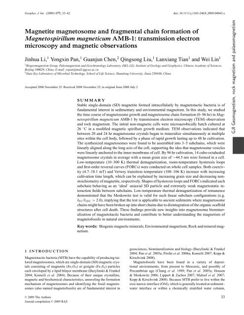
Figure 1. Representative transmission electron microscopy (TEM) images <strong>of</strong> whole AMB-1 cells cultivated at different times. (a) Initial non-magnetic cell;<br />
(b) 24-hr cultivated cell with fine-grained <strong>magnetosome</strong>s (marked by arrows); (c) 28-hr cultivated cell; (d) 36-hr cultivated cell; (e) 96-hr cultivated cell showing<br />
<strong>magnetosome</strong> sub<strong>chain</strong>s (marked by Sn); (f) extracted <strong>magnetosome</strong>s from cells at 96-hr cultivation.<br />
300 to 10 K in a 5-T field (FC). The Verwey transition temperature<br />
(T v ) is defined as the temperature for the maximum <strong>of</strong> the first-order<br />
derivative <strong>of</strong> dM/dT from the FC curve. The δ ratio (δ FC /δ ZFC )was<br />
calculated after Moskowitz et al. (1993).<br />
2.4 Room-temperature hysteresis loops <strong>and</strong> FORCs<br />
Room-temperature hysteresis loops <strong>and</strong> FORCs were measured using<br />
an Alternating Gradient Force Magnetometer Model Micro-<br />
Mag 2900 (Princeton Measurements Corporation, sensitivity is<br />
1.0 × 10 −11 Am 2 ). The hysteresis loops were measured between<br />
±1.5 T with an averaging time <strong>of</strong> 100 ms. Saturation magnetization<br />
(M s ), saturation remanence (M rs ) <strong>and</strong> coercivity (B c ) were determined<br />
after correction for linear contributions from the diamagnetic<br />
<strong>and</strong> paramagnetic phases. Subsequently, the SIRM acquired<br />
at 1.5 T was demagnetized in a backfield to obtain the coercivity <strong>of</strong><br />
remanence (B cr ). Each FORC was measured by saturating the cell<br />
sample at 1.2 T, decreasing the field to a value <strong>of</strong> H a , <strong>and</strong> reversing<br />
the field sweep to the saturated state in a series <strong>of</strong> steps following<br />
the protocol described in Roberts et al. (2000). FORC diagrams<br />
were calculated using the FORCO-BELLO MATLAB code version<br />
0.99c with a smoothing factor (SF) <strong>of</strong> 3 (Winklh<strong>of</strong>er & Zimanyi<br />
2006). In a FORC diagram, the horizontal <strong>and</strong> vertical axes usually<br />
are referred to as microcoercivity (H c ) <strong>and</strong> interaction field, respectively.<br />
Strictly speaking, this assignment is only true for a system in<br />
which each particle is characterized by a rectangular hysteresis loop<br />
(Winklh<strong>of</strong>er & Zimanyi 2006). At the other extreme, in a noninteracting<br />
assemblage <strong>of</strong> r<strong>and</strong>omly oriented Stoner–Wohlfarth type<br />
particles, the vertical axis cannot have the meaning <strong>of</strong> an interaction<br />
field, <strong>and</strong> should rather be referred to as bias field (H b ), which<br />
C○ 2009 The Authors, GJI, 177, 33–42<br />
Journal compilation C○ 2009 RAS
36 J. Li et al.<br />
Table 1. Data <strong>of</strong> cell growth, <strong>magnetosome</strong> <strong>and</strong> sub<strong>chain</strong> <strong>formation</strong> <strong>of</strong> AMB-1 at different growth stages.<br />
Growth phase<br />
Time (hr)<br />
Cells Magnetosomes Magnetosome <strong>chain</strong>s<br />
OD 600 N m L m (nm) W m (nm) Shape factor Sub<strong>chain</strong> number d sc (nm) d cc (nm)<br />
0 0.000 0 – – – – – –<br />
Lag phase 10 0.000 0 – – – – – –<br />
20 0.008 0 – – – – – –<br />
24 0.017 6 ± 4 24.3 ± 4.5 19.1 ± 5.2 0.79 – – –<br />
28 0.018 6 ± 3 36.0 ± 11.7 30.1 ± 10.5 0.84 3 ± 1 221.3 ± 124.2 57.9 ± 10.4<br />
32 0.032 9 ± 4 42.8 ± 10.7 35.8 ± 10.4 0.83 4 ± 2 245.1 ± 153.7 60.5 ± 9.1<br />
Exponential phase 36 0.036 11 ± 3 43.9 ± 12.1 36.1 ± 11.2 0.82 4 ± 1 273.6 ± 131.8 59.8 ± 9.6<br />
40 0.041 9 ± 3 42.9 ± 10.4 37.2 ± 10.4 0.86 4 ± 1 286.4 ± 132.1 62.5 ± 9.6<br />
44 0.083 12 ± 3 43.8 ± 12.4 36.8 ± 11.3 0.84 5 ± 2 243.4 ± 134.9 61.5 ± 9.6<br />
48 0.107 12 ± 5 44.4 ± 12.1 37.5 ± 11.3 0.84 4 ± 1 239.0 ± 127.3 61.8 ± 10.0<br />
52 0.107 12 ± 3 45.7 ± 10.7 39.9 ± 10.1 0.87 4 ± 1 290.0 ± 158.6 61.9 ± 10.3<br />
56 0.105 11 ± 3 44.2 ± 12.3 37.2 ± 11.5 0.84 4 ± 2 283.1 ± 162.3 60.6 ± 9.4<br />
Stationary phase 60 0.098 12 ± 4 47.2 ± 12.5 39.6 ± 11.6 0.84 4 ± 1 283.1 ± 198.6 57.9 ± 11.7<br />
68 0.095 13 ± 4 46.1 ± 13.1 38.4 ± 11.7 0.83 5 ± 1 273.6 ± 185.6 55.9 ± 12.1<br />
76 0.081 14 ± 3 45.5 ± 13.1 39.2 ± 12.6 0.86 4 ± 2 276.7 ± 157.5 58.6 ± 9.6<br />
96 0.082 14 ± 3 48.2 ± 13.8 40.8 ± 12.9 0.85 4 ± 1 247.0 ± 125.5 56.9 ± 10.3<br />
Note: OD 600 refers optical density <strong>of</strong> cell suspensions; N m indicates the averaged <strong>magnetosome</strong> number per cell; L m <strong>and</strong> W m indicate the mean grain length<br />
<strong>and</strong> width <strong>of</strong> <strong>magnetosome</strong>, respectively; d sc indicates the inter-sub<strong>chain</strong> distance within cells, d cc indicates the centre-to-centre distance between adjacent<br />
<strong>magnetosome</strong>s within sub<strong>chain</strong>s.<br />
reflects the history-dependence <strong>of</strong> magnetization changes (Newell<br />
2005). Also, for non-interacting particles, the contributions to the<br />
FORC diagram for negative values <strong>of</strong> H b do not reflect irreversible<br />
switching events, but history-dependent reversible magnetization<br />
changes, as shown theoretically by Newell (2005) <strong>and</strong> experimentally<br />
by Winklh<strong>of</strong>er et al. (2008).<br />
3 RESULTS<br />
3.1 Cell <strong>and</strong> <strong>magnetosome</strong> growth, <strong>and</strong> <strong>magnetosome</strong><br />
sub<strong>chain</strong><br />
BasedontheOD 600 data, the time-course cell growth can be divided<br />
into three phases (Table 1). The cell numbers increase very slowly<br />
during a lag phase (0–20 hr), increase rapidly during an exponential<br />
phase (20–48 hr) <strong>and</strong> decrease slowly during a stationary phase<br />
(48–96 hr). The death phase, which is characteristic <strong>of</strong> exponential<br />
decrease in cell numbers, did not occur until 96 hr in this study.<br />
The number <strong>and</strong> the grain size <strong>of</strong> <strong>magnetosome</strong>s <strong>and</strong> <strong>chain</strong> arrangements<br />
are summarized in Table 1. No <strong>magnetosome</strong>s were<br />
visually observed in the AMB-1 cells during the lag phase. At<br />
24 hr, about 10 per cent <strong>of</strong> cells produced very fine-grained <strong>magnetosome</strong>s<br />
which were detected at multiple positions within the cell<br />
body (Fig. 1b, arrows). After 32 hr cultivation <strong>and</strong> later, almost all<br />
cells formed <strong>magnetosome</strong>s; <strong>magnetosome</strong>s in a bacterium were<br />
arranged in the form <strong>of</strong> several short <strong>chain</strong>s along the long axis <strong>of</strong><br />
the cell (see Figs 1c–e).<br />
Under the used growth conditions, the vast majority <strong>of</strong> cells<br />
(>90 per cent) formed <strong>fragmental</strong> <strong>chain</strong>s. The <strong>fragmental</strong> <strong>chain</strong><br />
commonly consists <strong>of</strong> 3–5 short <strong>chain</strong>s with large gaps (hereafter referred<br />
to as sub<strong>chain</strong>s). Each sub<strong>chain</strong> contains 3–6 closely aligned<br />
<strong>magnetosome</strong>s. Smaller <strong>magnetosome</strong>s are usually formed at the<br />
end <strong>of</strong> a sub<strong>chain</strong>. Nevertheless, the sub<strong>chain</strong>s were linearly aligned<br />
along the long axis <strong>of</strong> the cell. The centre-to-centre distance d cc<br />
varies from 55.9 to 62.5 nm <strong>and</strong> is nearly constant during the whole<br />
time course (Table 1). The distance between adjacent sub<strong>chain</strong>s d sc<br />
varies from ∼221 to ∼290 nm (Table 1). These snapshots at different<br />
stages <strong>of</strong> cell growth suggest that the relative position <strong>of</strong> a<br />
<strong>magnetosome</strong> within a sub<strong>chain</strong> is likely fixed.<br />
To quantify the <strong>magnetosome</strong> growth, the averaged <strong>magnetosome</strong><br />
number per cell (N m ), length (L m ) <strong>and</strong> width (W m ) <strong>of</strong> <strong>magnetosome</strong>,<br />
<strong>and</strong> shape factor were analysed (see Table 1). Statistically,<br />
the N m , L m <strong>and</strong> W m increase rapidly during the early exponential<br />
phase. Between 32 <strong>and</strong> 48 hr, the cells divide faster, while the N m<br />
still increases, indicating that <strong>magnetosome</strong>s were synthesized fast<br />
enough to compensate for the accelerated cell division. The slight<br />
decrease <strong>of</strong> N m at 40 hr suggests that <strong>magnetosome</strong> <strong>formation</strong> lags<br />
behind cell division at the end <strong>of</strong> the exponential phase. During the<br />
stationary phase, the cell number decreases with time, but the N m<br />
<strong>and</strong> grain sizes still increase slightly. After 96 hr cultivation, in average,<br />
the cell produced 14 cubo-octahedral <strong>magnetosome</strong> crystals<br />
with a mean grain size <strong>of</strong> 44.5 nm (Figs 1e <strong>and</strong> f).<br />
Fig. 2 shows the grain size <strong>and</strong> shape factor distributions <strong>of</strong> <strong>magnetosome</strong>s<br />
at 28 , 32 <strong>and</strong> 68 hr. The grain size distribution evolves<br />
gradually from positively skewed (skewness <strong>of</strong> 0.17 at 28 hr) to<br />
negatively skewed shape (skewness <strong>of</strong> −0.29 at 32 hr <strong>and</strong> −0.52 at<br />
68 hr). The positively skewed shape <strong>of</strong> the crystal size distribution<br />
represents the unconstrained growth (i.e. not limited by vesicles<br />
size). With the <strong>magnetosome</strong> growing, the spatial limitation <strong>of</strong><br />
vesicles possibly results in forming a negatively skewed distribution.<br />
The shape factor distributions <strong>of</strong> <strong>magnetosome</strong>s throughout<br />
the cell cultivation were very narrow <strong>and</strong> asymmetric, also indicating<br />
a strictly biochemical control on <strong>magnetosome</strong> <strong>formation</strong> (Eberl<br />
et al. 1998; Arató et al. 2005).<br />
3.2 The Verwey transition <strong>and</strong> the Moskowitz test<br />
Fig. 3 shows representative low-temperature magnetization curves<br />
<strong>and</strong> room-temperature hysteresis loops <strong>of</strong> the cell samples. Both<br />
FC <strong>and</strong> ZFC warming curves show a sharp drop <strong>of</strong> SIRM 5T−10K<br />
intensity around 100–106 K (Figs 3a, c <strong>and</strong> e), indicative <strong>of</strong> magnetite<br />
<strong>magnetosome</strong>s. Specifically, the T v is <strong>of</strong> 100, 104, 104, 104<br />
<strong>and</strong> 106 K for the cell samples at 28, 36, 48, 68 <strong>and</strong> 96 hr, respectively.<br />
On the warming curves after FC, those samples showed about<br />
90.9, 58.6, 49.8, 44.2 <strong>and</strong> 51.7 per cent <strong>of</strong> SIRM 5T−10K loss at 300 K,<br />
C○ 2009 The Authors, GJI, 177, 33–42<br />
Journal compilation C○ 2009 RAS
<strong>Magnetite</strong> <strong>magnetosome</strong> <strong>and</strong> <strong>fragmental</strong> <strong>chain</strong> <strong>formation</strong> <strong>of</strong> Magnetospirillum magneticum AMB-1 37<br />
Figure 2. Crystal size distributions (left-h<strong>and</strong> side) <strong>and</strong> shape factor distributions (right-h<strong>and</strong> side) <strong>of</strong> <strong>magnetosome</strong>s at 28, 32, <strong>and</strong> 68 hr. Size equals<br />
(W + L)/2.<br />
respectively. The decreasing demagnetization through warming <strong>and</strong><br />
increasing T v with cultivation time suggest a decreasing contribution<br />
<strong>of</strong> SP particles <strong>and</strong> an evolution towards more stoichiometric<br />
magnetite (Aragón et al. 1993; Muxworthy & McClell<strong>and</strong> 2000).<br />
The observed remanence losses at 300 K by low-temperature demagnetization<br />
are much bigger than those <strong>of</strong> other cultured <strong>and</strong><br />
uncultured MTB (see table 2 in Pan et al. 2005b). It is likely due to<br />
the smaller grain size formed <strong>and</strong> much shorter <strong>chain</strong>s in this study.<br />
However, the T v values (100–106 K) are comparable to other values<br />
published (see Pan et al. 2005b), suggesting that grain size effect<br />
on the T v value is limited (Prozorov et al. 2007).<br />
The calculated δ FC /δ ZFC values increase from 1.3 at 28 hr to ≥2.0<br />
after 36 hr. For the cell samples at 36, 48, 68 <strong>and</strong> 96 hr, their δ ratios<br />
are 2.2, 2.1, 2.3 <strong>and</strong> 2.0, respectively.<br />
3.3 Hysteresis loops <strong>and</strong> FORCs<br />
The room-temperature hysteresis loop for the cell sample at 28 hr<br />
is slightly wasp-waisted with the lowest B c , B cr <strong>and</strong> M rs /M s ,but<br />
the highest B cr /B c , falling into the region with higher SP content<br />
(Fig. 3b). This is well consistent with the TEM observation <strong>and</strong><br />
low-temperature magnetic measurement, which indicate a mixture<br />
<strong>of</strong> SP <strong>and</strong> SD particles. Other cell samples cultivated for longer time<br />
showed nearly classic Stoner–Wohlfarth type hysteresis loops with<br />
increasing coercivity values (Figs 3d <strong>and</strong> f). On the modified Day<br />
plot, the cell sample at 28 hr falls within the SD + SP region, while<br />
the other cell samples are located well in the SD grain size region<br />
(Fig. 4). The M rs /M s values are close to 0.5, which indicates that the<br />
magnetization within the cells is dominated by uniaxial anisotropy<br />
(imparted by the <strong>magnetosome</strong> <strong>chain</strong> or sub<strong>chain</strong> structure, which<br />
outweighs the intrinsic cubic magnetocrystalline anisotropy; Stoner<br />
& Wohlfarth 1948). The B c increases from 4.7 mT at 28 hr to<br />
18.1 mT at 96 hr; <strong>and</strong> the B cr increases from 11.2 mT at 28 hr to<br />
23.3 mT at 96 hr.<br />
FORC diagrams may provide detailed in<strong>formation</strong> about the distributions<br />
<strong>of</strong> coercivity <strong>and</strong> magnetostatic interaction fields, <strong>and</strong> the<br />
domain state <strong>of</strong> magnetic components (Pike et al. 1999; Roberts<br />
et al. 2000). FORC diagrams for the representative whole cell samples<br />
are shown in Fig. 5. Consistent with TEM observations, the<br />
C○ 2009 The Authors, GJI, 177, 33–42<br />
Journal compilation C○ 2009 RAS
38 J. Li et al.<br />
Figure 3. Low-temperature magnetic measurements <strong>and</strong> room-temperature hysteresis loops for the cell sample at 28, 36 <strong>and</strong> 96 hr. Solid (open) squares<br />
indicate the FC (ZFC) data. Thin line st<strong>and</strong>s for the dM/dT <strong>of</strong> FC data, its peak reflects the T v . The calculation <strong>of</strong> δ FC /δ ZFC ratio sees the text.<br />
FORC diagrams for the whole AMB-1 cells show a very clear<br />
evolution from a mainly SP pattern to an SD pattern. Specifically,<br />
as expected for very small grain size <strong>and</strong> less <strong>magnetosome</strong>s, the<br />
FORC diagram for the 28 hr sample is ‘noise’ (Fig. 5a). The FORC<br />
distribution <strong>of</strong> cell sample at 36 hr already indicates the presence<br />
<strong>of</strong> stable SD particles with a small fraction <strong>of</strong> viscous SD (close to<br />
H c = 0; Fig. 5b). Obviously, the viscous SD fraction is too small to<br />
influence the shape <strong>of</strong> the hysteresis loop, which is not constricted<br />
(Fig. 3d). And finally, the FORC diagram for the cell sample at 96 hr<br />
shows a typical SD behaviour (Fig. 5c).<br />
The FORC diagrams for the whole AMB-1 cells after 36 hr cultivation<br />
show rather narrow vertical distribution along the H c axis<br />
(Figs 5b <strong>and</strong> c), very different from the ones obtained for interacting<br />
magnetite particles (Muxworthy et al. 2004; Pan et al. 2005b).<br />
The latter shows a large vertical spread, while the FORC diagrams<br />
obtained here are confined to within a few mT <strong>of</strong> either side <strong>of</strong> the<br />
H c axis. The little vertical spread appears to be due to processingrelated<br />
smoothing, as the diagrams become increasingly concentrated<br />
along the H c axis for decreasing values <strong>of</strong> SF. Interestingly,<br />
the FORC diagram in Fig. 5(c) has a pronounced negative region<br />
in the lower-left domain, centred about the point (H c , H b ) = (0,<br />
−30) mT, which is connected to the maximum <strong>of</strong> the distribution<br />
at (H c , H b ) = (−30, 0) mT by a line with slope 45 ◦ . That is exactly<br />
what Newell predicted for a non-interacting, isotropically oriented<br />
Stoner–Wohlfarth assemblage (Newell 2005). This observation supports<br />
the interpretation <strong>of</strong> the corresponding major hysteresis loop<br />
(Fig. 3f) in terms <strong>of</strong> a ‘Stoner–Wohlfarth’ loop. From this analogy,<br />
we may conclude that magnetostatic interactions are too small to<br />
noticeably affect the magnetic hysteresis properties.<br />
4 DISCUSSION<br />
4.1 Magnetosome synthesis in AMB-1<br />
In the present study, no <strong>magnetosome</strong>s were detected during the lag<br />
phase despite non-magnetic AMB-1 cells growing in iron-rich conditions.<br />
The temporal variations <strong>of</strong> average <strong>magnetosome</strong> numbers,<br />
mean grain sizes <strong>and</strong> cell numbers suggest that the AMB-1 cells synthesize<br />
<strong>magnetosome</strong>s during both the exponential <strong>and</strong> stationary<br />
growth phases, but dominantly in the former phase (Table 1). This<br />
is different from the growth <strong>of</strong> AMB-1 under anaerobic conditions,<br />
which produced <strong>magnetosome</strong>s only in the exponential growth<br />
phase (Matsunaga et al. 1996; Yang et al. 2001). Komeili et al.<br />
(2004) showed that non-magnetic AMB-1 cells formed full-sized<br />
<strong>magnetosome</strong>s within 21 hr after transited into iron-rich medium.<br />
This suggests that both the initial cells <strong>and</strong> the culture environments<br />
affect the <strong>magnetosome</strong> <strong>formation</strong> in AMB-1. For instance,<br />
C○ 2009 The Authors, GJI, 177, 33–42<br />
Journal compilation C○ 2009 RAS
<strong>Magnetite</strong> <strong>magnetosome</strong> <strong>and</strong> <strong>fragmental</strong> <strong>chain</strong> <strong>formation</strong> <strong>of</strong> Magnetospirillum magneticum AMB-1 39<br />
Figure 4. Measured M rs /M s <strong>and</strong> B cr /B c data for the AMB-1 cells (solid<br />
symbols, this study) compared to the other MTB (open symbols, data from<br />
(Moskowitz et al. 1993), the mixture <strong>of</strong> MV1H + 10 nm ferr<strong>of</strong>luid (Carter-<br />
Stiglitz et al. 2001) <strong>and</strong> the predictions <strong>of</strong> linear mixing theory (Dunlop<br />
2002). Samples <strong>of</strong> the AMB-1 cells at 28 hr (square), 36 hr (circle),<br />
48 hr (inverse triangle), 68 hr (triangle) <strong>and</strong> 96 hr (asterisk); the wet MV-1<br />
<strong>and</strong> MV-2 (squares), freeze-dried MV-1 (triangles) <strong>and</strong> freeze-dried MS-1<br />
(inverse triangles) cells.<br />
Komeili et al. (2004) prepared their initial non-magnetic cells by<br />
pre-culturing AMB-1 strains anaerobically in iron-poor medium,<br />
<strong>and</strong> the time-course experiments were carried out under anaerobic<br />
conditions. Therefore, rapid <strong>magnetosome</strong> <strong>formation</strong> may be<br />
ubiquitous in optimal natural conditions.<br />
The reduced T v <strong>of</strong> <strong>magnetosome</strong> magnetite has been observed in<br />
other cultured <strong>and</strong> uncultured MTB cell samples (Moskowitz et al.<br />
1993, 2008; Carter-Stiglitz et al. 2004; Weiss et al. 2004; Pan et al.<br />
2005b; Kopp et al. 2006; Pósfai et al. 2006b; Prozorov et al. 2007).<br />
Generally speaking, cation substitution, oxidation <strong>and</strong> cation vacancies<br />
can reduce the T v (Muxworthy & McClell<strong>and</strong> 2000; Walz<br />
2002). Previous experiments showed that MTB formed magnetite<br />
has highly chemical purity even with titanium, chromium, cobalt,<br />
copper, nickel, mercury or lead in growth medium (Gorby 1989).<br />
Oxidation caused by sample h<strong>and</strong>ling in this study should be slight<br />
because fresh cells were quickly centrifuged at low temperature<br />
(4 ◦ C) for short time (15 min) <strong>and</strong> taken for the low-temperature<br />
measurements immediately. Cation vacancies (Frankel et al. 1979)<br />
or crystal defects (Devouard et al. 1998) cannot be excluded at this<br />
stage. Although the exact mechanism for the lower T v <strong>of</strong> <strong>magnetosome</strong>s<br />
is not determined, our data support the idea that the lower T v<br />
may be an intrinsic property <strong>of</strong> <strong>magnetosome</strong> magnetite (Pan et al.<br />
2005b; Fischer et al. 2008; Moskowitz et al. 2008).<br />
4.2 Mechanism <strong>of</strong> sub<strong>chain</strong> assemblage<br />
Recently, Komeili (2007) proposed a new mechanism for <strong>magnetosome</strong><br />
<strong>formation</strong> <strong>of</strong> AMB-1 involving in three steps: (1) invagination<br />
<strong>of</strong> <strong>magnetosome</strong> membrane from the inner membrane <strong>of</strong><br />
cell; (2) integration <strong>of</strong> the membrane invaginations into a intact<br />
<strong>chain</strong> with the help <strong>of</strong> cytoskeletal filaments <strong>and</strong> (3) biomineralization<br />
<strong>of</strong> magnetite particles within the empty membrane invaginations<br />
pre-aligned within <strong>and</strong> anchored to the inner membrane <strong>of</strong><br />
the cell. TEM observations <strong>of</strong> this study clearly showed that the<br />
<strong>magnetosome</strong> <strong>formation</strong> in AMB-1 starts simultaneously at multiple<br />
sites within cell body along the long axis <strong>of</strong> the cell. Gradually,<br />
Figure 5. FORC diagrams for the AMB-1 cell sample at 28 (a), 36 (b)<br />
<strong>and</strong> 96 hr (c). A lower-left ‘negative region’ can be observed for the 96-hr<br />
sample.<br />
the adjacent <strong>magnetosome</strong>s were assembled into 3–5 sub<strong>chain</strong>s,<br />
<strong>and</strong> the newly formed <strong>magnetosome</strong>s are added to the end <strong>of</strong> the<br />
sub<strong>chain</strong>s. The sub<strong>chain</strong> pattern was observed till the final stage.<br />
Although the empty membrane invaginations <strong>and</strong> the cytoskeletal<br />
filaments were not detected in this study, the observations <strong>of</strong> linear<br />
arrangement <strong>of</strong> <strong>magnetosome</strong>s along the long axis <strong>of</strong> the cell <strong>and</strong><br />
constant d cc values strongly support Komeili’s model. The physical<br />
connection <strong>of</strong> <strong>magnetosome</strong> vesicles to inner membrane <strong>and</strong> to cytoskeletal<br />
filaments within cell may maintain the linear arrangement<br />
<strong>of</strong> sub<strong>chain</strong>s. A different mechanism for the <strong>chain</strong> arrangement in<br />
Magnetospirillum gryphiswaldense MSR-1 was proposed, in which<br />
<strong>magnetosome</strong> biomineralization initiates simultaneously at multiple<br />
discrete sites along the entire length <strong>of</strong> the cell; <strong>and</strong> then the growing<br />
<strong>magnetosome</strong>s shift towards the mid-cell to form a straight, tightly<br />
C○ 2009 The Authors, GJI, 177, 33–42<br />
Journal compilation C○ 2009 RAS
40 J. Li et al.<br />
packed <strong>chain</strong> (Scheffel et al. 2006; Faivre et al. 2007). This different<br />
dynamics <strong>of</strong> <strong>magnetosome</strong>-<strong>chain</strong> assemblage might result from<br />
fundamental differences in the molecular mechanisms <strong>of</strong> different<br />
MTB species.<br />
A significant observation in this study is AMB-1 formed 3–<br />
5 sub<strong>chain</strong>s under the used culture conditions. Several factors<br />
should be considered. Previous study revealed that no <strong>magnetosome</strong>s<br />
were formed by AMB-1 when oxygen in gas phase exceeded<br />
35 per cent (Yang et al. 2001). We grew AMB-1 in microaerobic<br />
condition (∼21 per cent oxygen in the gas phase at initial time),<br />
which could inhibit <strong>magnetosome</strong> <strong>formation</strong>. Second, the used batch<br />
way culture (without subsequent iron-feeding) may lead to insufficient<br />
supply <strong>of</strong> available iron during stationary growth phase. Third,<br />
the retrogression <strong>of</strong> the initial non-magnetic cells (aerobic culture)<br />
possibly deteriorates the <strong>magnetosome</strong> <strong>formation</strong> ability <strong>of</strong> cells. To<br />
fully underst<strong>and</strong> the sub<strong>chain</strong> <strong>formation</strong>, further experiments (i.e.<br />
iron-feeding <strong>and</strong> various growth conditions) are needed.<br />
4.3 Magnetostatic interaction <strong>of</strong> <strong>magnetosome</strong> sub<strong>chain</strong>s<br />
The magnetostatic interactions in MTB cell samples could arise<br />
from the intra<strong>chain</strong>, inter<strong>chain</strong> <strong>and</strong> intercell interactions. Previously<br />
theoretical <strong>and</strong> experimental studies have revealed that magnetostatic<br />
interactions inside <strong>magnetosome</strong> <strong>chain</strong> along the direction <strong>of</strong><br />
the <strong>chain</strong> lead to the entire <strong>chain</strong> to behave in a magnetically coherent<br />
fashion (Penninga et al. 1995; Hanzlik et al. 2002), <strong>and</strong> to act<br />
as an ‘ideal‘ uniaxial Stoner–Wohlfarth particle (Dunin-Borkowski<br />
et al. 1998; Simpson et al. 2005; Pósfai et al. 2006a). The sub<strong>chain</strong><br />
configuration <strong>of</strong> AMB-1 cells in present study provides an opportunity<br />
to study the inter-sub<strong>chain</strong> <strong>and</strong> intra-sub<strong>chain</strong> interactions. As<br />
clearly seen in hysteresis loops (Figs 3d <strong>and</strong> f) <strong>and</strong> FORCs (Figs 5b<br />
<strong>and</strong> c), these sub<strong>chain</strong>s behave as uniaxial Stoner–Wohlfarth particles.<br />
There are significant intra-sub<strong>chain</strong> interactions <strong>and</strong> nearly<br />
negligible interactions between sub<strong>chain</strong>s. The large d sc (>200 nm)<br />
<strong>and</strong> good separation <strong>of</strong> whole cells also cause weak or negligible<br />
inter-sub<strong>chain</strong> <strong>and</strong> intercell interactions.<br />
The absence <strong>of</strong> interaction characteristics also corroborate the<br />
earlier studies that FORC diagram cannot detect strong intra<strong>chain</strong><br />
interactions <strong>of</strong> <strong>magnetosome</strong>s because each <strong>magnetosome</strong> <strong>chain</strong><br />
acts as an individual elongated SD particle (Pan et al. 2005b; Chen<br />
et al. 2007). This idea was independently supported by magnetic<br />
induction mapping <strong>of</strong> multiple <strong>magnetosome</strong> <strong>chain</strong>s in MTB using<br />
electron holography (Simpson et al. 2005; Pósfai et al. 2006a).<br />
4.4 More geological significance<br />
In nature, MTB commonly inhabit in OAI in aquatic environments<br />
with a high cell concentration <strong>of</strong> 10 5 –10 6 cells mL –1 .TheOAI<br />
system can provide MTB with beneficial vertical redox <strong>and</strong> chemical<br />
gradients, <strong>and</strong> relatively high concentration <strong>of</strong> dissolved iron<br />
(Simmons et al. 2004; Simmons & Edwards 2006; Moskowitz et al.<br />
2008). Thus, the natural environments have advantages for <strong>magnetosome</strong><br />
<strong>formation</strong> compared with laboratory culture conditions.<br />
So far, the direct observation <strong>of</strong> MTB growth in natural environment<br />
is not practically feasible. Detailed studies on the cultivated<br />
MTB strains are still crucial for underst<strong>and</strong>ing the mechanism <strong>of</strong><br />
<strong>magnetosome</strong> <strong>formation</strong> <strong>and</strong> their biogeochemical significances,<br />
for example, nitrogen fixation, nitrogen denitrification <strong>and</strong> iron cycling,<br />
despite that there could be dramatic differences between the<br />
laboratory condition <strong>and</strong> natural environment. If <strong>fragmental</strong> <strong>chain</strong><br />
were found to exist in other cultivated or uncultivated MTB, the sub<strong>chain</strong><br />
pattern <strong>and</strong> experimental conditions used in this study may<br />
provide very useful reference to account for the <strong>fragmental</strong> <strong>chain</strong><br />
<strong>formation</strong>.<br />
Importantly, the present study shows for the first time that the<br />
Moskowitz test is still valid for sub<strong>chain</strong>s <strong>of</strong> <strong>magnetosome</strong>s (Figs 1,<br />
3c <strong>and</strong> e). The failure <strong>of</strong> Moskowitz test for the cell sample at<br />
28 hr is obviously due to the presence <strong>of</strong> a high-fraction SP particles<br />
<strong>and</strong> very poor <strong>chain</strong> <strong>formation</strong>, as indicated by the shape<br />
<strong>of</strong> warming curve (Fig. 3a), hysteresis loop (Fig. 3b) <strong>and</strong> FORC<br />
diagram (Fig. 5a). All other four measured samples (36, 48, 68<br />
<strong>and</strong> 96 hr) successfully passed the test. This finding significantly<br />
exp<strong>and</strong>s the practicality <strong>of</strong> the test to identify magnet<strong>of</strong>ossils in ancient<br />
sediments. Besides, the observed lower T v (100–106 K) for the<br />
cultivated AMB-1 suggests a reduced stoichiometry <strong>of</strong> magnetite<br />
<strong>magnetosome</strong>s.<br />
It is realized that magnetic minerals in sediments, usually a mixture<br />
<strong>of</strong> biogenic <strong>and</strong> abiogenic phases, are much more complicated<br />
than expected previously, <strong>and</strong> we have little knowledge how well<br />
<strong>magnetosome</strong>s are preserved in bulk sediments. To establish effective<br />
criteria for identification <strong>of</strong> magnet<strong>of</strong>ossils in sediment sample,<br />
rock magnetic studies in combination <strong>of</strong> electron microscopy analyses<br />
<strong>and</strong> ferromagnetic resonance (FMR) spectroscopy are required.<br />
Kopp & Kirschvink (2008) recently proposed six-criteria scoring<br />
schemes for evaluating identifications, <strong>and</strong> stressed the FMR spectroscopy<br />
as a tool for investigating both the narrowness <strong>of</strong> grain<br />
size distributions <strong>and</strong> magnetic anisotropy. With these new comprehensive<br />
approaches, it is expected that future studies <strong>of</strong> sediments<br />
<strong>and</strong> sedimentary rocks will unravel more palaeoenvironmental <strong>and</strong><br />
magnetic signals carried by magnet<strong>of</strong>ossils.<br />
5 CONCLUSIONS<br />
The combination <strong>of</strong> multiparameter rock magnetic analyses <strong>and</strong><br />
TEM observations consistently featured the processes <strong>of</strong> the <strong>magnetosome</strong><br />
<strong>formation</strong> in AMB-1 under a controlled microaerobic<br />
growth condition. It was found that AMB-1 form magnetite <strong>magnetosome</strong>s<br />
during both the exponential <strong>and</strong> stationary growth phases,<br />
but dominantly in the former one. The growing <strong>magnetosome</strong>s are<br />
organized into 3–5 sub<strong>chain</strong>s, linearly aligned along the long axis<br />
<strong>of</strong> the cell. Together with the constant d cc values, observations <strong>of</strong><br />
this study provide lines <strong>of</strong> evidence that <strong>magnetosome</strong>s are physically<br />
connected to the cell <strong>and</strong> linearly assembled. The sub<strong>chain</strong>s<br />
behave as uniaxial SD particles with strong intra-sub<strong>chain</strong> magnetostatic<br />
interactions <strong>and</strong> rather weak inter-sub<strong>chain</strong> <strong>and</strong> intercell<br />
magnetostatic interactions as shown by highly suppressed vertical<br />
distributions in FORC diagrams. The Moskowitz test was proven to<br />
be valid for <strong>magnetosome</strong> arranged in sub<strong>chain</strong>s.<br />
ACKNOWLEDGMENTS<br />
We are grateful to the instructive comments from C.L. Deng, D.<br />
Schüler, <strong>and</strong> M.J. Dekkers on an earlier manuscript. We thank C.G.<br />
Langereis, M. Winklh<strong>of</strong>er <strong>and</strong> anonymous for their useful review to<br />
improve the manuscript. We also thank R.J. Harrison <strong>and</strong> H.F. Qin<br />
for discussions on the FORC diagrams. This work was supported by<br />
the Chinese Academy <strong>of</strong> Sciences (grant KZCX-3-sw-150), the National<br />
Natural Science Foundation <strong>of</strong> China (grants 40325011 <strong>and</strong><br />
40821091) <strong>and</strong> the Natural Science Fund <strong>of</strong> Sh<strong>and</strong>ong Province,<br />
China (grant 2006ZRB01973). Y.X. Pan <strong>and</strong> Q.S. Liu were<br />
supported by the ‘100 Talent Program <strong>of</strong> the Chinese Academy<br />
<strong>of</strong> Sciences’.<br />
C○ 2009 The Authors, GJI, 177, 33–42<br />
Journal compilation C○ 2009 RAS
<strong>Magnetite</strong> <strong>magnetosome</strong> <strong>and</strong> <strong>fragmental</strong> <strong>chain</strong> <strong>formation</strong> <strong>of</strong> Magnetospirillum magneticum AMB-1 41<br />
REFERENCES<br />
Aragón, R., Gehring, P.M. & Shapiro, S.M., 1993. Stoichiometry, percolation,<br />
<strong>and</strong> Verwey ordering in magnetite, Phys. Rev. Lett., 70, 1635–1638.<br />
Arató, B., Szanyi, Z., Flies, C., Schüler, D., Frankel, R.B., Buseck, P.R. &<br />
Pósfai, M., 2005. Crystal-size <strong>and</strong> shape distributions <strong>of</strong> magnetite from<br />
uncultured magnetotactic bacteria as a potential biomarker, Am. Mineral.,<br />
90, 1233–1240.<br />
Bazylinski, D.A. & Frankel, R.B., 2004. Magnetosome <strong>formation</strong> in prokaryotes,<br />
Nat. Rev. Microbiol., 2, 217–230.<br />
Carter-Stiglitz, B., Moskowitz, B.M. & Jackson, M., 2001. Unmixing<br />
magnetic assemblages <strong>and</strong> the magnetic behavior <strong>of</strong> bimodal mixtures,<br />
J. geophys. Res., 106, 26 397–26 412.<br />
Carter-Stiglitz, B., Moskowitz, B.M. & Jackson, M., 2004. More on<br />
the low-temperature magnetism <strong>of</strong> stable single domain magnetite: reversibility<br />
<strong>and</strong> non-stoichiometry, Geophys. Res. Lett., 31, L06606,<br />
doi:10.1029/02003GL019155.<br />
Chang, S.-B.R., Stolz, J.F., Kirschvink, J.L. & Awramik, S.M., 1989. Biogenic<br />
magnetite in stromatolites. II. Occurrence in ancient sedimentray<br />
environments, Precambrian Res., 43, 305–315.<br />
Chen, A.P., Egli, R. & Moskowitz, B.M., 2007. First-order reversal curve<br />
(FORC) diagrams <strong>of</strong> natural <strong>and</strong> cultured biogenic magnetic particles,<br />
J. geophys. Res., 112, B08S90, doi:10.1029/2006JB004575.<br />
Devouard, B., Pósfai, M., Hua, X., Bazylinski, D.A., Frankel, R.B. & Buseck,<br />
P.R., 1998. <strong>Magnetite</strong> from magnetotactic bacteria: size distributions <strong>and</strong><br />
twinning, Am. Mineral., 83, 1387–1398.<br />
Dunin-Borkowski, R.E., McCartney, M.R., Frankel, R.B., Bazylinski, D.A.,<br />
Pósfai, M. & Buseck, P.R., 1998. Magnetic microstructure <strong>of</strong> magnetotactic<br />
bacteria by electron holography, Science, 282, 1868–1870.<br />
Dunlop, D.J., 2002. Theory <strong>and</strong> application <strong>of</strong> the Day plot (M rs /M s versus<br />
H cr /H c ) 1. Theoretical curves <strong>and</strong> tests using titanomagnetite data,<br />
J. geophys. Res., 107, 2056, doi:10.1029/2001JB000486.<br />
Eberl, D.D., Drits, V.A. & Srodon, J., 1998. Deducing growth mechanisms<br />
for minerals from the shapes <strong>of</strong> crystal size distributions, Am.J.Sci.,298,<br />
499–533.<br />
Egli, R., 2004. Characterization <strong>of</strong> individual rock magnetic components by<br />
analysis <strong>of</strong> remanence curves. 3. Bacterial magnetite <strong>and</strong> natural processes<br />
in lakes, Phys. Chem. Earth, 29, 869–884.<br />
Faivre, D., Böttger, L.H., Matzanke, B.F. & Schüler, D., 2007. Intracellular<br />
magnetite biomineralization in bacteria proceeds by a distinct pathway<br />
involving membrane-bound ferritin <strong>and</strong> an iron(II) species, Angew. Chem.<br />
Int. Ed. Engl., 46, 8495–8499.<br />
Fischer, H., Mastrogiacomo, G., Löffler, J.F., Warthmann, R.J.,<br />
Weidler, P.G. & Gehring, A.U., 2008. Ferromagnetic resonance <strong>and</strong> magnetic<br />
characteristics <strong>of</strong> intact <strong>magnetosome</strong> <strong>chain</strong>s in Magnetospirillum<br />
gryphiswaldense, Earth planet. Sci. Lett., 270, 200–208.<br />
Frankel, R.B., Bazylinski, D.A., Johnson, M.S. & Taylor, B.L., 1997.<br />
Magneto-aerotaxis in marine coccoid bacteria, Biophys. J., 73, 994–1000.<br />
Frankel, R.B., Blakemore, R.P. & Wolfe, R.S., 1979. <strong>Magnetite</strong> in freshwater<br />
magnetotactic bacteria, Science, 203, 1355–1356.<br />
Gorby, Y.A., 1989. Regulation <strong>of</strong> <strong>magnetosome</strong> biogenesis by oxygen <strong>and</strong><br />
nitrogen, PhD thesis. University <strong>of</strong> New Hampshire, New Hampshire.<br />
Hanzlik, M., Winklh<strong>of</strong>er, M. & Petersen, N., 2002. Pulsed-field-remanence<br />
measurements on individual magnetotactic bacteria, J. Magn. Magn.<br />
Mater., 248, 258–267.<br />
Hesse, P.P., 1994. Evidence for bacterial paleoecological origin <strong>of</strong> mineral<br />
magnetic cycles in oxic <strong>and</strong> sub-oxic tasman sea sediments, Mar. Geol.,<br />
117, 1–17.<br />
Housen, B.A. & Moskowitz, B.M., 2006. Depth distribution <strong>of</strong> magnet<strong>of</strong>ossils<br />
in near-surface sediments from the Blake/Bahama Outer Ridge, western<br />
North Atlantic Ocean, determined by low-temperature magnetism,<br />
J. geophys. Res., 111, G01005, doi:10.1029/02005JG000068.<br />
Kim, B., Kodama, K. & Moeller, R., 2005. Bacterial magnetite produced<br />
in water column dominates lake sediment mineral magnetism: Lake Ely,<br />
USA, Geophys. J. Int., 163, 26–37.<br />
Kobayashi, A., Kirschvink, J.L., Nash, C.Z., Kopp, R.E., Sauer, D.A.,<br />
Bertani, L.E., Voorhout, W.F. & Taguchi, T., 2006. Experimental observation<br />
<strong>of</strong> <strong>magnetosome</strong> <strong>chain</strong> collapse in magnetotactic bacteria: sedimentological,<br />
paleomagnetic, <strong>and</strong> evolutionary implications, Earth planet. Sci.<br />
Lett., 245, 538–550.<br />
Komeili, A., 2007. Molecular mechanisms <strong>of</strong> <strong>magnetosome</strong> <strong>formation</strong>,<br />
Annu. Rev. Biochem., 76, 351–366.<br />
Komeili, A., Li, Z., Newman, D.K. & Jensen, G.J., 2006. Magnetosomes are<br />
cell membrane invaginations organized by the actin-like protein MamK,<br />
Science, 311, 242–245.<br />
Komeili, A., Vali, H., Beveridge, T.J. & Newman, D.K., 2004. Magnetosome<br />
vesicles are present before magnetite <strong>formation</strong>, <strong>and</strong> MamA is required<br />
for their activation, Proc. Natl. Acad. Sci. USA, 101, 3839–3844.<br />
Kopp, R.E. & Kirschvink, J.L., 2008. The identification <strong>and</strong> biogeochemical<br />
interpretation <strong>of</strong> fossil magnetotactic bacteria, Earth Sci. Rev., 86, 42–61.<br />
Kopp, R.E., Nash, C.Z., Kobayashi, A., Weiss, B.P., Bazylinski, D.A. &<br />
Kirschvink, J.L., 2006. Ferromagnetic resonance spectroscopy for assessment<br />
<strong>of</strong> magnetic anisotropy <strong>and</strong> magnetostatic interactions: a case<br />
study <strong>of</strong> mutant magnetotactic bacteria, J. geophys. Res., 111, B12S25,<br />
doi:10.1029/2006JB004529.<br />
Linford, N., Linford, P. & Platzman, E., 2005. Dating environmental change<br />
using magnetic bacteria in archaeological soils from the upper Thames<br />
Valley, UK, J. Archaeol. Sci., 32, 1037–1043.<br />
Lippert, P.C. & Zachos, J.C., 2007. A biogenic origin for anomalous<br />
fine-grained magnetic material at the Paleocene-Eocene boundary<br />
at Wilson Lake, New Jersey, Paleoceanography, 22, PA4104,<br />
doi:10.1029/2007PA001471.<br />
Malo<strong>of</strong>, A.C. et al., 2007. Sedimentary iron cycling <strong>and</strong> the origin <strong>and</strong><br />
preservation <strong>of</strong> magnetization in platform carbonate muds, Andros Isl<strong>and</strong>,<br />
Bahamas, Earth planet. Sci. Lett., 259, 581–598.<br />
Matsunaga, T., Sakaguchi, T. & Tadokoro, F., 1991. <strong>Magnetite</strong> <strong>formation</strong> by<br />
a magnetic bacterium capable <strong>of</strong> growing aerobically, Appl. Microbiol.<br />
Biotechnol., 35, 651–655.<br />
Matsunaga, T., Tsujimura, N. & Kamiya, S., 1996. Enhancement <strong>of</strong> magnetic<br />
particle production by nitrate <strong>and</strong> succinate fed-batch culture <strong>of</strong><br />
Magnetospirillum sp. AMB-1, Biotechnol. Tech., 10, 495–500.<br />
Moskowitz, B.M., Bazylinski, D.A., Egli, R., Frankel, R.B. & Edwards,<br />
K.J., 2008. Magnetic properties <strong>of</strong> marine magnetotactic bacteria in a<br />
seasonally stratified coastal pond (Salt Pond, MA, USA), Geophys. J. Int.,<br />
174, 75–92.<br />
Moskowitz, B.M., Frankel, R.B. & Bazylinski, D.A., 1993. Rock magnetic<br />
criteria for the detection <strong>of</strong> biogenic magnetite, Earth planet. Sci. Lett.,<br />
120, 283–300.<br />
Muxworthy, A., Heslop, D. & Williams, W., 2004. Influence <strong>of</strong> magnetostatic<br />
interactions on first-order-reversal-curve (FORC) diagrams: a micromagnetic<br />
approach, Geophys. J. Int., 158, 888–897.<br />
Muxworthy, A.R. & McClell<strong>and</strong>, E., 2000. Review <strong>of</strong> the low-temperature<br />
magnetic properties <strong>of</strong> magnetite from a rock magnetic perspective, Geophys.<br />
J. Int., 140, 101–114.<br />
Newell, A.J., 2005. A high-precision model <strong>of</strong> first-order reversal<br />
curve (FORC) functions for single-domain ferromagnets with<br />
uniaxial anisotropy, Geochem. Geophys. Geosyst., 6, Q05010,<br />
doi:10.1029/02004GC000877.<br />
Pósfai, M., Kasama, T. & Dunin-Borkowski, R.E., 2006a. Characterization<br />
<strong>of</strong> bacterial magnetic nanostructures using high-resolution transmission<br />
electron microscopy <strong>and</strong> <strong>of</strong>f-axis electron holography, in Magnetoreception<br />
<strong>and</strong> Magnetosomes in Bacteria, pp. 197–225, ed. Schüler, D.,<br />
Springer-Verlag, Berlin.<br />
Pósfai, M., Moskowitz, B.M., Arató, B., Schüler, D., Flies, C., Bazylinski,<br />
D.A. & Frankel, R.B., 2006b. Properties <strong>of</strong> intracellular magnetite crystals<br />
produced by Desulfovibrio magneticus strain RS-1, Earth planet. Sci.<br />
Lett., 249, 444–455.<br />
Paasche, O., Lovlie, R., Dahl, S., Bakke, J. & Nesje, A., 2004. Bacterial magnetite<br />
in lake sediments: late glacial to Holocene climate <strong>and</strong> sedimentary<br />
changes in northern Norway, Earth planet. Sci. Lett., 223, 319–333.<br />
Pan, Y.X., Petersen, N., Davila, A.F., Zhang, L.M., Winklh<strong>of</strong>er, M., Liu, Q.S.,<br />
Hanzlik, M. & Zhu, R.X., 2005a. The detection <strong>of</strong> bacterial magnetite in<br />
recent sediments <strong>of</strong> Lake Chiemsee (southern Germany), Earth planet.<br />
Sci. Lett., 232, 109–123.<br />
C○ 2009 The Authors, GJI, 177, 33–42<br />
Journal compilation C○ 2009 RAS
42 J. Li et al.<br />
Pan, Y.X., Petersen, N., Winklh<strong>of</strong>er, M., Davila, A.F., Liu, Q.S., Frederichs,<br />
T., Hanzlik, M. & Zhu, R.X., 2005b. Rock magnetic properties <strong>of</strong> uncultured<br />
magnetotactic bacteria, Earth planet. Sci. Lett., 237, 311–325.<br />
Penninga, I., Dewaard, H., Moskowitz, B.M., Bazylinski, D.A. & Frankel,<br />
R.B., 1995. Remanence measurements on individual magnetotactic bacteria<br />
using a pulsed magnetic-field, J. Magn. Magn. Mater., 149, 279–286.<br />
Pike, C.R., Roberts, A.P. & Verosub, K.L., 1999. Characterizing interactions<br />
in fine magnetic particle systems using first order reversal curves, J. Appl.<br />
Phys., 85, 6660–6667.<br />
Prozorov, R., Prozorov, T., Williams, T.J., Bazylinski, D.A., Mallapragada,<br />
S.K. & Narasimhan, B., 2007. Magnetic irreversibility <strong>and</strong> Verwey transition<br />
in nano-crystalline bacterial magnetite, Phys. Rev. B, 76, 054406,<br />
doi:10.1103/physRevB054476.054406.<br />
Roberts, A.P., Pike, C.R. & Verosub, K.L., 2000. First-order reversal curve<br />
diagrams: a new tool for characterizing the magnetic properties <strong>of</strong> natural<br />
samples, J. geophys. Res., 105, 28 461–28 476.<br />
Scheffel, A., Gruska, M., Faivre, D., Linaroudis, A., Plitzko, J.M. & Schüler,<br />
D., 2006. An acidic protein aligns <strong>magnetosome</strong>s along a filamentous<br />
structure in magnetotactic bacteria, Nature, 440, 110–114.<br />
Simmons, S.L. & Edwards, K.J., 2006. Geobiology <strong>of</strong> magnetotactic bacteria,<br />
in Magnetoreception <strong>and</strong> <strong>magnetosome</strong>s in bacteria, pp. 77–102, ed.<br />
Schüler, D., Springer-Verlag, Berlin.<br />
Simmons, S.L., Sievert, S.M., Frankel, R.B., Bazylinski, D.A. & Edwards,<br />
K.J., 2004. Spatiotemporal distribution <strong>of</strong> marine magnetotactic bacteria<br />
in a seasonally stratified coastal salt pond, Appl. Environ. Microbiol., 70,<br />
6230–6239.<br />
Simpson, E.T., Kasama, T., Pósfai, M., Buseck, P.R., Harrison, R.J. & Dunin-<br />
Borkowski, R.E., 2005. Magnetic induction mapping <strong>of</strong> magnetite <strong>chain</strong>s<br />
in magnetotactic bacteria at room temperature <strong>and</strong> close to the Verwey<br />
transition using electron holography, J. Phys.: Conf. Ser., 17, 108–121.<br />
Snowball, I., Zillen, L. & S<strong>and</strong>gren, P., 2002. Bacterial magnetite in Swedish<br />
varved lake-sediments: a potential bio-marker <strong>of</strong> environmental change,<br />
Quat. Int., 88, 13–19.<br />
Stoner, E.C. & Wohlfarth, E.P., 1948. A mechanism <strong>of</strong> magnetic hysteresis<br />
in heterogeneous alloys, Phil. Trans. R. Soc. Lond. Ser. A., 240, 599–642.<br />
Walz, F., 2002. The Verwey transition—a topical review, J. Phys., 14, R285–<br />
R340.<br />
Weiss, B.P., Kim, S.S., Kirschvink, J.L., Kopp, R.E., Sankaran, M.,<br />
Kobayashi, A. & Komeili, A., 2004. Ferromagnetic resonance <strong>and</strong> lowtemperature<br />
magnetic tests for biogenic magnetite, Earth planet. Sci. Lett.,<br />
224, 73–89.<br />
Winklh<strong>of</strong>er, M., Dumas, R.K. & Liu, K., 2008. Identifying reversible <strong>and</strong><br />
irreversible magnetization changes in prototype patterned media using<br />
first- <strong>and</strong> second-order reversal curves, J. Appl. Phys., 103, 07C518,<br />
doi:10.1063/1061.2837888.<br />
Winklh<strong>of</strong>er, M. & Zimanyi, G.T., 2006. Extracting the intrinsic switching<br />
field distribution in perpendicular media: a comparative analysis, J. Appl.<br />
Phys., 99, 08E710, doi:10.1063/1061.2176598.<br />
Yang, C.D., Takeyama, H., Tanaka, T. & Matsunaga, T., 2001. Effects <strong>of</strong><br />
growth medium composition, iron sources <strong>and</strong> atmospheric oxygen concentrations<br />
on production <strong>of</strong> luciferase-bacterial magnetic particle complex<br />
by a recombinant Magnetospirillum magneticum AMB-1, Enzyme<br />
Microb. Technol., 29, 13–19.<br />
APPENDIX<br />
A.1 Chemical composition <strong>of</strong> growth medium<br />
The modified MSGM used in this study contains the following<br />
ingredients (in grams per litre): 0.74 g <strong>of</strong> succinic acid, 0.68 g <strong>of</strong><br />
potassium dihydrogen phosphate, 0.12 g <strong>of</strong> sodium nitrate <strong>and</strong> 0.1 g<br />
<strong>of</strong> sodium thioglycolate. The medium was supplemented with 10 mL<br />
<strong>of</strong> Wolfe’s vitamin solution <strong>and</strong> 10 mL <strong>of</strong> Wolfe’s mineral solution.<br />
The pH was adjusted to 7.0 with NaOH solution prior to sterilization<br />
at 115 ◦ C for 30 min. Compared with the normal MSGM, our<br />
modified MSGM contains three-fold more ferric quinate (60 μM)<br />
<strong>and</strong> is enriched by the addition <strong>of</strong> 0.1 g yeast extract <strong>and</strong> 0.2 g<br />
<strong>of</strong> polypeptone. The addition <strong>of</strong> yeast extract <strong>and</strong> polypeptone was<br />
proven to be able to increase the cell growth <strong>and</strong> <strong>magnetosome</strong><br />
production (Yang et al. 2001).<br />
A.2 Preparation <strong>and</strong> inoculation <strong>of</strong> non-magnetic<br />
AMB-1 cells<br />
First, the pre-cultured cells were inoculated into 200 mL MSGM<br />
contained in a 300-mL flask. The initial cell density is about<br />
2.0 × 10 6 cells mL –1 (measured by bacterial counting chamber).<br />
The flask was then put in a shaking incubator with free gas exchange<br />
<strong>and</strong> was agitated at 80 rpm at 26 ◦ C. After 36 hr shaking<br />
cultivation, the cells reached an exponential growth phase with cell<br />
density <strong>of</strong> about 5.0 × 10 8 cells mL –1 . The newly divided cells<br />
did not contain intracellular magnetic particles based on TEM observations.<br />
The non-magnetic AMB-1 cells were obtained through<br />
a 36-hr aerobic incubation <strong>of</strong> AMB-1 strains. About 2-mL nonmagnetic<br />
AMB-1 cell suspension was inoculated into each <strong>of</strong> the<br />
96 250-mL serum vials (the initial density <strong>of</strong> cells is approximately<br />
2.0 × 10 6 cells mL –1 ). To generate a microaerobic condition, the<br />
vials were sealed with butyl-rubber stopper after the inoculation to<br />
avoid further gas exchange with ambient air.<br />
C○ 2009 The Authors, GJI, 177, 33–42<br />
Journal compilation C○ 2009 RAS