Fine Needle Aspiration Cytology (FNAC) as a Diagnostic ... - ijapbc
Fine Needle Aspiration Cytology (FNAC) as a Diagnostic ... - ijapbc
Fine Needle Aspiration Cytology (FNAC) as a Diagnostic ... - ijapbc

You also want an ePaper? Increase the reach of your titles
YUMPU automatically turns print PDFs into web optimized ePapers that Google loves.
www.<strong>ijapbc</strong>.com IJAPBC – Vol. 2(2), Apr-Jun, 2013 ISSN: 2277 - 4688<br />
________________________________________________________________________________<br />
(swollen pale nucleus) or karyorrhexis (nuclear<br />
fragmentation). Karyolytic neutrophils in an<br />
effusion warrant suspicion of sepsis, but definitive<br />
identification relies on the presence of intracellular<br />
organism. Extracellular bacteria may also be<br />
observed, but care must be taken to ensure that<br />
these organisms are not contaminants, normal flora,<br />
or present in used staining solutions. Numerous<br />
bacterial types have been <strong>as</strong>sociated with septic<br />
exudates in the dog and cat. Most septic exudates,<br />
particularly in the cat, involve anaerobes or<br />
facultative anaerobe. In general, identification of<br />
bacterial species and appropriate microbial therapy<br />
should be determined by culture techniques. The<br />
exception may be Actinomyces and Nocardia,<br />
which appear microscopically <strong>as</strong> characteristic<br />
long, filamentous, beaded rods along with the<br />
presence of ‘‘sulfur granules,’’ which are<br />
microcolonies of bacteria .Many septic exudates<br />
occur by introducing organisms into the body<br />
cavity via traumatic puncture wounds; bite wounds;<br />
perforation of the intestinal tract; migrating foreign<br />
bodies; ruptured pulmonary, hepatic, or prostatic<br />
abscesses; pyometra; pneumonia; or pleuritis. Most<br />
septic exudates involve bacterial sepsis; however,<br />
infections with Mycopl<strong>as</strong>ma, rickettsial agents,<br />
fungal agents, and par<strong>as</strong>ites may occur less<br />
frequently.<br />
Hemorrhagic effusions<br />
Hemorrhagic effusions can result from ruptured<br />
vessels or alterations in v<strong>as</strong>cular endothelial<br />
integrity that is normally maintained by the<br />
interaction of platelets and various clotting factors.<br />
Hemorrhagic effusions grossly and microscopically<br />
contain a certain amount of blood, and the PCV of<br />
the fluid should be at le<strong>as</strong>t 10% to 25% of the<br />
peripheral blood. These fluids must be<br />
distinguished from the iatrogenic blood<br />
contamination that might occur during any<br />
sampling procedure. Several factors may help in<br />
distinguishing between these two processes, but<br />
peracute hemorrhage occurring less than 45<br />
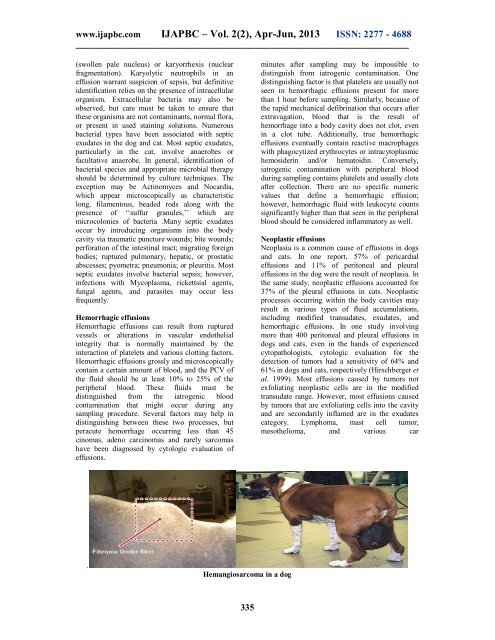
cinom<strong>as</strong>, adeno carcinom<strong>as</strong> and rarely sarcom<strong>as</strong><br />
have been diagnosed by cytologic evaluation of<br />
effusions.<br />
minutes after sampling may be impossible to<br />
distinguish from iatrogenic contamination. One<br />
distinguishing factor is that platelets are usually not<br />
seen in hemorrhagic effusions present for more<br />
than 1 hour before sampling. Similarly, because of<br />
the rapid mechanical defibrination that occurs after<br />
extravagation, blood that is the result of<br />
hemorrhage into a body cavity does not clot, even<br />
in a clot tube. Additionally, true hemorrhagic<br />
effusions eventually contain reactive macrophages<br />
with phagocytized erythrocytes or intracytopl<strong>as</strong>mic<br />
hemosiderin and/or hematoidin. Conversely,<br />
iatrogenic contamination with peripheral blood<br />
during sampling contains platelets and usually clots<br />
after collection. There are no specific numeric<br />
values that define a hemorrhagic effusion;<br />
however, hemorrhagic fluid with leukocyte counts<br />
significantly higher than that seen in the peripheral<br />
blood should be considered inflammatory <strong>as</strong> well.<br />
Neopl<strong>as</strong>tic effusions<br />
Neopl<strong>as</strong>ia is a common cause of effusions in dogs<br />
and cats. In one report, 57% of pericardial<br />
effusions and 11% of peritoneal and pleural<br />
effusions in the dog were the result of neopl<strong>as</strong>ia. In<br />
the same study, neopl<strong>as</strong>tic effusions accounted for<br />
37% of the pleural effusions in cats. Neopl<strong>as</strong>tic<br />
processes occurring within the body cavities may<br />
result in various types of fluid accumulations,<br />
including modified transudates, exudates, and<br />
hemorrhagic effusions. In one study involving<br />
more than 400 peritoneal and pleural effusions in<br />
dogs and cats, even in the hands of experienced<br />
cytopathologists, cytologic evaluation for the<br />
detection of tumors had a sensitivity of 64% and<br />
61% in dogs and cats, respectively (Hirschberger et<br />
al. 1999). Most effusions caused by tumors not<br />
exfoliating neopl<strong>as</strong>tic cells are in the modified<br />
transudate range. However, most effusions caused<br />
by tumors that are exfoliating cells into the cavity<br />
and are secondarily inflamed are in the exudates<br />
category. Lymphoma, m<strong>as</strong>t cell tumor,<br />
mesothelioma, and various car<br />
.<br />
Hemangiosarcoma in a dog<br />
335