Onychomycosis and Its Treatment - ijapbc
Onychomycosis and Its Treatment - ijapbc
Onychomycosis and Its Treatment - ijapbc

Create successful ePaper yourself
Turn your PDF publications into a flip-book with our unique Google optimized e-Paper software.
www.<strong>ijapbc</strong>.com IJAPBC – Vol. 2(1), Jan- Mar, 2013 ISSN: 2277 - 4688<br />
___________________________________________________________________________<br />
INTERNATIONAL JOURNAL OF ADVANCES IN PHARMACY,<br />
BIOLOGY AND CHEMISTRY<br />
<strong>Onychomycosis</strong> <strong>and</strong> <strong>Its</strong> <strong>Treatment</strong><br />
Amartya De, NN. Bala <strong>and</strong> Abu Taher<br />
Review Article<br />
B.C.D.A College of Pharmacy & Technology, 78, Jessore Road(S), Hridaypur, Barasat,<br />
Kolkata, West Bengal, India.<br />
ABSTRACT<br />
<strong>Onychomycosis</strong> is a very common problem affected many people more much in rural area. This condition<br />
may affect toenails or fingernails, but toenail infections are particularly common. In this article we discuss<br />
about etiology,pathophysiology,prevention <strong>and</strong> treatment of the disease.<br />
Keywords: <strong>Onychomycosis</strong>, subungual onychomycosis, c<strong>and</strong>idiasis, Laser <strong>Treatment</strong>.<br />
INTRODUCTION<br />
<strong>Onychomycosis</strong> (also known as "dermatophytic<br />
onychomycosis," "ringworm of the nail," <strong>and</strong> "tinea<br />
unguium") 1 means fungal infection of the nail 2 . It<br />
is the most common disease of the nails <strong>and</strong><br />
constitutes about a half of all nail abnormalities 3 .<br />
This condition may affect toenails or fingernails,<br />
but toenail infections are particularly common. The<br />
prevalence of onychomycosis is about 6-8% in the<br />
adult population 4 .<br />
the Finnish study, only 2 of the 91 patients with<br />
dermatophyte-related onychomycosis of the<br />
toenails also had fingernail involvement. Toenail<br />
infections were approximately 20 times more<br />
common than fingernail infections in the Ohio<br />
cohort The increased frequency of toenail in<br />
comparison to fingernail infections probably<br />
reflects the greater incidence of tinea pedis than of<br />
tinea manuum 5 .<br />
CLASSIFICATION OF ONYCHOMYCOSIS<br />
Four types of onychomycosis, characterized<br />
according to clinical presentation <strong>and</strong> the route of<br />
invasion, are recognized.<br />
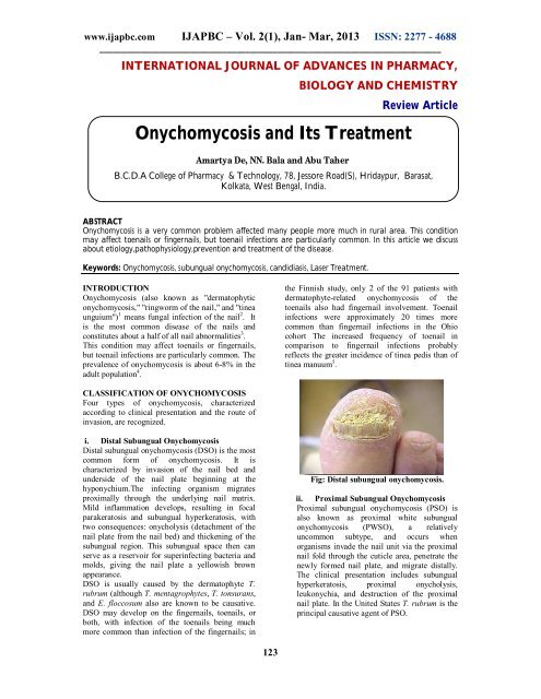
i. Distal Subungual <strong>Onychomycosis</strong><br />
Distal subungual onychomycosis (DSO) is the most<br />
common form of onychomycosis. It is<br />
characterized by invasion of the nail bed <strong>and</strong><br />
underside of the nail plate beginning at the<br />
hyponychium.The infecting organism migrates<br />
proximally through the underlying nail matrix.<br />
Mild inflammation develops, resulting in focal<br />
parakeratosis <strong>and</strong> subungual hyperkeratosis, with<br />
two consequences: onycholysis (detachment of the<br />
nail plate from the nail bed) <strong>and</strong> thickening of the<br />
subungual region. This subungual space then can<br />
serve as a reservoir for superinfecting bacteria <strong>and</strong><br />
molds, giving the nail plate a yellowish brown<br />
appearance.<br />
DSO is usually caused by the dermatophyte T.<br />
rubrum (although T. mentagrophytes, T. tonsurans,<br />
<strong>and</strong> E. floccosum also are known to be causative.<br />
DSO may develop on the fingernails, toenails, or<br />
both, with infection of the toenails being much<br />
more common than infection of the fingernails; in<br />
Fig: Distal subungual onychomycosis.<br />
ii. Proximal Subungual <strong>Onychomycosis</strong><br />
Proximal subungual onychomycosis (PSO) is<br />
also known as proximal white subungual<br />
onychomycosis (PWSO), a relatively<br />
uncommon subtype, <strong>and</strong> occurs when<br />
organisms invade the nail unit via the proximal<br />
nail fold through the cuticle area, penetrate the<br />
newly formed nail plate, <strong>and</strong> migrate distally.<br />
The clinical presentation includes subungual<br />
hyperkeratosis, proximal onycholysis,<br />
leukonychia, <strong>and</strong> destruction of the proximal<br />
nail plate. In the United States T. rubrum is the<br />
principal causative agent of PSO.<br />
123
www.<strong>ijapbc</strong>.com IJAPBC – Vol. 2(1), Jan- Mar, 2013 ISSN: 2277 - 4688<br />
___________________________________________________________________________<br />
Fig: Proximal subungual onychomycosis.<br />
The pattern of growth in PSO is from the proximal<br />
nail fold on the lunula area distally to involve all<br />
layers of the nail Although PSO is the most<br />
infrequently occurring form of onychomycosis in<br />
the general population, it is common in AIDS<br />
patients <strong>and</strong> is considered an early clinical marker<br />
of HIV infection (.In one study of 62 patients with<br />
AIDS or AIDS-related complex <strong>and</strong><br />
onychomycosis, 54 patients (88.7%) had PSO, with<br />
T. rubrum being the etiologic agent in more than<br />
half of these patients. In 54 patients, the feet were<br />
affected, <strong>and</strong> in 5 patients, the h<strong>and</strong>s were infected;<br />
infections of both toenails <strong>and</strong> fingernails were<br />
present in 3 patients 6 . Infection may also<br />
occasionally arise secondary to trauma.<br />
iii. White Superficial <strong>Onychomycosis</strong><br />
White superficial onychomycosis (WSO) is less<br />
common than DSO (estimated proportion of<br />
onychomycosis cases, 10%) <strong>and</strong> occurs when<br />
certain fungi invade the superficial layers of the<br />
nail plate directly (Later, the infection may move<br />
through the nail plate to infect the cornified layer of<br />
the nail bed <strong>and</strong> hyponychium.) It can be<br />
recognized by the presence of well-delineated<br />
opaque “white isl<strong>and</strong>s” on the external nail plate,<br />
which coalesce <strong>and</strong> spread as the disease<br />
progresses. At this point, the nail becomes rough,<br />
soft, <strong>and</strong> crumbly. Inflammation is usually minimal<br />
in patients with WSO, because viable tissue is not<br />
involved (WSO) occurs primarily in the toenails.<br />
iv. C<strong>and</strong>ida Infections of the Nail<br />
C<strong>and</strong>ida nail infections occur in patients with<br />
chronic mucocutaneous c<strong>and</strong>idiasis, <strong>and</strong> are caused<br />
by C. albicans .The organism invades the entire nail<br />
plate. C<strong>and</strong>ida spp. may cause other syndromes,<br />
including onycholysis <strong>and</strong> paronychia. These forms<br />
occur more commonly in women than in men <strong>and</strong><br />
often affect the middle finger, which may come<br />
into contact with C<strong>and</strong>ida organisms that reside in<br />
the intestine or vagina .C<strong>and</strong>ida onychomycosis<br />
can therefore be divided into three general<br />
categories.<br />
(i) Infection beginning as a paronychia (infection of<br />
the structures surrounding the nail; also called a<br />
“whitlow”), the most common type of C<strong>and</strong>ida<br />
onychomycosis, first appears as an edematous,<br />
reddened pad surrounding the nail plate. Invasion<br />
by C<strong>and</strong>ida spp., unlike dermatophytic invasion,<br />
penetrates the nail plate only secondarily after it<br />
has attacked the soft tissue around the nail .After<br />
infection of the nail matrix occurs, transverse<br />
depressions (Beau’s lines) may appear in the nail<br />
plate, which becomes convex, irregular, <strong>and</strong> rough<br />
<strong>and</strong>, ultimately, dystrophic .<br />
(ii) Patients with chronic mucocutaneous<br />
c<strong>and</strong>idiasis are at risk for the second type of , called<br />
C<strong>and</strong>ida granuloma, which accounts for fewer than<br />
1% of onychomycosis cases .This condition is seen<br />
in immunocompromised patients <strong>and</strong> involves<br />
direct invasion of the nail plate .The organism<br />
invades the nail plate directly <strong>and</strong> may affect the<br />
entire thickness of the nail, resulting, in advanced<br />
cases, in swelling of the proximal <strong>and</strong> lateral nail<br />
folds until the digit develops a pseudo-clubbing or<br />
“chicken drumstick” appearance . 8<br />
(iii) Finally, C<strong>and</strong>ida onycholysis can occur when<br />
the nail plate has separated from the nail bed. This<br />
form is more common on the h<strong>and</strong>s than the feet<br />
.Distal subungual hyperkeratosis can be seen as a<br />
yellowish gray mass lifts off the nail plate. The<br />
lesion resembles that seen in patients with DSO.<br />
v. Total Dystrophic <strong>Onychomycosis</strong><br />
Total dystrophic onychomycosis is used to describe<br />
end-stage nail disease, although some clinicians<br />
consider it a distinct subtype. It may be the end<br />
result of any of the four main patterns of<br />
onychomycosis. The entire nail unit becomes thick<br />
<strong>and</strong> dystrophic . 9<br />
Fig: White superficial onychomycosis<br />
The most common etiologic agent in WSO is T.<br />
mentagrophytes .In addition, several<br />
nondermatophyte molds, including Aspergillus<br />
terreus, Acremonium roseogrisum (later confirmed<br />
to be Acremonium potronii), <strong>and</strong> Fusarium<br />
oxysporum, have been implicated by Zaias et al. 7<br />
ANATOMY OF THE NAIL<br />
To have a better underst<strong>and</strong>ing of how<br />
onychomycosis affects the nail, a general<br />
knowledge of the anatomy of the nail is helpful.<br />
The nail, or nail unit, consists of the following<br />
parts:<br />
<br />
The nail matrix (where the nail starts) is<br />
where nail cells multiply <strong>and</strong> keratinize<br />
(harden <strong>and</strong> form into nail material) before<br />
being incorporated into the fingernail or<br />
124
www.<strong>ijapbc</strong>.com IJAPBC – Vol. 2(1), Jan- Mar, 2013 ISSN: 2277 - 4688<br />
___________________________________________________________________________<br />
toenail. Most of the matrix is not visible.<br />
The matrix starts under the skin 5 mm<br />
below the nail fold (the area of the cuticle<br />
where the finger or toe skin meets the nail)<br />
<strong>and</strong> covers the area called the lunula, or<br />
half moon (the white half moon-shaped<br />
area at the bottom of the nail).<br />
<br />
<br />
<br />
The cuticle is a fold of modified skin<br />
where the finger or toe meets the nail. The<br />
cuticle protects the matrix from infection.<br />
The nail plate is the nail itself.<br />
The nail bed is the soft tissue underneath<br />
the nail, anchoring the nail plate. The nail<br />
plate protects the nail bed. 9<br />
CAUSES OF ONYCHOMYCOSIS<br />
<strong>Onychomycosis</strong> is caused by three main classes of<br />
organisms: dermatophytes (fungi that infect hair,<br />
skin, <strong>and</strong> nails <strong>and</strong> feed on nail tissue), yeasts, <strong>and</strong><br />
nondermatophyte molds. All three classes cause the<br />
very similar early <strong>and</strong> chronic symptoms or<br />
appearances, so the visual appearance of the<br />
infection may not reveal which class is responsible<br />
for the infection. Dermatophytes (including<br />
Epidermophyton, Microsporum, <strong>and</strong> Trichophyton<br />
species) are, by far, the most common causes of<br />
onychomycosis worldwide. Yeasts cause 8% of<br />
cases, <strong>and</strong> nondermatophyte molds cause 2% of<br />
onychomycosis cases.<br />
<br />
The dermatophyte Trichophyton rubrum is<br />
the most common fungus causing distal<br />
lateral subungual onychomycosis (DLSO)<br />
<strong>and</strong> proximal subungual onychomycosis<br />
(PSO).<br />
The dermatophyte Trichophyton<br />
mentagrophytes commonly causes white<br />
superficial onychomycosis (WSO), <strong>and</strong><br />
<br />
<br />
more rarely, WSO can be caused by<br />
species of nondermatophyte molds.<br />
The yeast C<strong>and</strong>ida albicans is the most<br />
common cause of chronic mucocutaneous<br />
c<strong>and</strong>idiasis (disease of mucous membrane<br />
<strong>and</strong> regular skin) of the nail<br />
c<strong>and</strong>idiasis (disease of mucous membrane<br />
<strong>and</strong> regular skin) of the nail.<br />
EPIDEMIOLOGY<br />
Frequency<br />
United States:-The recent proliferation of fungal<br />
infections in the United States can be traced to the<br />
large immigration of dermatophytes, especially<br />
Trichophyton rubrum, from West Africa <strong>and</strong><br />
Southeast Asia to North America <strong>and</strong> Europe.<br />
International:-The incidence of onychomycosis<br />
has been reported to be 2-13% in North America. A<br />
multicenter survey in Canada showed the<br />
prevalence of onychomycosis at 6.5%.<br />
<strong>Onychomycosis</strong> accounts for half of all nail<br />
disorders, <strong>and</strong> onychomycosis is the most common<br />
125
www.<strong>ijapbc</strong>.com IJAPBC – Vol. 2(1), Jan- Mar, 2013 ISSN: 2277 - 4688<br />
___________________________________________________________________________<br />
nail disease in adults. Toenails are much more<br />
likely to be infected than fingernails. Thirty percent<br />
of patients with a cutaneous fungal infection also<br />
have onychomycosis. The incidence of<br />
onychomycosis has been increasing, owing to such<br />
factors as diabetes, immunosuppression, <strong>and</strong><br />
increasing age.<br />
Studies in the United Kingdom, Spain, <strong>and</strong> Finl<strong>and</strong><br />
found prevalence rates of onychomycosis to be 3-<br />
8%.<br />
Race<br />
<strong>Onychomycosis</strong> affects persons of all races.<br />
Sex<br />
<strong>Onychomycosis</strong> affects males more commonly than<br />
females. However, c<strong>and</strong>idal infections are more<br />
common in women than in men.<br />
Age<br />
Studies indicate that adults are 30 times more likely<br />
to have onychomycosis than children.<br />
<strong>Onychomycosis</strong> has been reported to occur in 2.6%<br />
of children younger than 18 years but as many as<br />
90% of elderly people.<br />
SIGNS AND SYMPTOMS<br />
PATHOPHYSIOLOGY<br />
The pathogenesis of onychomycosis depends on the<br />
clinical subtype. In distal lateral subungual<br />
onychomycosis, the most common form of<br />
onychomycosis, the fungus spreads from plantar<br />
skin <strong>and</strong> invades the nail bed via the hyponychium.<br />
Inflammation occurring in these areas of the nail<br />
apparatus causes the typical physical signs of distal<br />
lateral subungual onychomycosis. In contrast,<br />
white superficial onychomycosis is a rarer<br />
presentation caused by direct invasion of the<br />
surface of the nail plate. In proximal subungual<br />
onychomycosis, the least common subtype, fungi<br />
penetrate the nail matrix via the proximal nail fold<br />
<strong>and</strong> colonize the deep portion of proximal nail<br />
plate. Endonyx onychomycosis is a variant of distal<br />
lateral subungual onychomycosis in which the<br />
fungi infect the nail via the skin <strong>and</strong> directly invade<br />
the nail plate. Total dystrophic onychomycosis<br />
involves the entire nail unit.<br />
Nail invasion by C<strong>and</strong>ida is not common because<br />
the yeast needs an altered immune response as a<br />
predisposing factor to be able to penetrate the nails.<br />
Despite the frequent isolation of C<strong>and</strong>ida from the<br />
proximal nail fold or the subungual space of<br />
patients with chronic paronychia or onycholysis, in<br />
these patients C<strong>and</strong>ida is only a secondary<br />
colonizer. In chronic mucocutaneous c<strong>and</strong>idiasis,<br />
the yeast infects the nail plate <strong>and</strong> eventually the<br />
proximal <strong>and</strong> lateral nail folds. 13<br />
The most common symptom of a fungal nail<br />
infection is the nail becoming thickened <strong>and</strong><br />
discoloured: white, black, yellow or green. As the<br />
infection progresses the nail can become brittle,<br />
with pieces breaking off or coming away from the<br />
toe or finger completely. If left untreated, the skin<br />
can become inflamed <strong>and</strong> painful underneath <strong>and</strong><br />
around the nail. There may also be white or yellow<br />
patches on the nail bed or scaly skin next to 10 the<br />
nail. 11 . There is usually no pain or other bodily<br />
symptoms, unless the disease is severe People with<br />
onychomycosis may experience significant<br />
psychosocial problems due to the appearance of the<br />
nail, particularly when fingers – which are always<br />
visible – rather than toenails are affected. 12<br />
Dermatophytids are fungus-free skin lesions that<br />
sometimes form as a result of a fungus infection in<br />
another part of the body. This could take the form<br />
of a rash or itch in an area of the body that is not<br />
infected with the fungus. Dermatophytids can be<br />
thought of as an allergic reaction to the fungus.<br />
DIAGNOSIS OF ONYCHOMYCOSIS<br />
<strong>Onychomycosis</strong> (OM) can be identified by its<br />
appearance. However, other conditions <strong>and</strong><br />
infections can cause problems in the nails that look<br />
like onychomycosis. OM must be confirmed by<br />
laboratory tests before beginning treatment,<br />
because treatment is long, expensive, <strong>and</strong> does<br />
have some risks. 14<br />
A sample of the nail can be examined<br />
under a microscope to detect fungi. See<br />
Anatomy of the Nail for information on<br />
the parts of the nail.<br />
The nails must be clipped <strong>and</strong> cleaned<br />
with an alcohol swab to remove bacteria<br />
<strong>and</strong> dirt so the fungal structures can be<br />
more easily visualized with a microscope.<br />
If the doctor suspects distal lateral<br />
subungual onychomycosis (DLSO), a<br />
sample (specimen) should be taken from<br />
the nail bed to be examined. The sample<br />
should be taken from a site closest to the<br />
cuticle, where the concentration of fungi is<br />
the greatest.<br />
If proximal subungual onychomycosis<br />
(PSO) is suspected, the sample is taken<br />
from the underlying nail bed close to the<br />
lunula.<br />
126
www.<strong>ijapbc</strong>.com IJAPBC – Vol. 2(1), Jan- Mar, 2013 ISSN: 2277 - 4688<br />
___________________________________________________________________________<br />
<br />
<br />
<br />
<br />
A piece of the nail surface is taken for<br />
examination if white superficial<br />
onychomycosis (WSO) is suspected.<br />
To detect c<strong>and</strong>idal onychomycosis, the<br />
doctor should take a sample from the<br />
affected nail bed edges closest to the<br />
cuticle <strong>and</strong> sides of the nail.<br />
In the laboratory, the sample may be<br />
treated with a solution made from 20%<br />
potassium hydroxide (KOH) in dimethyl<br />
sulfoxide (DMSO) to help rule out or<br />
more easily verify the presence of fungi by<br />
reducing debris <strong>and</strong> human tissue in the<br />
sample. The specimen may also be treated<br />
with dyes (a process called staining) to<br />
make it easier to see the fungal structure<br />
through the microscope that help identify<br />
the precise species of the pathogen.<br />
If fungi are present in the infected nail,<br />
they can be seen through a microscope,<br />
but the exact type (species) cannot be<br />
determined by simply looking through a<br />
microscope. To identify what exactly is<br />
causing onychomycosis, a fungal culturing<br />
is used. Using a fungal culture to identify<br />
the particular fungus is important because<br />
regular therapy may not work on<br />
nondermatophyte molds.<br />
The infected nail is scraped or<br />
clipped.<br />
The scrapings or clippings are<br />
crushed <strong>and</strong> put into containers.<br />
Any fungus in the samples can<br />
grow in the laboratory in these<br />
special containers. This is true for<br />
most molds <strong>and</strong> yeast also.<br />
The species of pathogen (usually<br />
a fungus) can be identified from<br />
the cultures grown in the lab by<br />
technicians trained to recognize<br />
the microscopic structures that<br />
are identifiers of the fungal<br />
species.<br />
RISK FACTORS<br />
Aging is the most common risk factor for<br />
onychomycosis due to diminished blood<br />
circulation, longer exposure to fungi, <strong>and</strong> nails<br />
which grow more slowly <strong>and</strong> thicken, increasing<br />
susceptibility to infection. Nail fungus tends to<br />
affect men more often than women, <strong>and</strong> is<br />
associated with a family history of this infection. 14<br />
Other risk factors include perspiring heavily, being<br />
in a humid or moist environment, psoriasis,<br />
wearing socks <strong>and</strong> shoes that hinder ventilation <strong>and</strong><br />
do not absorb perspiration, going barefoot in damp<br />
public places such as swimming pools, gyms <strong>and</strong><br />
shower rooms, having athlete's foot (tinea pedis),<br />
minor skin or nail injury, damaged nail, or other<br />
infection, <strong>and</strong> having diabetes, circulation problems<br />
or a weakened immune system.<br />
ONYCHOMYCOSIS PREVENTION<br />
Although it may be impossible to prevent<br />
onychomycosis infections in everyone, there are<br />
ways to reduce a person's chance to get infected.<br />
The following are some of the methods to avoid<br />
nail infections:<br />
Remember that nail infections can be<br />
passed from person to person so washing<br />
h<strong>and</strong>s (<strong>and</strong> feet) after contacting another<br />
person with nail infections is a good<br />
pratice.<br />
Do not go barefoot in public showers or<br />
locker rooms.<br />
Use antifungal spray or powder in shoes,<br />
especially gym shoes.<br />
Be sure that if a manicure or pedicure is<br />
done, instruments are sterilized before<br />
each person is exposed to them.<br />
Keep feet dry <strong>and</strong> clean as possible.<br />
Keep finger <strong>and</strong> toe nails trimmed; do not pick at or<br />
chew on fingernails or the skin around them.<br />
TREATMENT OF ONYCHOMYCOSIS<br />
Medications<br />
In the past, medicines used to treat onychomycosis<br />
(OM) were not very effective. OM is difficult to<br />
treat because nails grow slowly <strong>and</strong> receive very<br />
little blood supply. However, recent advances in<br />
treatment options, including oral (taken by mouth)<br />
<strong>and</strong> topical (applied on the skin or nail surface)<br />
medications, have been made. Newer oral<br />
medicines have revolutionized treatment of<br />
onychomycosis. However, the rate of recurrence is<br />
high, even with newer medicines. <strong>Treatment</strong> is<br />
expensive, has certain risks, <strong>and</strong> recurrence is<br />
possible. 15<br />
Topical antifungals are medicines applied<br />
to the skin <strong>and</strong> nail area that kill fungi <strong>and</strong><br />
some other pathogens.<br />
These topical agents should only<br />
be used if less than half the nail is<br />
involved or if the person with<br />
onychomycosis cannot take the<br />
oral medicines. Medicines<br />
include amorolfine (approved for<br />
use outside the United States),<br />
ciclopirox olamine (Penlac,<br />
which is applied like nail polish),<br />
sodium<br />
pyrithione,<br />
bifonazole/urea (available outside<br />
the United States), propylene<br />
glycol-urea-lactic acid,<br />
imidazoles, such as ketoconazole<br />
(Nizoral Cream), <strong>and</strong><br />
allylamines, such as terbinafine<br />
(Lamisil Cream).<br />
127
www.<strong>ijapbc</strong>.com IJAPBC – Vol. 2(1), Jan- Mar, 2013 ISSN: 2277 - 4688<br />
___________________________________________________________________________<br />
<br />
<br />
Topical treatments are limited<br />
because they cannot penetrate the<br />
nail deeply enough, so they are<br />
generally unable to cure<br />
onychomycosis. Topical<br />
medicines may be useful as<br />
additional therapy in combination<br />
with oral medicines. This results<br />
in treatment medicine<br />
concentrations that come from<br />
two directions, topically <strong>and</strong> from<br />
within the body via oral<br />
medicine.<br />
Newer oral medicines are available. These<br />
antifungal medicines are more effective<br />
because they go through the body to<br />
penetrate the nail plate within days of<br />
starting therapy.<br />
Newer oral antifungal drugs<br />
terbinafine (Lamisil Tablets) <strong>and</strong><br />
itraconazole (Sporanox Capsules)<br />
have replaced older therapies,<br />
such as griseofulvin, in the<br />
treatment of onychomycosis.<br />
They offer shorter treatment<br />
periods (oral antifungal<br />
medications usually are<br />
administered over a three-month<br />
period), higher cure rates, <strong>and</strong><br />
fewer side effects. These<br />
medications are fairly safe, with<br />
few contraindications (conditions<br />
that make taking the medicine<br />
inadvisable), but they should not<br />
be taken by patients with liver<br />
disease or heart failure. Before<br />
prescribing one of these<br />
medications, doctors often order<br />
a blood test to make sure the liver<br />
is functioning properly. Common<br />
side effects include nausea <strong>and</strong><br />
stomach pain.<br />
Fluconazole (Diflucan) is not<br />
approved by the Food <strong>and</strong> Drug<br />
Administration (FDA) for<br />
treatment of onychomycosis, but<br />
it may be used by some clinicians<br />
as an alternative to itraconazole<br />
<strong>and</strong> terbinafine.<br />
To decrease the side effects <strong>and</strong> duration<br />
of oral therapy, topical <strong>and</strong> surgical<br />
treatments (see below) may be combined<br />
with oral antifungal management. 17<br />
Surgery<br />
Surgical approaches to onychomycosis treatment<br />
include surgically or chemically removing the<br />
nail. 18<br />
Thick nails may be chemically removed<br />
by using a urea compound. This technique<br />
128<br />
<br />
<br />
usually should be deferred to a surgeon or<br />
dermatologist.<br />
Surgically removing the nail plate is not<br />
effective treatment of onychomycosis<br />
without additional therapy. This procedure<br />
should be considered an adjunctive<br />
(additional) treatment combined with oral<br />
medical therapy.<br />
A combination of oral, topical, <strong>and</strong><br />
surgical therapy may increase the<br />
effectiveness of treatment <strong>and</strong> reduce the<br />
cost of ongoing treatments.<br />
Laser <strong>Treatment</strong><br />
One of the newest treatments to kill pathogens<br />
infecting the nails is laser therapy. The laser beam<br />
can penetrate the nail tissue <strong>and</strong> disrupt fungal <strong>and</strong><br />
other pathogens enough to kill them. Some patients<br />
may experience some mild discomfort during the<br />
procedure. Reports suggest that laser therapy is<br />
about as effective as medical therapy 19 . Some<br />
patients may require more than one treatment. This<br />
treatment can be very expensive.<br />
Alternative <strong>Treatment</strong>s<br />
There are many claims made that home remedies<br />
can be used to treat a fungal nail infection. Products<br />
such as Listerine, VapoRub, beer soaks, peroxide,<br />
<strong>and</strong> others are purported to be effective.<br />
Unfortunately, there is little or no data to support<br />
these claims; some of the commercially available<br />
products do not promote their use for nail<br />
infections although some individuals may use them<br />
for alternative treatments 19 .<br />
CONCLUSION<br />
Although regional <strong>and</strong> temporal variability exists<br />
among the microorganisms that are pathogenic in<br />
onychomycosis, this disease is caused primarily by<br />
dermatophytes. After decades of frustration <strong>and</strong><br />
disappointment with this stubborn infection,<br />
dermatologists <strong>and</strong> other clinicians now have<br />
access to drugs with high cure rates <strong>and</strong> excellent<br />
safety profiles. Moreover, short treatment times<br />
increase patient compliance, reduce treatment<br />
costs, <strong>and</strong> allow patients to feel hopeful that their<br />
unsightly infections will be ended.<br />
Perhaps the most important task of the clinician is<br />
accurate diagnosis of the causal agent. Direct<br />
microscopy <strong>and</strong> culture are both necessary to<br />
ensure this. Selection of an optimal antifungal drug<br />
whose spectrum of activity encompasses the<br />
infecting microorganism can proceed only with<br />
accurate diagnosis.<br />
In the last decade, there have been significant<br />
advances in the development of effective <strong>and</strong> safe<br />
drugs for onychomycosis. What remains to be<br />
achieved? Unfortunately, onychomycosis is likely<br />
to remain a disease of modern civilization 20 . The<br />
environmental conditions that foster it, longer life
www.<strong>ijapbc</strong>.com IJAPBC – Vol. 2(1), Jan- Mar, 2013 ISSN: 2277 - 4688<br />
___________________________________________________________________________<br />
expectancies, <strong>and</strong> the increasing numbers of<br />
immunocompromised individuals have combined<br />
to increase its prevalence. Against this trend are the<br />
new antifungal drugs itraconazole, terbinafine, <strong>and</strong><br />
fluconazole. But perhaps the single area most<br />
deserving of our attention in the near future is that<br />
of improving diagnostic methods. Diagnostic<br />
methodology <strong>and</strong> fungal susceptibility testing lag<br />
behind therapeutic advances. We should turn our<br />
attention to these problems.<br />
REFERENCES<br />
1. Rapini, Ronald P.; Bolognia, Jean L.;<br />
Jorizzo, Joseph L. (2007). Dermatology:<br />
2-Volume Set. St. Louis: Mosby. p. 1135.<br />
ISBN 1-4160-2999-0. 16262878.<br />
2. "onychomycosis" at Dorl<strong>and</strong>'s Medical<br />
Dictionary.<br />
3. Szepietowski JC, Salomon J (2007). "Do<br />
fungi play a role in psoriatic nails?".<br />
Mycoses 50 (6): 437–42.<br />
doi:10.1111/j.1439-0507.2007.01405.x.<br />
PMID 17944702.<br />
4. "Impact 07 - Dermatology" (PDF). Bay<br />
Bio. 2007. Retrieved 2007-06-13.<br />
5. James, William D.; Berger, Timothy G.<br />
(2006). Andrews' Diseases of the Skin:<br />
clinical Dermatology. Saunders Elsevier.<br />
ISBN 0-7216-2921-0.<br />
6. Cmecorner.com. Retrieved 2010-08-05.<br />
7. Szepietowski JC, Reich A (September<br />
2008). "Stigmatisation in onychomycosis<br />
patients: a population-based study".<br />
Mycoses 52 (4): 343. doi:10.1111/j.1439-<br />
0507.2008.01618.x. PMID 18793262.<br />
8. Chi CC, Wang SH, Chou MC (2005).<br />
"The causative pathogens of<br />
onychomycosis in southern Taiwan".<br />
Mycoses 48 (6): 413–20.<br />
doi:10.1111/j.1439-0507.2005.01152.x.<br />
PMID.<br />
9. Clayton, Y. M., <strong>and</strong> R. J. Hay. 1993.<br />
Epidemiology of fungal skin <strong>and</strong> nail<br />
disease: roundtable discussion held at<br />
Dermatology 2000, Vienna, 17 May 1993.<br />
Br. J. Dermatol. 130(Suppl. 4):9–11.<br />
10. Clinical Courier. New strategies for the<br />
effective management of superficial<br />
fungal infections. Clin Courier.<br />
1997;16:2–3.<br />
11. http://www.emedicinehealth.com/onycho<br />
mycosis/discussion_em-739.htm.<br />
12. Goodfield, M. J. D. 1992. Short-duration<br />
therapy with terbinafine for dermatophyte<br />
onychomycosis: a multicentre trial. Br. J.<br />
Dermatol. 126(Suppl. 39):33–35.<br />
13. Ellis, D. H., J. E. Marley, A. B. Watson,<br />
<strong>and</strong> T. G. Williams. 1997. Significance of<br />
non-dermatophyte molds <strong>and</strong> yeasts in<br />
onychomycosis. Dermatology 194(Suppl.<br />
1):40–42.<br />
14. De Doncker P. Pharmacokinetics of oral<br />
antifungal agents. Dermatol Ther.<br />
997;3:46– 57.<br />
15. Winn W C., Jr . Mycotic diseases. In:<br />
Henry J B, editor. Clinical diagnosis <strong>and</strong><br />
management. Philadelphia, Pa: The W. B.<br />
Saunders Co.; 1996. pp. 1210–1251.<br />
16. Summerbell R C, Kane J, Krajden S.<br />
<strong>Onychomycosis</strong>, tinea pedis, <strong>and</strong> tinea<br />
manuum caused by non-dermatophytic<br />
filamentous fungi. Mycoses. 1989;32:609–<br />
619.<br />
17. Zaias N, Tosti A, Rebell G, Morelli R,<br />
Bardazzi F, Bieley H, Zaiac N, Glick B,<br />
Paley B, Allevato M, Baran R.<br />
Autosomal dominant pattern of distal<br />
subungual onychomycosis caused by<br />
Trichophyton rubrum. J Am Acad<br />
Dermatol. 1996;34(2 Pt 1):302–304.<br />
18. http://emedicine.medscape.com/article/11<br />
05828-treatment#a1127.<br />
19. Del Rosso J Q. Advances in the treatment<br />
of superficial fungal infections: focus on<br />
onychomycosis <strong>and</strong> dry tinea pedis. J Am<br />
Osteopath Assoc. 1997;97:339–345.<br />
20. Gupta AK, Shear NJ. The new oral<br />
antifungal agents for onychomycosis of<br />
the toenails. J Eur Acad Dermatol<br />
Venereol 1999; 13:1–3.<br />
129