Formulation and Evaluation of Floating Microspheres of Captopril for ...
Formulation and Evaluation of Floating Microspheres of Captopril for ...
Formulation and Evaluation of Floating Microspheres of Captopril for ...

You also want an ePaper? Increase the reach of your titles
YUMPU automatically turns print PDFs into web optimized ePapers that Google loves.
Indian Journal <strong>of</strong> Novel Drug delivery 3(1), Jan-Mar, 2011, 17-23<br />
Indian Journal <strong>of</strong> Novel Drug Delivery<br />
IJNDD<br />
An Official Publication <strong>of</strong><br />
Karnataka Education <strong>and</strong><br />
Scientific Society<br />
Research Article<br />
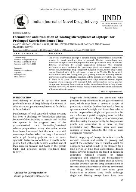
<strong>Formulation</strong> <strong>and</strong> <strong>Evaluation</strong> <strong>of</strong> <strong>Floating</strong> <strong>Microspheres</strong> <strong>of</strong> <strong>Captopril</strong> <strong>for</strong><br />
Prolonged Gastric Residence Time<br />
ANAND GADAD*, CHIRAG NAVAL, KRUNAL PATEL,PANCHAXARI DANDAGI AND VINAYAK<br />
MASTIHOLIMATH<br />
Department <strong>of</strong> Pharmaceutics, KLE University’s College <strong>of</strong> Pharmacy, Belgaum-590010, INDIA<br />
A R T I C L E D E T A I L S<br />
A B S T R A C T<br />
Article history:<br />
Received on 31 January 2011<br />
Modified on 17 March 2011<br />
Accepted on 24 March 2011<br />
Keywords:<br />
<strong>Floating</strong> microspheres,<br />
<strong>Captopril</strong>,<br />
Eudragit S-100,<br />
Ethyl cellulose<br />
The present study was an attempt to develop floating microspheres <strong>of</strong> captopril to<br />
prolong its gastric residence time in stomach. <strong>Floating</strong> microspheres was<br />
<strong>for</strong>mulated using biocompatible polymers like Eudragit S100 <strong>and</strong> Ethyl cellulose in<br />
different proportions by solvent evaporation technique. The prepared<br />
microspheres were evaluated <strong>for</strong> percentage yield, micromeritic properties,<br />
particle size, morphology, drug entrapment, buoyancy studies, In vitro drug release<br />
studies. Practical yield <strong>of</strong> the microspheres was up to 76.40%. The <strong>for</strong>mulated<br />
microspheres were free flowing with good packing properties. Scanning electron<br />
microscopy confirmed spherical structure <strong>and</strong> the particles were <strong>of</strong> the size range<br />
<strong>of</strong> 57.66 to 93.21µm. The microspheres with Ethyl cellulose showed higher<br />
buoyancy when compared with Eudragit S-100. All <strong>for</strong>mulation showed good in<br />
vitro percent buoyancy. In vitro release studies showed cumulative % drug release<br />
between 75.95-88.27%. In vitro release studies demonstrated non-Fickian diffusion<br />
<strong>of</strong> drug from the microsphere.<br />
© KESS All rights reserved<br />
INTRODUCTION<br />
Oral delivery <strong>of</strong> drugs is by far the most<br />
preferable route <strong>of</strong> drug delivery due to ease <strong>of</strong><br />
administration, patient compliance <strong>and</strong> flexibility<br />
in <strong>for</strong>mulation [1] .<br />
Development <strong>of</strong> oral controlled-release systems<br />
has been a challenge to <strong>for</strong>mulation scientists<br />
because <strong>of</strong> their inability to restrain <strong>and</strong> localize<br />
the system in the targeted area <strong>of</strong> the<br />
gastrointestinal tract. Controlled/sustained<br />
release preparations using alternative routes<br />
have been <strong>for</strong>mulated but the oral route still<br />
remains preferable. When the drug is <strong>for</strong>mulated<br />
with a gel <strong>for</strong>ming polymer such as semisynthetic<br />
derivatives <strong>of</strong> cellulose, it swells in the<br />
gastric fluid with a bulk density less than one. It<br />
then remains buoyant <strong>and</strong> floats in the gastric<br />
fluid, <strong>and</strong> prolongs gastric residence time<br />
(GRT) [2] .<br />
*Author <strong>for</strong> Correspondence:<br />
Email: gadadap@rediffmail.com<br />
Single-unit <strong>for</strong>mulations are associated with<br />
problem being obstructed in the gastrointestinal<br />
tract, which may have a potential danger <strong>of</strong><br />
producing irritation. On the other h<strong>and</strong>, a floating<br />
system made <strong>of</strong> multiple unit <strong>for</strong>ms has relative<br />
merits compared to a single unit preparation. On<br />
each subsequent gastric emptying, sunk particles<br />
will spread out over a large area <strong>of</strong> absorption<br />
sites, increasing the opportunity <strong>for</strong> drug release<br />
pr<strong>of</strong>ile <strong>and</strong> absorption in a more or less<br />
predictable way. Moreover, since each dose<br />
consists <strong>of</strong> many subunits, the risk <strong>of</strong> dose<br />
dumping is reduced [1] .<br />
Gastric emptying <strong>of</strong> dosage <strong>for</strong>m is extremely<br />
variable process <strong>and</strong> ability to prolong <strong>and</strong><br />
control the emptying time is valuable asset <strong>for</strong><br />
dosage <strong>for</strong>ms, which reside in the stomach <strong>for</strong> a<br />
long period <strong>of</strong> time than conventional dosage<br />
<strong>for</strong>ms. Several difficulties are faced in designing<br />
controlled released systems <strong>for</strong> better<br />
absorption <strong>and</strong> enhanced the bioavailability.<br />
Conventional oral dosage <strong>for</strong>ms such as tablets,<br />
capsules provide specific drug concentration in<br />
systemic circulation without <strong>of</strong>fering any control<br />
over drug delivery <strong>and</strong> also cause great<br />
fluctuations in plasma drug levels. Although<br />
single unit floating dosage <strong>for</strong>ms have been<br />
17
An<strong>and</strong> Gadad et al / Indian Journal <strong>of</strong> Novel Drug Delivery 3(1), Jan-Mar, 2011, 17-23<br />
extensively studied, these single unit dosage<br />
<strong>for</strong>ms have the disadvantage <strong>of</strong> a release all or<br />
nothing during emptying process while the<br />
multiple unit particulate system pass through the<br />
GIT to avoid the vagaries <strong>of</strong> gastric emptying <strong>and</strong><br />
thus release the drug more uni<strong>for</strong>mly. The<br />
uni<strong>for</strong>m distribution <strong>of</strong> these multiple unit<br />
dosage <strong>for</strong>ms along the GIT could result in more<br />
reproducible drug absorption <strong>and</strong> reduced risk<br />
<strong>of</strong> local irritation; This floating dosage <strong>for</strong>m<br />
enhance bioavailability, having a dissolution<br />
<strong>and</strong>/or stability problem in the small intestine<br />
fluids, being locally effective in the stomach,<br />
being absorbed only in the stomach <strong>and</strong>/or<br />
upper part <strong>of</strong> the intestine [3,4] .<br />
<strong>Captopril</strong>, an antihypertensive agent, has been<br />
widely used <strong>for</strong> the treatment <strong>of</strong> hypertension<br />
<strong>and</strong> congestive heart failure. It has been<br />
reported, however, that the duration <strong>of</strong><br />
antihypertensive action after a single oral dose <strong>of</strong><br />
captopril is only 6–8 h, so clinical use requires a<br />
daily dose <strong>of</strong> 37.5–75 mg to be taken three times.<br />
It is most stable at pH 1.2 <strong>and</strong> as the pH<br />
increases; it becomes unstable <strong>and</strong> undergoes a<br />
degradation reaction [2] .<br />
Thus a sustained <strong>and</strong> controlled release dosage<br />
<strong>for</strong>m <strong>of</strong> captopril is desirable. Hence an attempt<br />
has been made to design a floating microsphere<br />
system <strong>of</strong> captopril using Eudragit S100 <strong>and</strong><br />
Ethyl cellulose <strong>for</strong> prolonged gastric residence<br />
time <strong>and</strong> improve the release pr<strong>of</strong>ile <strong>of</strong> drug.<br />
MATERIALS AND METHODS<br />
<strong>Captopril</strong> was obtained as a gift sample from<br />
Wochardt Ltd., Aurangabad. Eudragit S-100 was<br />
gift sample from Evonik Degussa India Pvt. Ltd.,<br />
Mumbai. Ethyl cellulose was purchased from<br />
Himedia Laboratories Pvt. Ltd., Mumbai. All<br />
chemicals <strong>and</strong> reagents used were <strong>of</strong> analytical<br />
grade.<br />
Preparation <strong>of</strong> floating microspheres<br />
<strong>Floating</strong> microspheres were prepared by the<br />
solvent evaporation method [5] using 500 mg <strong>of</strong><br />
<strong>Captopril</strong> <strong>and</strong> with different proportion <strong>of</strong><br />
polymer as shown in Table 1 were dissolved in<br />
dichloromethane (5 ml) <strong>and</strong> this pasty, flowable<br />
mass was introduced into 50 ml <strong>of</strong> aqueous<br />
saline phase(0.9% Nacl) containing 0.04 %<br />
polyvinyl alcohol (20 mg) <strong>and</strong> 10% methanol (5<br />
ml). The system is stirred using propeller at 300<br />
rpm at room temperature <strong>for</strong> 2-3 h. The drug<br />
loaded floating microspheres <strong>for</strong>med were<br />
filtered, washed <strong>and</strong> dried in a hot air oven at<br />
60 0 C. The detailed composition <strong>of</strong> each<br />
<strong>for</strong>mulation is given in Table 1.<br />
Table 1: <strong>Formulation</strong> Design <strong>of</strong> <strong>Captopril</strong><br />
<strong>Floating</strong> <strong>Microspheres</strong><br />
Batch<br />
Code<br />
F1<br />
F2<br />
F3<br />
F4<br />
F5<br />
F6<br />
Polymer<br />
Eudragit<br />
S-100<br />
Eudragit<br />
S-100<br />
Eudragit<br />
S-100<br />
Ethyl<br />
cellulose<br />
Ethyl<br />
cellulose<br />
Ethyl<br />
cellulose<br />
<strong>Evaluation</strong> <strong>of</strong> floating microsphere<br />
Percentage yield<br />
The prepared microspheres <strong>of</strong> all batches were<br />
accurately weighed. The weight quantity <strong>of</strong><br />
prepared microspheres was divided by the total<br />
amount <strong>of</strong> all the excipients <strong>and</strong> drug used in the<br />
preparation <strong>of</strong> the microspheres, which give the<br />
total percentage yield <strong>of</strong> floating microspheres. It<br />
was calculated by using following equation [6] ,<br />
Percentage yield =<br />
Drug:<br />
Polymer<br />
1:1 1:1<br />
1:2 1:1<br />
1:4 1:1<br />
1:1 1:1<br />
1:2 1:1<br />
1:4 1:1<br />
Actual yield <strong>of</strong> product<br />
Total weight <strong>of</strong> excipients <strong>and</strong> drug<br />
X 100<br />
Micromeritic properties<br />
Micromeritic properties such as Carr's index %<br />
(% I c) <strong>and</strong> Hausner's ratio (H R) were<br />
characterized by using the following equations:<br />
H R = ρ t / ρ b<br />
% I c = (ρ t - ρ b / ρ t) x 100<br />
Where, ρ t = tapped density, ρ b = bulk density<br />
The angle <strong>of</strong> repose (θ) <strong>of</strong> the microspheres,<br />
which measures the resistance to particle flow,<br />
was determined by the fixed funnel method,<br />
using the following equation:<br />
tan θ = H/R<br />
Dichlorometha<br />
ne: Methanol<br />
Where, H is the height <strong>of</strong> the heap that <strong>for</strong>med<br />
after making the microspheres flow from the<br />
glass funnel <strong>and</strong> R is the radius [7,8] .<br />
18
An<strong>and</strong> Gadad et al / Indian Journal <strong>of</strong> Novel Drug Delivery 3(1), Jan-Mar, 2011, 17-23<br />
Particle size determination<br />
Microsphere size was determined by using an<br />
optical microscope under regular polarized light,<br />
<strong>and</strong> the mean microsphere size was calculated by<br />
measuring 100 particles with the help <strong>of</strong> a<br />
calibrated ocular micrometer [9] .<br />
Morphological study using SEM<br />
The morphological study was carried out by<br />
Scanning Electron Microscope (SEM).<br />
<strong>Microspheres</strong> were scanned <strong>and</strong> examined under<br />
Electron Microscope HITACHI SU 1500, Japan<br />
connected with fine coat, JEOL JFC-1100E Ion<br />
sputter. The sample was loaded on copper<br />
sample holder <strong>and</strong> sputter coated with carbon<br />
followed by gold [9] .<br />
Drug entrapment <strong>and</strong> drug loading<br />
<strong>Microspheres</strong> equivalent to 50 mg <strong>of</strong> the drug<br />
were taken <strong>for</strong> evaluation. The amount <strong>of</strong> drug<br />
entrapped was estimated by crushing the<br />
microspheres <strong>and</strong> extracting with aliquots <strong>of</strong><br />
0.1N HCl (pH-1.2) repeatedly. The extract was<br />
transferred to a 100ml volumetric flask <strong>and</strong> the<br />
volume was made up using 0.1N HCl. The<br />
solution was filtered <strong>and</strong> the absorbance was<br />
measured after suitable dilution<br />
spectrophotometrically (UV 1700, Shimadzu,<br />
Japan) at 212 nm against appropriate blank. The<br />
amount <strong>of</strong> drug loaded <strong>and</strong> entrapped in the<br />
microspheres was calculated by the following<br />
<strong>for</strong>mulae [6,10] :<br />
Percentage drug loading =<br />
Weight <strong>of</strong> the drug loaded in the<br />
microspheres X 100<br />
Total weight <strong>of</strong> the microspheres<br />
Percentage drug entrapment =<br />
Amount <strong>of</strong> drug actually present<br />
Theoretical drug load expected<br />
X 100<br />
In vitro buoyancy study<br />
<strong>Microspheres</strong> (300mg) were spread over the<br />
surface <strong>of</strong> a USP XXIV dissolution apparatus type<br />
II filled with 900 ml <strong>of</strong> 0.1 N HCl containing<br />
0.02% Tween 80. The medium was agitated with<br />
a paddle rotating at 100 rpm <strong>for</strong> 12 h. The<br />
floating <strong>and</strong> the settled portions <strong>of</strong> microspheres<br />
were recovered separately. The microspheres<br />
were dried <strong>and</strong> weighed. Buoyancy percentage<br />
was calculated as the ratio <strong>of</strong> the mass <strong>of</strong> the<br />
microspheres that remained floating <strong>and</strong> the<br />
total mass <strong>of</strong> the microspheres.<br />
% Buoyancy = Q f / (Q f + Q s)<br />
Where Q f <strong>and</strong> Q s are the weight <strong>of</strong> the floating<br />
<strong>and</strong> the settled microspheres respectively.<br />
At predetermined time intervals the radiograph<br />
<strong>of</strong> the abdomen was taken using an X-ray<br />
machine [1,11] .<br />
In-vitro release study<br />
The drug release study from microsphere was<br />
per<strong>for</strong>med using USP dissolution apparatus Type<br />
I in 900 ml <strong>of</strong> 0.1 N HCl dissolution media (pH-<br />
1.2) at 100 rpm <strong>and</strong> 37 0 C. 10 ml <strong>of</strong> sample was<br />
withdrawn at predetermined time interval <strong>for</strong> 12<br />
h <strong>and</strong> same volume <strong>of</strong> fresh medium was<br />
replaced to maintained sink condition.<br />
Withdrawn samples were assayed<br />
spectrophotometrically at 212 nm [6,11,12] .<br />
RESULTS AND DISCUSSION<br />
<strong>Captopril</strong>, an antihypertensive agent, with short<br />
half-life, low single dose administration <strong>and</strong> oral<br />
bioavailability is 65 %, was selected as a model<br />
drug to <strong>for</strong>mulate a controlled release<br />
<strong>for</strong>mulation with improved oral bioavailability<br />
by prolonging the gastric residence time.<br />
<strong>Formulation</strong> <strong>of</strong> floating microspheres<br />
<strong>Floating</strong> microspheres were successfully<br />
prepared <strong>for</strong> the delivery <strong>of</strong> <strong>Captopril</strong> to enhance<br />
absorption <strong>and</strong> bioavailability by increasing the<br />
gastric retention time. In concern to this<br />
approach, the primary necessity is to float the<br />
beads in gastric environment. In this study, six<br />
<strong>for</strong>mulations were prepared. In each<br />
<strong>for</strong>mulation, the polymer concentration was<br />
varied. Eudragit S-100 (a synthetic polymer) <strong>and</strong><br />
Ethyl cellulose (a cellulose-based derivative) are<br />
biocompatible, hydrophobic polymers, which<br />
prolong the release <strong>of</strong> water-soluble <strong>and</strong> water<br />
insoluble drugs from their matrices.<br />
Incorporation <strong>of</strong> NaCl to the aqueous phase was<br />
necessary to prevent the dispersed phase from<br />
settling due to high density <strong>of</strong> dichloromethane,<br />
thereby making the dispersion <strong>and</strong> stabilization<br />
<strong>of</strong> the droplets by stirring difficult.<br />
Percentage yield <strong>of</strong> all <strong>for</strong>mulation F1 to F6 were<br />
calculated <strong>and</strong> results are shown in Table 2. The<br />
percentage yield slightly decreased as the ratio <strong>of</strong><br />
polymer increased.<br />
19
An<strong>and</strong> Gadad et al / Indian Journal <strong>of</strong> Novel Drug Delivery 3(1), Jan-Mar, 2011, 17-23<br />
Table 2: Various <strong>Formulation</strong> Parameters <strong>for</strong> <strong>Captopril</strong> <strong>Floating</strong> <strong>Microspheres</strong>.<br />
<strong>Formulation</strong><br />
Code<br />
% Yield Average<br />
particle size<br />
(μm)<br />
%<br />
Drug<br />
loading<br />
%<br />
Entrapment<br />
efficiency<br />
%<br />
Buoyancy<br />
% Drug<br />
release after<br />
12 hrs.<br />
F1 76.40±2.06 57.66±7.27 38.36±0.39 76.72±2.57 86.67±1.65 84.20±0.62<br />
F2 73.13±1.39 87.45±5.30 27.50±0.73 82.48±3.61 83.00±1.23 81.11±0.53<br />
F3 68.84±0.84 93.20±9.63 18.18±0.61 90.90±1.07 79.33±1.41 75.95±0.78<br />
F4 74.70±0.93 62.46±6.58 39.44±0.45 78.88±4.05 90.33±1.27 88.25±0.53<br />
F5 72.33±2.37 66.30±8.43 28.30±0.36 84.89±3.84 87.33±1.72 84.42±0.48<br />
F6 65.64±1.12 85.52±6.32 18.60±0.27 93.00±1.26 85.67±1.19 78.29±0.47<br />
n=3, Mean ±SD<br />
(A)<br />
(B)<br />
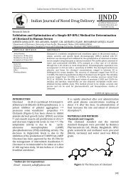
Figure 1: Scanning electron microphotographs <strong>of</strong> <strong>Captopril</strong> floating microspheres <strong>for</strong>mulation F3 (A)<br />
<strong>and</strong> F6 (B)<br />
20
An<strong>and</strong> Gadad et al / Indian Journal <strong>of</strong> Novel Drug Delivery 3(1), Jan-Mar, 2011, 17-23<br />
Micromeritic Properties<br />
All <strong>for</strong>mulations F1 to F6 <strong>of</strong> <strong>Floating</strong><br />
microspheres were evaluated <strong>for</strong> variable<br />
micromeritic parameters such as bulk density,<br />
tapped density, % Compressibility index,<br />
Hausner’s ratio <strong>and</strong> angle <strong>of</strong> repose.<br />
The % Compressibility index was in the range <strong>of</strong><br />
11-18 <strong>for</strong> all the <strong>for</strong>mulations F1 to F6 indicating<br />
good flow property.<br />
The value <strong>of</strong> Hausner’s ratio <strong>for</strong> the all<br />
<strong>for</strong>mulation F1to F6 was below 1.25 which<br />
indicates good flow property.<br />
The values <strong>of</strong> angle <strong>of</strong> repose <strong>for</strong> <strong>for</strong>mulations<br />
F1to F5 was found to be in the range <strong>of</strong> 25 o -30 o<br />
which indicated the good flow potential <strong>and</strong> the<br />
<strong>for</strong>mulation F6 showed below 25 o which<br />
indicated excellent flow.<br />
Particle size<br />
Average particle size <strong>of</strong> microspheres as<br />
determined by optical microscopy by using stage<br />
micrometer <strong>and</strong> ocular micrometer is shown in<br />
Table 2. With increase in Eudragit S-100 <strong>and</strong><br />
Ethyl cellulose concentration in the<br />
microspheres from F1 to F3 <strong>and</strong> F4 to F6, the<br />
particle size <strong>of</strong> microspheres increases<br />
respectively. This is because the viscosity <strong>of</strong> the<br />
polymer solution increases with increasing<br />
polymer concentration, which in turn decreases<br />
the stirring efficiency [5] . The polymer rapidly<br />
precipitates leading to hardening <strong>and</strong> avoiding<br />
further particle size reduction during solvent<br />
evaporation. The average particle size <strong>of</strong><br />
Eudragit S-100 was greater due to the greater<br />
viscosity than Ethyl cellulose.<br />
SEM <strong>of</strong> microspheres<br />
The microspheres <strong>of</strong> <strong>Captopril</strong> with Eudragit<br />
S100 were smooth, spherical <strong>and</strong> slightly<br />
aggregated when compared with the<br />
microspheres <strong>of</strong> <strong>Captopril</strong> with ethyl cellulose<br />
which were porous, rough, grossly, discrete<br />
spherical. Scanning electron photomicrographs<br />
<strong>of</strong> the <strong>for</strong>mulations F3 <strong>and</strong> F6 are shown in Fig 1.<br />
Drug loading <strong>and</strong> Entrapment efficiency<br />
The values <strong>of</strong> drug loading % <strong>and</strong> entrapment<br />
efficiency % are as shown in Table 2. As the<br />
polymer concentration was increased the drug<br />
loading % decreased <strong>and</strong> entrapment efficiency<br />
% was increased due to increase in the viscosity<br />
<strong>of</strong> the solution. <strong>Microspheres</strong> with Ethyl cellulose<br />
showed higher incorporation efficiency than<br />
those with Eudragit S-100.<br />
In vitro buoyancy studies<br />
To assess the floating properties, the<br />
microspheres were placed in 0.1N HCl containing<br />
0.02% Tween 80, to simulate gastric conditions.<br />
The use <strong>of</strong> Tween 80 was to account <strong>for</strong> the<br />
wetting effect <strong>of</strong> the natural surface-active agents<br />
in the GIT. The microspheres floated <strong>for</strong><br />
prolonged time over the surface <strong>of</strong> the<br />
dissolution medium without any apparent<br />
gelation. Buoyancy percentage <strong>of</strong> the<br />
microspheres was in the range <strong>of</strong> 79.33% -<br />
86.66% <strong>for</strong> <strong>for</strong>mulation F1 to F3 <strong>and</strong> 85.66% -<br />
90.33% <strong>for</strong> <strong>for</strong>mulation F4 to F6 at the end <strong>of</strong> 12<br />
h. The nature <strong>of</strong> the polymer influenced the<br />
floating behavior <strong>of</strong> the microspheres.<br />
The floating behavior <strong>of</strong> the <strong>for</strong>mulations <strong>of</strong><br />
Eudragit S-100 <strong>and</strong> Ethyl cellulose was studied in<br />
dissolution apparatus filled with 900 ml <strong>of</strong> 0.1 N<br />
HCl containing 0.02% Tween 80. The results <strong>of</strong> in<br />
vitro % buoyancy <strong>for</strong> <strong>for</strong>mulations F1to F6 are<br />
given in Table 2. The microspheres prepared by<br />
using higher polymer concentrations shows high<br />
density. So the microspheres having higher<br />
polymer concentrations were less buoyant than<br />
those with lower polymers concentrations [5] .<br />
<strong>Captopril</strong>-Eudragit S-100 microspheres showed<br />
lesser buoyancies when compared to the<br />
<strong>Captopril</strong>-Ethyl cellulose microspheres because<br />
<strong>of</strong> the lesser number <strong>of</strong> pores in the <strong>for</strong>mer. The<br />
floating abilities persisted until disintegration <strong>of</strong><br />
the microspheres began.<br />
In vitro drug release study<br />
Dissolution studies on all the six <strong>for</strong>mulations <strong>of</strong><br />
<strong>Captopril</strong> floating microspheres were carried out<br />
using a USP dissolution apparatus Type I. 0.1N<br />
HCl was used as the dissolution medium.<br />
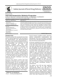
The In vitro drug release data <strong>for</strong> pure drug <strong>and</strong><br />
<strong>for</strong>mulations is shown in Table 2 <strong>and</strong> Fig 2. The<br />
cumulative percent drug release after 12 h was<br />
found to be 84.20, 81.11 <strong>and</strong> 75.95% <strong>for</strong> the<br />
<strong>for</strong>mulations F1, F2 <strong>and</strong> F3 respectively whereas<br />
cumulative percent drug release after 12 h was<br />
88.25, 84.42 <strong>and</strong> 78.30 <strong>for</strong> <strong>for</strong>mulations F4, F5<br />
<strong>and</strong> F6 respectively. The cumulative drug release<br />
significantly decreased with increase in polymer<br />
concentration. The increased density <strong>of</strong> the<br />
polymer matrix at higher concentrations results<br />
in an increased diffusional pathlength. This may<br />
decrease the overall drug release from the<br />
polymer matrix. Furthermore, smaller<br />
microspheres are <strong>for</strong>med at a lower polymer<br />
concentration <strong>and</strong> have a larger surface area<br />
exposed to dissolution medium, giving rise to<br />
faster drug release.<br />
21
An<strong>and</strong> Gadad et al / Indian Journal <strong>of</strong> Novel Drug Delivery 3(1), Jan-Mar, 2011, 17-23<br />
% Cum. Drug Release<br />
110<br />
100<br />
90<br />
80<br />
70<br />
60<br />
50<br />
40<br />
30<br />
20<br />
10<br />
0<br />
0 1 2 3 4 5 6 7 8 9 10<br />
Figure 2: In vitro drug release pr<strong>of</strong>ile <strong>of</strong> pure<br />
drug <strong>and</strong> <strong>for</strong>mulations F1 to F6.<br />
The data obtained from in vitro dissolution<br />
studies were fitted to zero-order, first-order <strong>and</strong><br />
Korsemeyer-Peppas equations. Fitting <strong>of</strong> the<br />
release rate data to the various models revealed<br />
that the <strong>for</strong>mulation F1, F2, F4 <strong>and</strong> F5 followed<br />
first order release kinetics <strong>and</strong> the <strong>for</strong>mulations<br />
F3 <strong>and</strong> F6 followed Higuchi model.<br />
In the case <strong>of</strong> the Fickian release mechanism, the<br />
rate <strong>of</strong> drug release is much less than that <strong>of</strong><br />
polymer relaxation (erosion). So the drug release<br />
is chiefly dependent on the diffusion through the<br />
matrix. In the non-Fickian (anomalous) case, the<br />
rate <strong>of</strong> drug release is due to the combined effect<br />
<strong>of</strong> drug diffusion <strong>and</strong> polymer relaxation. Case II<br />
release generally refers to the polymer<br />
relaxation. The n values <strong>for</strong> <strong>for</strong>mulations F1 to<br />
F6 ranged from 0.674 to 0.709, indicating that<br />
the release mechanism was non-Fickian or<br />
anomalous release (0.5 < n < 1). Based on the n<br />
values, F1 to F6, drug release from microsphere<br />
were controlled by polymer relaxation (erosion)<br />
as well as diffusion.<br />
CONCLUSION<br />
In this study sustained release <strong>Captopril</strong> <strong>Floating</strong><br />
<strong>Microspheres</strong> were prepared successfully using<br />
solvent evaporation method. It may be concluded<br />
that <strong>Captopril</strong> microspheres would be promising<br />
drug delivery system <strong>for</strong> oral administration <strong>of</strong><br />
<strong>Captopril</strong> to sustained the drug release upto 12 h.<br />
The <strong>for</strong>mulation was found to be efficient with<br />
good recovery yield, percentage drug<br />
entrapment. The flow properties <strong>of</strong> all<br />
<strong>for</strong>mulations were within the acceptable range<br />
<strong>and</strong> there<strong>for</strong>e they could be easily filled into<br />
capsules dosage <strong>for</strong>m. So, Sustained release<br />
floating microspheres <strong>of</strong> <strong>Captopril</strong> may provide a<br />
convenient dosage <strong>for</strong>m <strong>for</strong> achieving best<br />
per<strong>for</strong>mance regarding flow, drug entrapment<br />
<strong>and</strong> release.<br />
ACKNOWLEDGEMENTS<br />
Authors are thankful to KLE University’s, College<br />
<strong>of</strong> Pharmacy, Belgaum <strong>for</strong> providing necessary<br />
facilities to conduct the work <strong>and</strong> Hindalco<br />
Industries Ltd., R & D Center, Belgaum <strong>for</strong><br />
providing the facility <strong>of</strong> SEM study at their<br />
organization.<br />
REFERENCES<br />
[1] Tanwar YS, Naruka PS, Ojha GR.<br />
Development <strong>and</strong> evaluation <strong>of</strong> floating<br />
microspheres <strong>of</strong> verapamil hydrochloride.<br />
Brazilian J Pharm Sci. 2007; 43(4):529-34.<br />
[2] Rahman Z, Ali M, Khar RK. Design <strong>and</strong><br />
evaluation <strong>of</strong> bilayer floating tablets <strong>of</strong><br />
captopril. Acta Pharm. 2006; 56(1):49-57.<br />
[3] Gaba P, Gaba M, Garg R, Gupta GD. <strong>Floating</strong><br />
microspheres: a review. 2008; 6(5).<br />
Available from URL:http://www.<br />
pharmainfo.net/reviews/floatingmicrospheres-review.<br />
[4] Uzdemir N, Ordu S, Ozkan Y. Studies <strong>of</strong><br />
floating dosage <strong>for</strong>ms <strong>of</strong> furosemide: in<br />
vitro <strong>and</strong> in vivo evaluation <strong>of</strong> bilayer tablet<br />
<strong>for</strong>mulations. Drug Dev Ind Pharm. 2000;<br />
26(8):857–66.<br />
[5] Umamaheshwari RB, Jain S, Bhadra D, Jain<br />
NK. <strong>Floating</strong> microsphere bearing<br />
acetohydroxamic acid <strong>for</strong> treatment <strong>of</strong><br />
H.pylori. J Pharm Pharmacol. 2003;<br />
55(12):1607-13.<br />
[6] Patel A, Ray S, Thakur RS. In vitro<br />
evaluation <strong>and</strong> optimization <strong>of</strong> controlled<br />
release floating drug delivery System <strong>of</strong><br />
met<strong>for</strong>min hydrochloride. DARU. 2006;<br />
14(2): 57-64.<br />
[7] El-Kamel AH, Sokar MS, Al Gamal SS,<br />
Naggar VF. Preparation <strong>and</strong> evaluation <strong>of</strong><br />
ketopr<strong>of</strong>en floating oral delivery system.<br />
Int J Pharm. 2001;220 (1-2):13-21.<br />
[8] Jain AK, Jain CP, Tanwar YS, Naruka PS.<br />
<strong>Formulation</strong>, characterization <strong>and</strong> in vitro<br />
evaluation <strong>of</strong> floating microspheres <strong>of</strong><br />
famotidine as a gastro retentive dosage<br />
<strong>for</strong>m. Asian J Pharm. 2009; 3(3): 222-6.<br />
[9] Saravanan M, Dhanaraju MD, Sridhar SK,<br />
Ramach<strong>and</strong>ran S, Sam SKG, An<strong>and</strong> P,<br />
Bhaskar K, Rao GS. Preparation,<br />
22
An<strong>and</strong> Gadad et al / Indian Journal <strong>of</strong> Novel Drug Delivery 3(1), Jan-Mar, 2011, 17-23<br />
characterization <strong>and</strong> in vitro release<br />
kinetics <strong>of</strong> ibupr<strong>of</strong>en polystyrene<br />
microspheres. Indian J Pharm Sci. 2004; 66<br />
(3):287-92.<br />
[10] Ma N, Xu L, Wang Q, Zhang X, Zhang W, Li Y,<br />
Jin L, Li S. Development <strong>and</strong> evaluation <strong>of</strong><br />
new sustained-release floating<br />
microspheres. Int J Pharm. 2008; 358(1-2):<br />
82-90.<br />
[11] Srivastava AK, Ridhurkar DN, Wadhwa S.<br />
<strong>Floating</strong> microspheres <strong>of</strong> cimetidine:<br />
<strong>for</strong>mulation, characterization <strong>and</strong> in vitro<br />
evaluation. Acta Pharm. 2005; 55(3):277-<br />
85.<br />
[12] Altaf MA, Sreedharan, Charyulu N. Ionic<br />
gelation controlled drug delivery systems<br />
<strong>for</strong> gastric-mucoadhesive microcapsules <strong>of</strong><br />
captopril. Indian J Pharm Sci. 2008;<br />
70(5):655-8.<br />
23