Low-resolution PDF - IMB
Low-resolution PDF - IMB
Low-resolution PDF - IMB

You also want an ePaper? Increase the reach of your titles
YUMPU automatically turns print PDFs into web optimized ePapers that Google loves.
30<br />
Microscopy<br />
Core Facility<br />
Andreas Vonderheit<br />
Education<br />
2000 Diploma in Molecular Biology, ZMBH, University of Heidelberg<br />
2004 PhD in Biochemistry, ETH, Zürich<br />
Positions held<br />
2005-2008 Postdoctoral Researcher, Institute of Nanotechnology, ETH, Zürich<br />
2008-2011 Screening Scientist at the RNAi Image-based Screening Center (RISC)<br />
at the Light Microscopy Centre (LMC), ETH, Zürich<br />
Since 2011 Head of Microscopy, Core Facilities and Technology,<br />
Institute of Molecular Biology (<strong>IMB</strong>), Mainz<br />
Core Facility Overview<br />
The Microscopy Core Facility not only provides hands-on training and<br />
access to various state-of-the-art microscopes, but also offers further<br />
education through the provision of a variety of practical courses and<br />
lectures. Training lectures provided range from a general “Introduction<br />
to Microscopy” to “Pitfalls in Image Acquisition” and “Ethics in Image<br />
Acquisition and Processing”. Lectures, courses and the scientific services<br />
available are all open to Mainz’ scientific community. To date the lectures<br />
have attracted a large number of non-<strong>IMB</strong> researchers, with about<br />
80% of the audience coming from Johannes Gutenberg University<br />
and the University Medical Center. The microscopes provided are<br />
utilized equally by <strong>IMB</strong> staff and external users.<br />
Services Offered<br />
The facility provides hands-on training and access to various ultramodern<br />
confocal laser scanning microscopes, super-<strong>resolution</strong> microscopes<br />
and motorized fast-flourescence microscopes for live-cell<br />
microscopy. There are also PCs for image processing equipped with<br />
software for deconvolution and 3D-rendering. The following microscopic<br />
equipment, a large part of which was financed by DFG, is available:<br />
• M80 Demonstration Stereo Microscope: Equipped with a camera<br />
and a 24“ monitor for teaching<br />
• M205 FA Fluorescence Stereo Microscope: Equipped with a<br />
camera, fluorescence light source and three filters for UV, green,<br />
and red fluorophores<br />
• DM2500 Fluorescence Upright Microscope: Equipped with a<br />
colour camera, fluorescence light source and three filters for UV,<br />
green, and red fluorophores and with five air objectives (this microscope<br />
is perfectly suited for histology)<br />
• AF7000 Widefield Fluorescence Microscope: This widefield microscope<br />
is equipped with an incubator box for live-microscopy, fast<br />
filter wheels and a fast camera<br />
• TCS LSI Macro Zoom Confocal: This microscope combines a stereo<br />
microscope with a confocal. Organisms like C. elegans or Drosophila<br />
larvae can be scanned as a whole or zoomed down to single<br />
cell level in confocal mode<br />
• TCS SPE Confocal Microscope: An upright confocal microscope<br />
with one detector<br />
• TCS SP5 Confocal Microscope: An inverse microscope with four<br />
PMTs, four lasers, and a fast resonance scanner<br />
• TCS STED CW Super-Resolution Microscope: A super-<strong>resolution</strong><br />
microscope which allows live microscopy and FCS. Equipped with<br />
an incubation box, two normal PMTs and two HyD detectors<br />
• SR GSD Super-Resolution Microscope (localization method):<br />
A super-<strong>resolution</strong> microscope on which TIRF is possible<br />
• Image Processing Station<br />
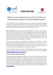
Figure 1. Mitochondria network in CHO cells labeled with Mitotracker® and imaged with<br />
a confocal microscope. (A) One plane of a z-stack. (B) Same plane after deconvolution. (C)<br />
Cutout of A. (D) Cutout of B. Scalebar: 4 µm. Imaged by A. Vonderheit.