View PDF - Australasian Plant Pathology Society
View PDF - Australasian Plant Pathology Society
View PDF - Australasian Plant Pathology Society

You also want an ePaper? Increase the reach of your titles
YUMPU automatically turns print PDFs into web optimized ePapers that Google loves.
Development of a soil DNA extraction and quantitative PCR method for detecting two<br />
Cylindrocarpon species in soil<br />
INTRODUCTION<br />
C.M. Probst{ XE "Probst, C.M." }, M.V. Jaspers, E.E. Jones and H.J. Ridgway<br />
Faculty of Agriculture and Life Sciences, Lincoln University, PO Box 84, Lincoln 8150, New Zealand<br />
Cylindrocarpon black foot disease has been identified worldwide<br />
as a common cause of vine death in nurseries and in young<br />
vineyards (1), especially in sites converted from orchards or<br />
replanted from grapes. This indicates that the existing soil<br />
conditions may have contributed to the disease, although very<br />
little information is available regarding the survival of the<br />
pathogens in soil. In New Zealand, the three Cylindrocarpon<br />
species equally responsible for black foot disease on grapevines<br />
are C. destructans, C. macrodidymum and C. liriodendri (1).<br />
Recently, quantitative PCR (qPCR) has begun to supersede soil<br />
dilution plating as a method for precise determination of soil<br />
inoculum levels (2). In this research program, qPCR was<br />
optimised to test large soil samples for presence of two<br />
Cylindrocarpon species.<br />
MATERIALS AND METHODS<br />
Fungal isolates. The Lincoln University Culture Collection<br />
provided isolates of the three Cylindrocarpon species, which had<br />
been obtained from symptomatic New Zealand grapevines.<br />
Single spore colonies of 10 randomly selected isolates per<br />
species were grown on potato dextrose agar (PDA) at 20°C in the<br />
dark.<br />
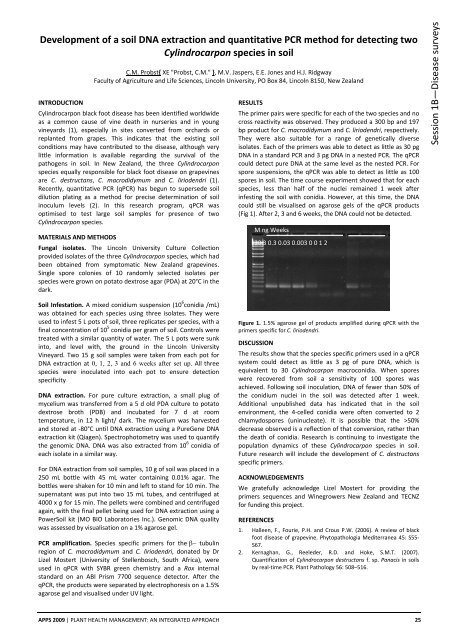
RESULTS<br />
The primer pairs were specific for each of the two species and no<br />
cross reactivity was observed. They produced a 300 bp and 197<br />
bp product for C. macrodidymum and C. liriodendri, respectively.<br />
They were also suitable for a range of genetically diverse<br />
isolates. Each of the primers was able to detect as little as 30 pg<br />
DNA in a standard PCR and 3 pg DNA in a nested PCR. The qPCR<br />
could detect pure DNA at the same level as the nested PCR. For<br />
spore suspensions, the qPCR was able to detect as little as 100<br />
spores in soil. The time course experiment showed that for each<br />
species, less than half of the nuclei remained 1 week after<br />
infesting the soil with conidia. However, at this time, the DNA<br />
could still be visualised on agarose gels of the qPCR products<br />
(Fig 1). After 2, 3 and 6 weeks, the DNA could not be detected.<br />
M ng Weeks<br />
30 3 0.3 0.03 0.003 0 0 1 2<br />
Session 1B—Disease surveys<br />
Soil Infestation. A mixed conidium suspension (10 6 conidia /mL)<br />
was obtained for each species using three isolates. They were<br />
used to infest 5 L pots of soil, three replicates per species, with a<br />
final concentration of 10 5 conidia per gram of soil. Controls were<br />
treated with a similar quantity of water. The 5 L pots were sunk<br />
into, and level with, the ground in the Lincoln University<br />
Vineyard. Two 15 g soil samples were taken from each pot for<br />
DNA extraction at 0, 1, 2, 3 and 6 weeks after set up. All three<br />
species were inoculated into each pot to ensure detection<br />
specificity<br />
DNA extraction. For pure culture extraction, a small plug of<br />
mycelium was transferred from a 5 d old PDA culture to potato<br />
dextrose broth (PDB) and incubated for 7 d at room<br />
temperature, in 12 h light/ dark. The mycelium was harvested<br />
and stored at ‐80°C until DNA extraction using a PureGene DNA<br />
extraction kit (Qiagen). Spectrophotometry was used to quantify<br />
the genomic DNA. DNA was also extracted from 10 6 conidia of<br />
each isolate in a similar way.<br />
For DNA extraction from soil samples, 10 g of soil was placed in a<br />
250 mL bottle with 45 mL water containing 0.01% agar. The<br />
bottles were shaken for 10 min and left to stand for 10 min. The<br />
supernatant was put into two 15 mL tubes, and centrifuged at<br />
4000 x g for 15 min. The pellets were combined and centrifuged<br />
again, with the final pellet being used for DNA extraction using a<br />
PowerSoil kit (MO BIO Laboratories Inc.). Genomic DNA quality<br />
was assessed by visualisation on a 1% agarose gel.<br />
PCR amplification. Species specific primers for the β− tubulin<br />
region of C. macrodidymum and C. liriodendri, donated by Dr<br />
Lizel Mostert (University of Stellenbosch, South Africa), were<br />
used in qPCR with SYBR green chemistry and a Rox internal<br />
standard on an ABI Prism 7700 sequence detector. After the<br />
qPCR, the products were separated by electrophoresis on a 1.5%<br />
agarose gel and visualised under UV light.<br />
Figure 1. 1.5% agarose gel of products amplified during qPCR with the<br />
primers specific for C. liriodendri.<br />
DISCUSSION<br />
The results show that the species specific primers used in a qPCR<br />
system could detect as little as 3 pg of pure DNA, which is<br />
equivalent to 30 Cylindrocarpon macroconidia. When spores<br />
were recovered from soil a sensitivity of 100 spores was<br />
achieved. Following soil inoculation, DNA of fewer than 50% of<br />
the conidium nuclei in the soil was detected after 1 week.<br />
Additional unpublished data has indicated that in the soil<br />
environment, the 4‐celled conidia were often converted to 2<br />
chlamydospores (uninucleate). It is possible that the >50%<br />
decrease observed is a reflection of that conversion, rather than<br />
the death of conidia. Research is continuing to investigate the<br />
population dynamics of these Cylindrocarpon species in soil.<br />
Future research will include the development of C. destructans<br />
specific primers.<br />
ACKNOWLEDGEMENTS<br />
We gratefully acknowledge Lizel Mostert for providing the<br />
primers sequences and Winegrowers New Zealand and TECNZ<br />
for funding this project.<br />
REFERENCES<br />
1. Halleen, F., Fourie, P.H. and Crous P.W. (2006). A review of black<br />
foot disease of grapevine. Phytopathologia Mediterranea 45: S55‐<br />
S67.<br />
2. Kernaghan, G., Reeleder, R.D. and Hoke, S.M.T. (2007).<br />
Quantification of Cylindrocarpon destructans f. sp. Panacis in soils<br />
by real‐time PCR. <strong>Plant</strong> <strong>Pathology</strong> 56: 508–516.<br />
APPS 2009 | PLANT HEALTH MANAGEMENT: AN INTEGRATED APPROACH 25


![[Compatibility Mode].pdf](https://img.yumpu.com/27318716/1/190x135/compatibility-modepdf.jpg?quality=85)