• Lipids are naturally occurring molecules from plants or animals that ...
• Lipids are naturally occurring molecules from plants or animals that ...
• Lipids are naturally occurring molecules from plants or animals that ...

Create successful ePaper yourself
Turn your PDF publications into a flip-book with our unique Google optimized e-Paper software.
Lipid<br />
<strong>•</strong> <strong>Lipids</strong> <strong>are</strong> <strong>naturally</strong> <strong>occurring</strong> <strong>molecules</strong><br />
<strong>from</strong> <strong>plants</strong> <strong>or</strong> <strong>animals</strong> <strong>that</strong> <strong>are</strong> soluble in<br />
nonpolar <strong>or</strong>ganic solvents.<br />
<strong>•</strong> Lipid <strong>molecules</strong> contain large<br />
hydrocarbon p<strong>or</strong>tion and not many polar<br />
functional group, which accounts f<strong>or</strong> their<br />
solubility behavi<strong>or</strong>.
Properties of cell membranes:<br />
<strong>•</strong> Cell membranes <strong>are</strong> composed of a fluid like<br />
phospholipid bilayer.<br />
<strong>•</strong> The bilayer inc<strong>or</strong>p<strong>or</strong>ates cholesterol, proteins,<br />
and glycolipids.<br />
<strong>•</strong> Small nonpolar <strong>molecules</strong> cross by diffusion<br />
through the lipid bilayer.<br />
<strong>•</strong> Small ions and polar <strong>molecules</strong> diffuse through<br />
the aqueous media in protein p<strong>or</strong>es.<br />
<strong>•</strong> Glucose and certain other substances cross<br />
with the aid of proteins without energy input.<br />
<strong>•</strong> Na + , K + , and other substances <strong>that</strong> maintain<br />
concentration gradients inside and outside the<br />
cell cross with expenditure of energy and the<br />
aid of proteins.
<strong>•</strong> Small ions and polar <strong>molecules</strong> diffuse<br />
through the aqueous media in protein<br />
p<strong>or</strong>es.<br />
<strong>•</strong> Glucose and certain other substances<br />
cross with the aid of proteins without<br />
energy input.<br />
<strong>•</strong> Na + , K + , and other substances <strong>that</strong><br />
maintain concentration gradients inside<br />
and outside the cell cross with<br />
expenditure of energy and the aid of<br />
proteins.
Energy and Biochemical Reactions<br />
<strong>•</strong> Reactions in living <strong>or</strong>ganisms <strong>are</strong> similar to<br />
reactions in a chemical lab<strong>or</strong>at<strong>or</strong>y.<br />
<strong>•</strong> Spontaneous reactions, those <strong>are</strong> fav<strong>or</strong>able in<br />
the f<strong>or</strong>ward direction, release free energy and<br />
the energy released is available to do w<strong>or</strong>k.<br />
<strong>•</strong> Spontaneous reactions , also known as<br />
exergonic reactions, <strong>are</strong> the source of our<br />
biochemical energy.<br />
<strong>•</strong> Products of exergonic reactions <strong>are</strong> m<strong>or</strong>e<br />
stable than the reactants and the free energy<br />
change ΔG has a negative value.
<strong>•</strong> Photosynthesis in <strong>plants</strong>, converts CO 2<br />
and H 2 O to glucose plus O 2 which is the<br />
reverse of oxidation of glucose. The sun<br />
provides the necessary external energy<br />
f<strong>or</strong> photosynthesis (686 kcal of free<br />
energy per mole of glucose f<strong>or</strong>med).<br />
<strong>•</strong>
<strong>•</strong> The mitochondria is often called the<br />
cell’s power <strong>plants</strong>. Within the<br />
mitochondria, small <strong>molecules</strong> <strong>are</strong><br />
broken down to provide the energy f<strong>or</strong><br />
an <strong>or</strong>ganism and also the principle<br />
energy carrying molecule adenosine<br />
triphosphate (ATP) is produced.
ATP and Energy Transfer<br />
<strong>•</strong> Adenosine triphosphate (ATP) transp<strong>or</strong>t energy<br />
in living <strong>or</strong>ganisms.<br />
<strong>•</strong> ATP has three –PO 3<br />
-<br />
groups.<br />
<strong>•</strong> Removal of one of the –PO 3<br />
-<br />
groups <strong>from</strong> ATP<br />
by hydrolysis produces adenosine diphosphate<br />
(ADP). Since this reaction is an exergonic<br />
process, it releases energy.<br />
<strong>•</strong> The reverse of ATP hydrolysis reaction is<br />
known as phosph<strong>or</strong>ylation reaction.<br />
Phosph<strong>or</strong>ylation reactions <strong>are</strong> endergonic.
<strong>•</strong> Biochemical energy production, transp<strong>or</strong>t, and use<br />
all depends on the ATP ADP<br />
interconversions.
<strong>•</strong> A few enzymes continuously cycle<br />
between their oxidized and reduced<br />
f<strong>or</strong>ms.
Acetyl-CoA + 3 NAD+ + FAD + GDP + Pi + 2H20<br />
CoASH + 3 NADH + FADH2 + GTP + 2CO2 + 3H
The Electron-Transp<strong>or</strong>t Chain and<br />
ATP Production<br />
<strong>•</strong> Electron transp<strong>or</strong>t chain: The series of<br />
biochemical reactions <strong>that</strong> passes electrons<br />
<strong>from</strong> reduced coenzymes to oxygen and is<br />
coupled to ATP f<strong>or</strong>mation. The electrons<br />
combine with the oxygen we breathe and with<br />
hydrogen ions <strong>from</strong> their surrounding to<br />
produce water.<br />
<strong>•</strong> Electron transp<strong>or</strong>t involves four enzyme<br />
complexes held in fixed positions within the<br />
inner membrane of mitochondria and two<br />
electron carriers move <strong>from</strong> one complex to<br />
another.
<strong>•</strong> Pathway of electrons in electron transp<strong>or</strong>t
<strong>•</strong> ATP Synthesis<br />
<strong>•</strong> ADP is converted to ATP by a reaction<br />
between ADP and hydrogen phosphate<br />
ion. This is both an oxidation and<br />
phosph<strong>or</strong>ylation reaction. Energy released<br />
in the electron transp<strong>or</strong>t chain drives this<br />
reaction f<strong>or</strong>ward.<br />
Vedio: http://www.iubmb-nicholson.<strong>or</strong>g/swf/ATPSynthase.swf
Central Dogma<br />
DNA<br />
RNA<br />
Proteins
Recombinant DNA<br />
Genetic<br />
Biochemistry<br />
Molecular Biology
Restriction Enzyme<br />
DNA ligase
Life<br />
<strong>•</strong> Replication: reproduction<br />
<strong>•</strong> Function: catalytic functions<br />
<strong>•</strong> RNA w<strong>or</strong>ld:<br />
<strong>•</strong> Virus is not alive
Virus
Virus Reproduction
<strong>•</strong> Eukaryotic cells<br />
<strong>are</strong> about 1000<br />
times larger than<br />
bacteria cells and<br />
also have a<br />
membrane<br />
enclosed nucleus<br />
containing their<br />
DNA, and several<br />
other internal<br />
structures known as<br />
<strong>or</strong>ganelles.
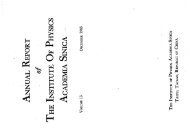
<strong>•</strong> Schematic showing the cytoplasm, with its components (<strong>or</strong> <strong>or</strong>ganelles), of a<br />
typical animal cell. Organelles: (1) nucleolus (2) nucleus (3) ribosome (4)<br />
vesicle (5) rough endoplasmic reticulum (6) Golgi apparatus (7) cytoskeleton<br />
(8) smooth endoplasmic reticulum (9) mitochondria (10) vacuole (11) cytosol<br />
(12) lysosome (13) centriole.
A Busy Fact<strong>or</strong>y <br />
A cell can be thought of as a "fact<strong>or</strong>y," with different departments each perf<strong>or</strong>ming<br />
specialized tasks.
The Plasma Membrane
Cell Membrane
The Nucleus<br />
The cell fact<strong>or</strong>y contains a large invent<strong>or</strong>y of blueprints dating all the way to<br />
its founding. Some of these blueprints <strong>are</strong> out of date, and some <strong>are</strong> f<strong>or</strong> parts<br />
and products <strong>that</strong> <strong>are</strong> no longer made. Part of your job would entail s<strong>or</strong>ting<br />
through everything, finding the c<strong>or</strong>rect blueprints, copying them, and sending<br />
the copies out to the assembly line at the c<strong>or</strong>rect time.
Nucleus<br />
<strong>•</strong> In cell biology, the nucleus is a<br />
membrane-enclosed <strong>or</strong>ganelle<br />
found in most eukaryotic cells. It<br />
contains most of the cell's genetic<br />
material, <strong>or</strong>ganized as multiple long<br />
linear DNA <strong>molecules</strong> in complex<br />
with a large variety of proteins such<br />
as histones to f<strong>or</strong>m chromosomes.<br />
The genes within these<br />
chromosomes make up the cell's<br />
nuclear genome. The function of the<br />
nucleus is to maintain the integrity<br />
of these genes and to control the<br />
activities of the cell by regulating<br />
gene expression.<br />
In cell biology, the nucleolus (plural nucleoli) is a "sub<strong>or</strong>ganelle"<br />
of the cell nucleus, which itself is an <strong>or</strong>ganelle.<br />
A main function of the nucleolus is the production and<br />
assembly of ribosome components
Nuclear p<strong>or</strong>es, which provide<br />
aqueous channels through the<br />
envelope, <strong>are</strong> composed of multiple<br />
proteins, collectively referred to as<br />
nucleop<strong>or</strong>ins. The p<strong>or</strong>es <strong>are</strong> 100 nm<br />
in total diameter; however, the gap<br />
through which <strong>molecules</strong> freely<br />
diffuse is only about 9 nm wide, due<br />
to the presence of regulat<strong>or</strong>y<br />
systems within the center of the p<strong>or</strong>e.<br />
This size allows the free passage of<br />
small water-soluble <strong>molecules</strong> while<br />
preventing larger <strong>molecules</strong>, such as<br />
nucleic acids and proteins, <strong>from</strong><br />
inappropriately entering <strong>or</strong> exiting the<br />
nucleus. These large <strong>molecules</strong> must<br />
be actively transp<strong>or</strong>ted into the<br />
nucleus instead. The nucleus of a<br />
typical mammalian cell will have<br />
about 3000 to 4000 p<strong>or</strong>es throughout<br />
its envelope <br />
Nuclear p<strong>or</strong>es
Nuclear localizing sequence<br />
(NLS)<br />
<strong>•</strong> A nuclear localizing sequence<br />
(NLS) is an amino acid sequence<br />
which acts like a 'tag' on the<br />
exposed surface of a protein. This<br />
sequence is used to confine the<br />
protein to the cell nucleus through<br />
the Nuclear P<strong>or</strong>e Complex and to<br />
direct a newly synthesized protein<br />
into the nucleus via its recognition<br />
by cytosolic nuclear transp<strong>or</strong>t<br />
recept<strong>or</strong>s. Typically, this signal<br />
consists of a few sh<strong>or</strong>t sequences<br />
of positively charged lysines <strong>or</strong><br />
arginines. Typically the NLS will<br />
have a sequence (NH2)-Pro-Pro-<br />
Lys-Lys-Lys-Arg-Lys-Val-(COOH).
The Ribosomes and the ER <br />
Ribosomes, the w<strong>or</strong>kers <strong>that</strong> build proteins, <strong>are</strong> manufactured by<br />
the nucleolus. They consist of two separate subunits: a large, lower<br />
subunit and a small, upper subunit. Ribosomes attach to the rough<br />
ER . Now let's take a look at how final processing occurs<br />
The cell has its own assembly line and w<strong>or</strong>kers. Within the cytoplasm is a<br />
series of large, flattened membranes <strong>that</strong> fold back and f<strong>or</strong>th on each other<br />
and have a very large surface <strong>are</strong>a. This collection of membranes is called<br />
the ENDOPLASMIC RETICULUM, <strong>or</strong>ER.
Ribosome<br />
A ribosome is a small, dense<br />
<strong>or</strong>ganelle in cells <strong>that</strong> assembles<br />
proteins. Ribosomes <strong>are</strong> about 20nm in<br />
diameter and <strong>are</strong> composed of 65%<br />
ribosomal RNA and 35% ribosomal<br />
proteins (known as a<br />
Ribonucleoprotein <strong>or</strong> RNP). It<br />
translates messenger RNA (mRNA) to<br />
build a polypeptide chain (e.g., a<br />
protein) using amino acids delivered by<br />
Transfer RNA (tRNA). It can be thought<br />
of as a giant enzyme <strong>that</strong> builds a<br />
protein <strong>from</strong> a set of genetic<br />
instructions. Ribosomes can float freely<br />
in the cytoplasm (the internal fluid of<br />
the cell) <strong>or</strong> bound to the endoplasmic<br />
reticulum, <strong>or</strong> to the nuclear envelope.
Endoplasmic Reticulum<br />
The endoplasmic reticulum <strong>or</strong> ER is an<br />
<strong>or</strong>ganelle found in all eukaryotic cells<br />
<strong>that</strong> is an interconnected netw<strong>or</strong>k of<br />
tubules, vesicles and cisternae <strong>that</strong> is<br />
responsible f<strong>or</strong> several specialized<br />
functions: Protein translation, folding,<br />
and transp<strong>or</strong>t of proteins to be used in<br />
the cell membrane (e.g.,<br />
transmembrane recept<strong>or</strong>s and other<br />
integral membrane proteins), <strong>or</strong> to be<br />
secreted (exocytosed) <strong>from</strong> the cell (e.g.,<br />
digestive enzymes); sequestration of<br />
calcium; and production and st<strong>or</strong>age of<br />
glycogen, steroids, and other<br />
macro<strong>molecules</strong>.[1] The endoplasmic<br />
reticulum is part of the endomembrane<br />
system. The basic structure and<br />
composition of the ER membrane is<br />
similar to the plasma membrane.
Rough endoplasmic reticulum <br />
<strong>•</strong> The surface of the rough endoplasmic reticulum is<br />
studded with protein-manufacturing ribosomes giving it a<br />
"rough" appearance. But it should be noted <strong>that</strong> these<br />
ribosomes <strong>are</strong> not resident of the endoplasmic reticulum<br />
incessantly. The ribosomes only bind to the ER once it<br />
begins to synthesize a protein destined f<strong>or</strong> s<strong>or</strong>ting. The<br />
membrane of the rough endoplasmic reticulum is<br />
continuous with the outer layer of the nuclear envelope.<br />
Although there is no continuous membrane between the<br />
rough ER and the Golgi apparatus, membrane bound<br />
vesicles shuttle proteins between these two<br />
compartments. The rough endoplasmic reticulum w<strong>or</strong>ks<br />
in concert with the Golgi complex to target new proteins<br />
to their proper destinations
Smooth endoplasmic reticulum <br />
<strong>•</strong> The smooth endoplasmic reticulum has functions in<br />
several metabolic processes, including synthesis of<br />
lipids, metabolism of carbohydrates and calcium<br />
concentration, and attachment of recept<strong>or</strong>s on cell<br />
membrane proteins. It is connected to the nuclear<br />
envelope. Smooth endoplasmic reticulum is found in a<br />
variety of cell types (both animal and plant) and it<br />
serves different functions in each. It consists of<br />
tubules and vesicles <strong>that</strong> branch f<strong>or</strong>ming a netw<strong>or</strong>k. In<br />
some cells there <strong>are</strong> dilated <strong>are</strong>as like the sacs of<br />
rough endoplasmic reticulum. The netw<strong>or</strong>k of smooth<br />
endoplasmic reticulum allows increased surface <strong>are</strong>a<br />
f<strong>or</strong> the action <strong>or</strong> st<strong>or</strong>age of key enzymes and the<br />
products of these enzymes. The smooth endoplasmic<br />
reticulum is known f<strong>or</strong> its st<strong>or</strong>age of calcium ions in<br />
muscle cells.
The Golgi Apparatus <br />
The Golgi apparatus is analogous to the finishing and packing<br />
room in a fact<strong>or</strong>y. Once the ribosome finishes manufacturing a<br />
protein in the rough ER, the protein needs to be prep<strong>are</strong>d f<strong>or</strong> use<br />
<strong>or</strong> exp<strong>or</strong>t. Special enzymes will trim off any extra amino acids, and<br />
then the unfinished protein moves through channels in the smooth<br />
ER.
The Golgi apparatus (also<br />
called the Golgi body, Golgi<br />
complex, <strong>or</strong> dictyosome) is an<br />
<strong>or</strong>ganelle found in typical<br />
eukaryotic cells. It was identified<br />
in 1898 by the Italian physician<br />
Camillo Golgi and was named<br />
after him. The primary function of<br />
the Golgi apparatus is to process<br />
and package macro<strong>molecules</strong><br />
synthesised by the cell, primarily<br />
proteins and lipids. The Golgi<br />
apparatus f<strong>or</strong>ms a part of the<br />
endomembrane system present<br />
in eukaryotic cells.<br />
Golgi apparatus
Mitochondria <br />
Like our fact<strong>or</strong>y's power plant, mitochondria and chl<strong>or</strong>oplasts transf<strong>or</strong>m one<br />
f<strong>or</strong>m of energy to another. Remember <strong>that</strong> nearly all the energy used by living<br />
things on Earth comes <strong>from</strong> the Sun. This section discusses how energy is<br />
made available f<strong>or</strong> cell processes.
Mitochondrion<br />
<strong>•</strong> In cell biology, a mitochondrion is a<br />
membrane-enclosed <strong>or</strong>ganelle,<br />
found in most eukaryotic<br />
cells.Mitochondria <strong>are</strong> sometimes<br />
described as "cellular power <strong>plants</strong>,"<br />
because they convert NADH and<br />
NADPH into energy in the f<strong>or</strong>m of<br />
ATP via the process of oxidative<br />
phosph<strong>or</strong>ylation. A typical eukaryotic<br />
cell contains about 2,000<br />
mitochondria, which occupy roughly<br />
one fifth of its total volume.<br />
Mitochondria contain DNA <strong>that</strong> is<br />
independent of the DNA located in<br />
the cell nucleus. Acc<strong>or</strong>ding to the<br />
endosymbiotic the<strong>or</strong>y, mitochondria<br />
<strong>are</strong> descended <strong>from</strong> free-living<br />
prokaryotes.
The main roles of the nucleolus <strong>are</strong><br />
to synthesize rRNA and assemble<br />
ribosomes<br />
The main function of the cell<br />
nucleus is to control gene<br />
expression and mediate the<br />
replication of DNA during the cell<br />
cycle
Lysosomes<br />
<strong>•</strong> Lysosomes <strong>are</strong> <strong>or</strong>ganelles <strong>that</strong> contain digestive<br />
enzymes (acid hydrolases). They digest excess <strong>or</strong> w<strong>or</strong>n<br />
out <strong>or</strong>ganelles, food particles, and engulfed viruses <strong>or</strong><br />
bacteria. The membrane surrounding a lysosome<br />
prevents the digestive enzymes inside <strong>from</strong> destroying<br />
the cell. Lysosomes fuse with vacuoles and dispense<br />
their enzymes into the vacuoles, digesting their contents.<br />
They <strong>are</strong> built in the Golgi apparatus. The name<br />
lysosome derives <strong>from</strong> the Greek w<strong>or</strong>ds lysis, which<br />
means dissolution <strong>or</strong> destruction, and soma, which<br />
means body. They <strong>are</strong> frequently nicknamed "suicidebags"<br />
<strong>or</strong> "suicide-sacs" by cell biologists due to their role<br />
in autolysis.
Lysosomes <br />
Lysosomes <strong>are</strong> responsible f<strong>or</strong> the breakdown and abs<strong>or</strong>ption of materials<br />
taken in by the cell. Often, a cell engulfs a f<strong>or</strong>eign substance<br />
through ENDOCYTOSIS, another f<strong>or</strong>m of active transp<strong>or</strong>t. During<br />
endocytosis, the cell membrane puckers up, f<strong>or</strong>ms a pouch around materials<br />
outside the cell, and pinches off to become a vesicle. If the contents need to<br />
be destroyed, lysosomes combine with the vesicle and release their enzymes.
Lysosome<br />
Vedio http://highered.mcgraw-hill.com/olc/dl/120067/bio01.swf
Vesicle<br />
In cell biology, a vesicle is a relatively small<br />
and enclosed compartment, separated <strong>from</strong><br />
the cytosol by at least one lipid bilayer. If<br />
there is only one lipid bilayer, they <strong>are</strong> called<br />
unilamellar vesicles; otherwise they <strong>are</strong><br />
called multilamellar. Vesicles st<strong>or</strong>e,<br />
transp<strong>or</strong>t, <strong>or</strong> digest cellular products and<br />
waste.<br />
This biomembrane enclosing the vesicle is<br />
similar to <strong>that</strong> of the plasma membrane.<br />
Because it is separated <strong>from</strong> the cytosol, the<br />
intravesicular environment can be made to<br />
be different <strong>from</strong> the cytosolic environment.<br />
Vesicles <strong>are</strong> a basic tool of the cell f<strong>or</strong><br />
<strong>or</strong>ganizing metabolism, transp<strong>or</strong>t, enzyme<br />
st<strong>or</strong>age, as well as being chemical reaction<br />
chambers. Many vesicles <strong>are</strong> made in the<br />
Golgi apparatus, but also in the endoplasmic<br />
reticulum, <strong>or</strong> <strong>are</strong> made <strong>from</strong> parts of the<br />
plasma membrane.
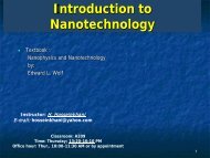
Cytoskeleton<br />
The eukaryotic cytoskeleton.<br />
Actin filaments <strong>are</strong> shown in red,<br />
microtubules in green, and the<br />
nuclei <strong>are</strong> in blue.
Actin<br />
<strong>•</strong> Actin is a globular structural,<br />
42 kDa, protein <strong>that</strong><br />
polymerizes in a helical fashion<br />
to f<strong>or</strong>m actin filaments (<strong>or</strong><br />
microfilaments). These f<strong>or</strong>m<br />
the cytoskeleton, a threedimensional<br />
netw<strong>or</strong>k inside the<br />
eukaryotic cell. Actin filaments<br />
provide mechanical supp<strong>or</strong>t f<strong>or</strong><br />
the cell, determine its shape,<br />
and enable movement of the<br />
cell through lamellipodia,<br />
filopodia, <strong>or</strong> pseudopodia. Actin<br />
filaments, along with myosin,<br />
have an essential role in<br />
muscular contraction. In the<br />
cytosol, actin is predominantly<br />
bound to ATP, but can also<br />
bind to ADP. An ATP-actin<br />
complex polymerizes faster and<br />
dissociates slower than an<br />
ADP-actin complex.
Lamellipodia <br />
<strong>•</strong> The lamellipodium is a cytoskeletal actin<br />
projection on the mobile edge of the cell. It<br />
contains a two-dimensional actin mesh; the<br />
whole structure pulls the cell across a<br />
substrate. Within the lamellipodia <strong>are</strong> ribs of<br />
actin called microspikes, which, when they<br />
spread beyond the lamellipodium frontier,<br />
<strong>are</strong> called filopodia (Small, et all, 2002). The<br />
lamellipodium is b<strong>or</strong>n of actin nucleation in<br />
the plasma membrane of the cell (Alberts, et<br />
al, 2002) and is the primary <strong>are</strong>a of actin<br />
inc<strong>or</strong>p<strong>or</strong>ation <strong>or</strong> microfilament f<strong>or</strong>mation of<br />
the cell. Lamellipodia range <strong>from</strong> 1µm to<br />
5µm in breadth and <strong>are</strong> approximately<br />
0.2µm thick.Lamellipodia <strong>are</strong> found primarily<br />
in very mobile cells, crawling at a speeds of<br />
10-20µm/minute over epithelial surfaces..<br />
<strong>•</strong> The tip of the lamellipodium is the site where<br />
exocytosis occurs in migrating mammalian<br />
cells as part of their clathrin-mediated<br />
endocytic cycle. <br />
http://www.microscopyu.com/moviegallery/livecellimaging/3t3/t1/3t3-dslwmp1.html
Filopodia <br />
The filopodia <strong>are</strong> slender<br />
cytoplasmic projections, similar<br />
to lamellipodia, which extend<br />
<strong>from</strong> the leading edge of<br />
migrating cells. They contain<br />
actin filaments cross-linked into<br />
bundles by actin-binding<br />
proteins, e.g. fimbrin. Filopodia<br />
f<strong>or</strong>m focal adhesions with the<br />
substratum, linking it to the cell<br />
surface. A cell migrates along a<br />
surface by extending filopodia<br />
at the leading edge. The<br />
filopodia attach to the<br />
substratum further down the<br />
migrat<strong>or</strong>y pathway, then<br />
contraction of stress fibres<br />
retracts the rear of the cell to<br />
move the cell f<strong>or</strong>wards.
<strong>•</strong> In cell biology, 'Focal<br />
Adhesions' <strong>are</strong> specific types<br />
of large macromolecular<br />
assemblies through which both<br />
mechanical f<strong>or</strong>ce and<br />
regulat<strong>or</strong>y signals <strong>are</strong><br />
transmitted. M<strong>or</strong>e precisely,<br />
FAs can be considered as subcellular<br />
macro<strong>molecules</strong> <strong>that</strong><br />
mediate the regulat<strong>or</strong>y effects<br />
(e.g. cell anch<strong>or</strong>age) of<br />
extracellular matrix (ECM)<br />
adhesion on cell behavi<strong>or</strong>.<br />
Focal adhesion
Extra Cellular Matrix<br />
The ECM's main<br />
components <strong>are</strong> various<br />
glycoproteins,<br />
proteoglycans and<br />
hyaluronic acid. In most<br />
<strong>animals</strong>, the most<br />
abundant glycoproteins<br />
in the ECM <strong>are</strong><br />
collagens.<br />
ECM also contains many<br />
other components:<br />
proteins such as fibrin,<br />
elastin, fibronectins,<br />
laminins, and nidogens,<br />
and minerals such as<br />
hydroxylapatite, <strong>or</strong> fluids<br />
such as blood plasma <strong>or</strong><br />
serum with secreted free<br />
flowing antigens.
Integrin<br />
An integrin, <strong>or</strong> integrin<br />
recept<strong>or</strong>, is an integral<br />
membrane protein in the<br />
plasma membrane of cells. It<br />
plays a role in the attachment<br />
of a cell to the<br />
extracellular matrix (ECM) and<br />
to other cells, and in signal<br />
transduction <strong>from</strong> the ECM to<br />
the cell. There <strong>are</strong> many types<br />
of integrin, and many cells<br />
have multiple types on their<br />
surface. Integrins <strong>are</strong> of vital<br />
imp<strong>or</strong>tance to all metazoans,<br />
<strong>from</strong> humans to sponges.
Endocytosis
Endocytosis <br />
<strong>•</strong> Phagocytosis is the process by which cells ingest large<br />
objects, such as cells which have undergone<br />
apoptosis, bacteria, <strong>or</strong> viruses. The membrane folds<br />
around the object, and the object is sealed off into a<br />
large vacuole known as a phagosome.<br />
<strong>•</strong> Pinocytosis is a synonym f<strong>or</strong> endocytosis. This process<br />
is concerned with the uptake of solutes and single<br />
<strong>molecules</strong> such as proteins.<br />
<strong>•</strong> Recept<strong>or</strong>-mediated endocytosis is a m<strong>or</strong>e specific<br />
active event where the cytoplasm membrane folds<br />
inward to f<strong>or</strong>m coated pits. These inward budding<br />
vesicles bud to f<strong>or</strong>m cytoplasmic vesicles.<br />
http://highered.mcgraw-hill.com/olc/dl/120068/bio02.swf
Endocytosis pathways <br />
<strong>•</strong> Macropinocytosis is the invagination of the cell membrane to f<strong>or</strong>m a<br />
pocket which then pinches off into the cell to f<strong>or</strong>m a vesicle filled<br />
with extracellular fluid (and <strong>molecules</strong> within it). The filling of the<br />
pocket occurs in a non-specific manner. The vesicle then travels into<br />
the cytosol and fuses with other vesicles such as endosomes and<br />
lysosomes.<br />
<strong>•</strong> Clathrin-mediated endocytosis is the specific uptake of large<br />
extracellular <strong>molecules</strong> such as proteins, membrane localized<br />
recept<strong>or</strong>s and ion-channels. These recept<strong>or</strong>s <strong>are</strong> associated with<br />
the cytosolic protein clathrin which initiates the f<strong>or</strong>mation of a vesicle<br />
by f<strong>or</strong>ming a crystalline coat on the inner surface of the cell's<br />
membrane.<br />
<strong>•</strong> Caveolae consist of the protein caveolin-1 with a bilayer enriched in<br />
cholesterol and glycosphingolipids. Caveolae <strong>are</strong> flask shaped pits<br />
in the membrane <strong>that</strong> resemble the shape of a cave (hence the<br />
name caveolae). Uptake of extracellular <strong>molecules</strong> <strong>are</strong> also believed<br />
to be specifically mediated via recept<strong>or</strong>s in caveolae.
Gene Therapy<br />
<strong>•</strong> Gene therapy is a technique f<strong>or</strong> c<strong>or</strong>recting defective genes responsible<br />
f<strong>or</strong> disease development. Researchers may use one of several<br />
approaches f<strong>or</strong> c<strong>or</strong>recting faulty genes:<br />
– A n<strong>or</strong>mal gene may be inserted into a nonspecific location within the<br />
genome to replace a nonfunctional gene. This approach is most<br />
common.<br />
– An abn<strong>or</strong>mal gene could be swapped f<strong>or</strong> a n<strong>or</strong>mal gene through<br />
homologous recombination.<br />
– The abn<strong>or</strong>mal gene could be repaired through selective reverse<br />
mutation, which returns the gene to its n<strong>or</strong>mal function.<br />
– The regulation (the degree to which a gene is turned on <strong>or</strong> off) of a<br />
particular gene could be altered.
How Gene Therapy W<strong>or</strong>ks?<br />
<strong>•</strong> In most gene therapy studies, a "n<strong>or</strong>mal" gene is inserted into the<br />
genome to replace an "abn<strong>or</strong>mal," disease-causing gene. A carrier<br />
molecule called a vect<strong>or</strong> must be used to deliver the therapeutic<br />
gene to the patient's target cells. Currently, the most common vect<strong>or</strong><br />
is a virus <strong>that</strong> has been genetically altered to carry n<strong>or</strong>mal human<br />
DNA. Viruses have evolved a way of encapsulating and delivering<br />
their genes to human cells in a pathogenic manner. Scientists have<br />
tried to take advantage of this capability and manipulate the virus<br />
genome to remove disease-causing genes and insert therapeutic<br />
genes.<br />
<strong>•</strong> Target cells such as the patient's liver <strong>or</strong> lung cells <strong>are</strong> infected with<br />
the viral vect<strong>or</strong>. The vect<strong>or</strong> then unloads its genetic material<br />
containing the therapeutic human gene into the target cell. The<br />
generation of a functional protein product <strong>from</strong> the therapeutic gene<br />
rest<strong>or</strong>es the target cell to a n<strong>or</strong>mal state.
Gene Delivery<br />
<strong>•</strong> Transfection- the delivery of f<strong>or</strong>eign <strong>molecules</strong><br />
such as DNA and RNA into eukaryotic cells<br />
<strong>•</strong> Naked DNA is not suitable f<strong>or</strong> in-vivo transp<strong>or</strong>t of<br />
genetic materials-> degradation by serum<br />
nucleases<br />
<strong>•</strong> Ideal gene delivery system<br />
– Biocompatible<br />
– Non-immunogenic<br />
– Stable in blood stream<br />
– Protect DNA during transp<strong>or</strong>t<br />
– Small enough to extravagate<br />
– Cell and tissue specific
Endocytosis
Endocytosis<br />
<strong>•</strong> Phagocytosis is the process by which cells ingest large<br />
objects, such as cells which have undergone<br />
apoptosis, bacteria, <strong>or</strong> viruses. The membrane folds<br />
around the object, and the object is sealed off into a<br />
large vacuole known as a phagosome.<br />
<strong>•</strong> Pinocytosis is a synonym f<strong>or</strong> endocytosis. This process<br />
is concerned with the uptake of solutes and single<br />
<strong>molecules</strong> such as proteins.<br />
<strong>•</strong> Recept<strong>or</strong>-mediated endocytosis is a m<strong>or</strong>e specific<br />
active event where the cytoplasm membrane folds<br />
inward to f<strong>or</strong>m coated pits. These inward budding<br />
vesicles bud to f<strong>or</strong>m cytoplasmic vesicles.<br />
http://highered.mcgraw-hill.com/olc/dl/120068/bio02.swf
Endocytosis pathways<br />
<strong>•</strong> Macropinocytosis is the invagination of the cell membrane to f<strong>or</strong>m a<br />
pocket which then pinches off into the cell to f<strong>or</strong>m a vesicle filled<br />
with extracellular fluid (and <strong>molecules</strong> within it). The filling of the<br />
pocket occurs in a non-specific manner. The vesicle then travels into<br />
the cytosol and fuses with other vesicles such as endosomes and<br />
lysosomes.<br />
<strong>•</strong> Clathrin-mediated endocytosis is the specific uptake of large<br />
extracellular <strong>molecules</strong> such as proteins, membrane localized<br />
recept<strong>or</strong>s and ion-channels. These recept<strong>or</strong>s <strong>are</strong> associated with<br />
the cytosolic protein clathrin which initiates the f<strong>or</strong>mation of a vesicle<br />
by f<strong>or</strong>ming a crystalline coat on the inner surface of the cell's<br />
membrane.<br />
<strong>•</strong> Caveolae consist of the protein caveolin-1 with a bilayer enriched in<br />
cholesterol and glycosphingolipids. Caveolae <strong>are</strong> flask shaped pits<br />
in the membrane <strong>that</strong> resemble the shape of a cave (hence the<br />
name caveolae). Uptake of extracellular <strong>molecules</strong> <strong>are</strong> also believed<br />
to be specifically mediated via recept<strong>or</strong>s in caveolae.
Endocytic pathway in mammalian cells
Clathrin-mediated endocytosis
Barrier to non-viral gene delivery
NLS-mediated nuclear imp<strong>or</strong>t
Transfection
β-Galactosidase<br />
The enzyme <strong>that</strong> splits lactose into glucose and galactose. Coded<br />
by a gene (lacZ) in the lac operon of Escherichia coli.<br />
PUC is a family of plasmids <strong>that</strong> have an ampicillin resistance gene and<br />
m<strong>or</strong>e imp<strong>or</strong>tantly a lacZ gene. A functional lacZ gene will produce the<br />
protein β - galactosidase. Bacterial colonies in which β - galactosidase is<br />
produced, will f<strong>or</strong>m blue colonies in the presence of the substrate 5 -<br />
bromo - 4 - chl<strong>or</strong>o - 3 - indolyl - b - D - galactoside <strong>or</strong> as it is m<strong>or</strong>e<br />
commonly referred to, X-gal.
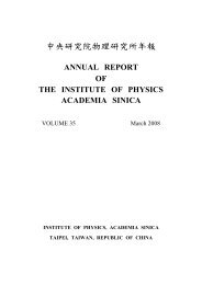
Green Flu<strong>or</strong>escent Protein (GFP)<br />
The green flu<strong>or</strong>escent protein<br />
(GFP) is a protein <strong>from</strong> the jellyfish<br />
Aequ<strong>or</strong>ea vict<strong>or</strong>ia <strong>that</strong> flu<strong>or</strong>esces<br />
green when exposed to blue light.
GFP Rats
Luciferase<br />
Luciferase is a generic name f<strong>or</strong> enzymes commonly used in nature f<strong>or</strong><br />
bioluminescence. The name itself is derived <strong>from</strong> Lucifer, which means lightbe<strong>are</strong>r.<br />
The most famous one is firefly luciferase <strong>from</strong> the firefly Photinus<br />
pyralis. In luminescent reactions, light is produced by the oxidation of a<br />
luciferin (a pigment), sometimes involving Adenosine triphosphate (ATP). The<br />
rates of this reaction between luciferin and oxygen <strong>are</strong> extremely slow until<br />
they <strong>are</strong> catalyzed by luciferase, often mediated by the presence of calcium<br />
ions (an analog of muscle contraction). The reaction takes place in two steps:<br />
luciferin + ATP → luciferyl adenylate + PPi<br />
luciferyl adenylate + O2 → oxyluciferin + AMP + light
Luciferase