Molecular Recognition by a Binary Code - ResearchGate
Molecular Recognition by a Binary Code - ResearchGate
Molecular Recognition by a Binary Code - ResearchGate

You also want an ePaper? Increase the reach of your titles
YUMPU automatically turns print PDFs into web optimized ePapers that Google loves.
doi:10.1016/j.jmb.2005.03.041 J. Mol. Biol. (2005) 348, 1153–1162<br />
<strong>Molecular</strong> <strong>Recognition</strong> <strong>by</strong> a <strong>Binary</strong> <strong>Code</strong><br />
Frederic A. Fellouse, Bing Li, Deanne M. Compaan, Andrew A. Peden<br />
Sarah G. Hymowitz and Sachdev S. Sidhu*<br />
Department of Protein<br />
Engineering, Genentech Inc<br />
1 DNA Way, South San<br />
Francisco, CA 94080, USA<br />
*Corresponding author<br />
Functional antibodies were obtained from a library of antigen-binding sites<br />
generated <strong>by</strong> a binary code restricted to tyrosine and serine. An antibody<br />
raised against human vascular endothelial growth factor recognized the<br />
antigen with high affinity (K D Z60 nM) and high specificity in cell-based<br />
assays. The crystal structure of another antigen binding fragment in<br />
complex with its antigen (human death receptor DR5) revealed the<br />
structural basis for this minimalist mode of molecular recognition. Natural<br />
antigen-binding sites are enriched for tyrosine and serine, and we show<br />
that these amino acid residues are intrinsically well suited for molecular<br />
recognition. Furthermore, these results demonstrate that molecular<br />
recognition can evolve from even the simplest chemical diversity.<br />
q 2005 Elsevier Ltd. All rights reserved.<br />
Keywords: phage display; protein engineering; combinatorial mutagenesis;<br />
antibody library; vascular endothelial growth factor<br />
Introduction<br />
The natural immune repertoire can generate<br />
antibodies that recognize essentially any antigen<br />
with high specificity and affinity. Antigen recognition<br />
is mediated <strong>by</strong> six complementarity determining<br />
regions (CDRs) 1,2 that present a large<br />
surface for contact with antigen. 3,4 CDR sequences<br />
are hypervariable, but it has become clear that the<br />
overall composition of functional CDRs is biased in<br />
favor of certain amino acid types. 5–11 In particular,<br />
tyrosine and serine residues are highly abundant,<br />
and tyrosine residues are involved in a disproportionate<br />
number of antigen contacts. 12–14<br />
The overabundance of tyrosine and serine<br />
residues may be a coincidental consequence of<br />
biases that exist in the sequences of antibody<br />
genes. 5,15–18 Alternatively, it is possible that these<br />
amino acid residues are particularly well suited for<br />
molecular recognition, and as a result, the immune<br />
system has evolved under selective pressure for the<br />
Abbreviations used: CDR, complementarity<br />
determining region; CDR-Hn (where nZ1,2 or 3), heavy<br />
chain CDR 1,2 or 3; CDR-L3, light chain CDR3; ECD,<br />
extracellular domain; ELISA, enzyme-linked<br />
immunosorbent assay; Fab, antigen-binding fragment;<br />
hDR5, human death receptor DR5; hVEGF, human<br />
vascular endothelial growth factor; GFP, green fluorescent<br />
protein.<br />
E-mail address of the corresponding author:<br />
sidhu@gene.com<br />
enrichment of tyrosine and serine in functional<br />
antigen-binding sites. 5,12,19,20<br />
We previously addressed this question <strong>by</strong> generating<br />
synthetic antibody libraries with solvent<br />
accessible CDR positions randomized with a<br />
degenerate codon that encodes for only four<br />
amino acid residues (tyrosine, serine, alanine and<br />
aspartate). 20 Surprisingly, we were able to generate<br />
high affinity antibodies against human vascular<br />
endothelial growth factor (hVEGF), an angiogenic<br />
hormone that has been implicated in tumorogenesis.<br />
21,22 Structural analyses suggested that it<br />
may be possible to simplify the code for antigen<br />
recognition even further, as most of the contacts<br />
with antigen were mediated <strong>by</strong> tyrosine sidechains,<br />
while aspartate was almost entirely<br />
excluded from the binding sites.<br />
We now show that a binary code, restricted to<br />
only tyrosine and serine, is sufficient to mediate<br />
antigen recognition with high affinity and<br />
specificity. Thus, it appears that these amino acid<br />
residues are particularly well suited for molecular<br />
recognition, and furthermore, naïve antigen recognition<br />
can evolve from even the simplest chemical<br />
diversity.<br />
Results<br />
Antibodies from a binary code<br />
We constructed two libraries of phage-displayed<br />
antigen-binding fragments (Fabs) <strong>by</strong> randomizing<br />
0022-2836/$ - see front matter q 2005 Elsevier Ltd. All rights reserved.
1154 <strong>Molecular</strong> <strong>Recognition</strong> <strong>by</strong> a <strong>Binary</strong> <strong>Code</strong><br />
CDR positions within a humanized Fab framework<br />
with a binary degenerate codon that encodes for<br />
equal proportions of tyrosine and serine. Solventaccessible<br />
positions in the first and second CDRs of<br />
the heavy chain (CDR-H1 and CDR-H2) were<br />
randomized, and random loops of variable lengths<br />
were inserted in place of seven residues in the third<br />
heavy chain CDR (CDR-H3). The libraries differed<br />
in that library A had a fixed light chain while the<br />
third CDR of the light chain (CDR-L3) was<br />
randomized in library B. Each library contained<br />
w10 10 unique members, and thus, the actual library<br />
diversities were comparable to the maximum<br />
number of unique sequences encoded <strong>by</strong> each<br />
library design (w4!10 9 ).<br />
The libraries were used to generate Fabs against a<br />
panel of six protein antigens. Specific binding<br />
clones were identified <strong>by</strong> phage enzyme-linked<br />
immunosorbent assay (ELISA) screening, and were<br />
defined as Fab-phage that bound to the cognate<br />
antigen but did not exhibit detectable binding to<br />
seven other proteins. Specific Fabs were isolated<br />
against all six antigens and DNA sequencing<br />
revealed diverse CDR sequences that contained<br />
only tyrosine and serine at the randomized sites<br />
(Figure 1). Competitive phage ELISAs were used to<br />
estimate affinities as IC 50 values. Fabs exhibited<br />
high affinity for four of the antigens (IC 50 !100 nM),<br />
moderate affinity for insulin (IC 50 Z1.4 mM) and low<br />
affinity for maltose binding protein (IC 50 O5 mM).<br />
Thus, the binary chemical diversity was sufficient to<br />
mediate specific recognition of all six antigens, and<br />
for four of the antigens the binding interactions<br />
were of high affinity. Fabs against two of the<br />
antigens (hVEGF and human death receptor DR5<br />
(hDR5)) were chosen for further functional and<br />
structural characterization.<br />
Functional characterization of anti-hVEGF Fabs<br />
DNA sequencing of 184 hVEGF-binding clones<br />
revealed 63 unique sequences, and some of these<br />
are shown in Figure 2(a). Most of the clones were<br />
from library B but some clones were also isolated<br />
from library A. Interestingly, the clones from library<br />
B exhibit homology within the selected CDR-L3 and<br />
CDR-H3 sequences. In contrast, the CDR-H3<br />
sequences from library A exhibit homology<br />
amongst themselves but are very different from<br />
the sequences from library B. Thus, it appears that<br />
the nature of the CDR-L3 sequence influenced the<br />
selection of CDR-H3 sequences, and as a result, two<br />
distinct classes of anti-hVEGF antibodies arose from<br />
the different libraries. The clones were screened <strong>by</strong><br />
competitive phage ELISA and exhibited IC 50 values<br />
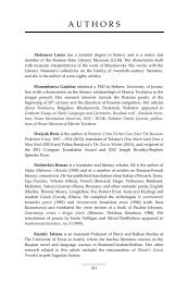
Figure 1. Sequences and specificity profiles of synthetic Fabs. (a) CDR sequences in the randomized regions. The CDR-<br />
L3 sequence is not shown for Fabs selected from library A in which the light chain was not randomized. Tyrosine and<br />
serine residues are shown in yellow or red, respectively. Residues in grey are buried residues that were not randomized<br />
in the libraries. The numbering is according to the nomenclature of Kabat et al. 1 (b) Fab-phage selected for binding to a<br />
particular antigen (cognate antigen, x-axis) were assayed for binding to various immobilized proteins <strong>by</strong> phage ELISA<br />
and bound phage were detected spectrophotometrically (optical density at 450 nm, y-axis). Affinities for cognate antigen<br />
were estimated as IC 50 values <strong>by</strong> competitive phage ELISA. 37,38 The following proteins were used: neutravidin (NAV, red<br />
bar), maltose binding protein (MBP, orange bar), Erbin PDZ domain (PDZ, yellow bar), insulin (green bar), human<br />
vascular endothelial growth factor (hVEGF, blue bar), human death receptor 5 (hDR5, pink bar), glutathione-Stransferase<br />
(purple bar), human immunoglobulin G (black bar), bovine serum albumin (white bar).
<strong>Molecular</strong> <strong>Recognition</strong> <strong>by</strong> a <strong>Binary</strong> <strong>Code</strong> 1155<br />
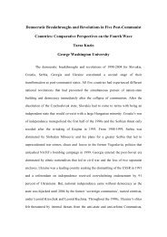
Figure 2. Affinity and specificity<br />
of anti-hVEGF Fabs. (a) CDR<br />
sequences of anti-hVEGF Fabs.<br />
The numbering is according to the<br />
nomenclature of Kabat et al. 1 and<br />
only positions that were randomized<br />
in the libraries are shown.<br />
Clones 1 through 7 were isolated<br />
from library B, while clones 8<br />
through 14 were isolated from<br />
library A. Dashes indicate gaps in<br />
the alignment. (b) Kinetic analysis<br />
of Fabs binding to immobilized<br />
hVEGF. The surface plasmon<br />
resonance traces are shown for<br />
Fab-YSv1 at four different concentrations<br />
(63 nM, 125 nM, 250 nM<br />
and 500 nM). Experimental data<br />
are represented <strong>by</strong> open circles<br />
and the global fits are represented<br />
<strong>by</strong> continuous lines. Kinetic parameters<br />
were determined for Fabs<br />
YSv1, YSv2 and YSv3. (c) Immunofluorescence<br />
staining with Fab-<br />
YSv1 (red) performed on A673<br />
cells expressing murine VEGF-<br />
GFP (green). VEGF-GFP and Fab-<br />
YSv1 staining co-localize in the<br />
extracellular space formed between<br />
cell-to-cell contacts (merge,<br />
yellow). The Fab-YSv1 staining<br />
was completely abolished <strong>by</strong><br />
pre-incubation of the antibody<br />
with excess recombinant hVEGF<br />
(C VEGF panels). The scale bar<br />
represents 20 mm. (d) Immunoprecipitations<br />
performed on media<br />
collected from metabolically<br />
labeled A673 cells. Fab-YSv1 and<br />
monoclonal antibody A4.6.1 show<br />
comparable specificity, as evidenced<br />
<strong>by</strong> identical patterns of<br />
bands for precipitated hVEGF isoforms.<br />
Anti-GFP polyclonal antibody<br />
was used as a negative<br />
control.<br />
ranging from approximately 50 nM to greater than<br />
5 mM.<br />
The three anti-hVEGF clones with the highest<br />
estimated affinities (top three sequences in<br />
Figure 2(a)) were purified as free Fab proteins.<br />
The binding kinetics of the purified Fabs<br />
(designated YSv1, YSv2 and YSv3) were studied<br />
<strong>by</strong> surface plasmon resonance (Figure 2(b)).<br />
Fab-YSv1 exhibited the highest affinity for hVEGF<br />
(K d Z60 nM), while the other two Fabs bound<br />
approximately fivefold less tightly due to faster off<br />
rates. It is notable that the sequences of Fab-YSv1<br />
and Fab-YSv2 differ in only three positions, and<br />
thus, these three differences account for the<br />
improved affinity of Fab-YSv1 in comparison with<br />
Fab-YSv2.<br />
We next investigated the specificity of Fab-YSv1<br />
<strong>by</strong> using the protein to visualize VEGF in<br />
mammalian cells transfected with a gene encoding<br />
for VEGF fused to green fluorescent protein (GFP).<br />
The immunohistochemical staining with Fab-YSv1<br />
precisely overlapped with the fluorescence signal<br />
from the VEGF-GFP fusion (Figure 2(c)). Furthermore,<br />
the signal was completely blocked <strong>by</strong><br />
incubating Fab-YSv1 with hVEGF prior to staining.<br />
We also conducted immunoprecipitations of<br />
endogenous hVEGF and compared the performance<br />
of Fab-YSv1 to that of a highly specific, natural<br />
anti-hVEGF monoclonal antibody (A4.6.1). 23 Both<br />
antibodies immunoprecipitated an identical set of<br />
bands (Figure 2(d)) that likely represent hVEGF<br />
variants generated <strong>by</strong> alternative mRNA splicing. 23<br />
Taken together, these results show that Fab-YSv1<br />
binds to hVEGF with high affinity and specificity<br />
comparable to that of a natural antibody, even in the<br />
complex cellular milieu.
1156 <strong>Molecular</strong> <strong>Recognition</strong> <strong>by</strong> a <strong>Binary</strong> <strong>Code</strong><br />
Structural characterization of an anti-hDR5 Fab<br />
To gain insights into the structural basis for<br />
antigen recognition <strong>by</strong> the binary code, we studied<br />
a Fab that was selected for binding to the<br />
extracellular domain of hDR5 (hDR5-ECD), a cellsurface<br />
receptor that mediates apoptotic cell<br />
death. 24 Nine unique anti-hDR5 clones were<br />
obtained, and the Fab that exhibited the highest<br />
estimated affinity <strong>by</strong> competitive phage ELISA<br />
(YSd1; Figure 1(a)) also bound to hDR5 with high<br />
affinity in surface plasmon resonance experiments<br />
(K d Z34 nM).<br />
The crystal structure of Fab-YSd1 in complex<br />
with the hDR5-ECD was solved and refined to<br />
3.35 Å resolution (Figure 3(a) and Table 1). While<br />
the structure is only of modest resolution, the<br />
quality of the structure is quite good with 98.9%<br />
Table 1. Data collection and refinement statistics for the<br />
Fab-YSd1:hDR5-ECD complex<br />
A. Diffraction data<br />
Space group P3 2 21<br />
Unit cell constants (Å)<br />
aZ147, cZ145<br />
Resolution (Å)<br />
50–3.35 (3.47–3.35) a<br />
b<br />
R sym 0.12 (0.34) a<br />
hI/sIi<br />
8.3 (4.5) a<br />
No. of reflections 149,117<br />
No. of unique reflections 26,539<br />
Completeness (%)<br />
99.7 (99.8) a<br />
B. Refinement<br />
Resolution (Å) 30–3.35<br />
No. of reflections 26,003<br />
c<br />
R work , R free 0.224, 0.280 (0.232, 0.281)<br />
No. of residues 1056<br />
No. of non-H atoms 8,026<br />
rmsd bond length (Å) 0.012<br />
rmsd angles (deg.) 1.3<br />
rmsd B (bonded atoms) (Å 2 ) 2.3<br />
Ramachandran plot (%) d 83.4, 15.5, 0.7, 0.4<br />
a Numbers in parentheses refer to the highest resolution shell.<br />
b R sym ZSjIKhIij=SI, where hIi is the average intensity of<br />
symmetry related observations of a unique reflection.<br />
c R work ZSjF o KF c j=SF o , where F o and F c are the observed and<br />
calculated structure factor amplitudes, respectively. R free is the<br />
R-factor for a randomly selected 10% of the reflections excluded<br />
from all refinement.<br />
d Percentage of residues in the most favored, additionally<br />
allowed, generously allowed, and disallowed regions of a<br />
Ramachandran plot.<br />
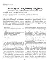
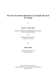
Figure 3. The structure of Fab-YSd1 bound to the hDR5-<br />
ECD. (a) The complex of the hDR5-ECD with Fab-YSd1.<br />
The hDR5-ECD is depicted as a white molecular surface.<br />
The main-chain of Fab-YSd1 is shown as sticks; the light<br />
and heavy chains are shown in green and blue,<br />
respectively, and CDR-H3 is shown in yellow.<br />
(b) Experimental density for CDR-H3. The F o KF c omit<br />
map contoured at 2.75s shows unbiased electron density<br />
(blue) for CDR-H3. Phase bias was removed <strong>by</strong> omitting<br />
CDR-H3 prior to refinement. The coordinates of the final<br />
refined model for CDR-H3 are shown as sticks, colored<br />
according to atom type (carbon, yellow; oxygen, red;<br />
nitrogen, blue), with side-chains rendered as sticks. The<br />
hDR5-ECD is shown as a white molecular surface.<br />
Despite the relatively modest resolution of the structure<br />
(3.35 Å), the electron density maps are of high quality and<br />
allow for accurate determination of backbone and sidechain<br />
positions.<br />
of residues in the most favored or additionally<br />
allowed parts of the Ramachandran plot. The data<br />
quality was limited <strong>by</strong> the availability of single<br />
crystals. All crystals showed evidence of more than<br />
one lattice, and thus, to collect a high quality data<br />
set, it was necessary to screen many crystals and to<br />
then find the most single portion of the best crystal.<br />
Nonetheless, the maps were of excellent clarity and<br />
the initial maps calculated with phases from the<br />
molecular replacement solution showed continuous<br />
density for CDR-H3 with side-chain density for<br />
many of the tyrosine residues (Figure 3(b)). The<br />
presence of two copies of the Fab:antigen complex<br />
in the asymmetric unit permitted the use of NCS<br />
restraints which aided the refinement process.<br />
Additionally, tyrosine is a large side-chain with a<br />
distinctive shape, and thus, it can be positioned<br />
accurately even in maps at a resolution of 3.35 Å.<br />
Other than the unusual CDR-H3 conformation,<br />
the structure of Fab-YSd1 is similar to that of the<br />
humanized Fab-4D5 variant that was used as the<br />
scaffold for the antibody library 20,25 (Figure 4(a)),<br />
although the elbow angles differ due to differences<br />
in crystal packing. Overall, the structure shows that<br />
Fab-YSd1 interacts with antigen in a normal<br />
manner, but tyrosine residues form most of the<br />
contact surface (Figure 4(b) and (c)). Antigen<br />
binding results in the burial of 460 Å 2 on the<br />
heavy chain and 270 Å 2 on the light chain. Tyrosine<br />
and serine residues within CDRs H2, H3 and L3<br />
account for 58% and 21% of the buried surface area,<br />
respectively. The remainder of the buried surface<br />
area (21%) is contributed <strong>by</strong> CDR-L1, which was not<br />
randomized and lies on the periphery of the
<strong>Molecular</strong> <strong>Recognition</strong> <strong>by</strong> a <strong>Binary</strong> <strong>Code</strong> 1157<br />
interface (Figure 4(b)). On the antigen side of the<br />
interface, the buried surface area is contributed <strong>by</strong> a<br />
variety of amino acid types, but aromatic residues<br />
only account for a small fraction (14%) of the<br />
structural epitope. Thus, tyrosine plays a dominant<br />
role in antigen recognition <strong>by</strong> Fab-YSd1, but it does<br />
so <strong>by</strong> utilizing interactions with a diverse array of<br />
functional groups on the antigen, and interactions<br />
with aromatic side-chains do not play a major role.<br />
The very long CDR-H3 of Fab-YSd1 protrudes<br />
from the framework and makes extensive contact<br />
with hDR5, contributing 39% of the surface area<br />
buried upon antigen binding (288 Å 2 ). Structures of<br />
antibodies with long CDR-H3 loops have been<br />
reported, 26–28 but Fab-YSd1 is unique in that CDR-<br />
H3 contains a “biphasic” helix with tyrosine and<br />
serine residues clustered on opposite faces<br />
(Figure 4(d)). The tyrosine face is buried against<br />
the surface of hDR5, and as a result, 99% of the<br />
CDR-H3 accessible surface buried upon binding to<br />
hDR5 is contributed <strong>by</strong> tyrosine, and serine<br />
residues likely play structural roles. In this regard,<br />
CDR-H3 exemplifies how even the most minimalist<br />
binary diversity can give rise to a structural solution<br />
that mediates antigen recognition with high affinity<br />
and specificity.<br />
Fab-YSd1 binds to the N-terminal portion of<br />
hDR5 and occludes the smaller of two binding sites<br />
for the natural hDR5 ligand Apo2L/TRAIL 29<br />
(Figure 5(a)). Intriguingly, one of the Apo2L/<br />
TRAIL residues that contributes the most binding<br />
energy to the interaction with hDR5 is Tyr216, as<br />
substitution of this residue <strong>by</strong> alanine causes a 320-<br />
fold loss in biological activity and a ninefold loss in<br />
affinity for hDR5. 30 Tyr216 binds in a hydrophobic<br />
pocket in cysteine-rich domain 2 of hDR5 29<br />
(Figure 5(b)), resulting in a snug fit that buries<br />
greater than 90% of the side-chain. Although Fab-<br />
YSd1 uses many tyrosine residues to bind to hDR5,<br />
none of them precisely mimics this interaction.<br />
Tyr56 of the heavy chain occupies a similar position<br />
as Tyr216 of Apo2L/TRAIL, but it lies along the<br />
antigen surface and only 60% of the side-chain is<br />
buried (Figure 5(b)).<br />
This structure is also the first for a tissue necrosis<br />
factor receptor (TNFR) family member bound to an<br />
antibody fragment, and as such, it provides insight<br />
into how an antibody recognizes antigens of this<br />
type. Overall, the structure of hDR5 bound to Fab-<br />
YSd1 is similar to that of hDR5 bound to Apo2L/<br />
TRAIL. 29 Cysteine-rich domains 1 and 2 (residues<br />
22–86) from both structures superimpose on each<br />
other with a pair-wise root-mean-square deviation<br />
(rmsd) of less than 0.30 Å 2 on C a atoms. This is the<br />
same level of structural conservation observed<br />
amongst independent structures of hDR5 bound<br />
to Apo2L/TRAIL. 29 As in the Apo2L:hDR5<br />
complex structure, which contains three crystallographically<br />
independent copies of hDR5, the<br />
orientation of cysteine-rich domain 3 in the two<br />
independent copies of hDR5 bound to Fab-YSd1<br />
varies somewhat with respect to cysteine-rich<br />
domains 1 and 2, although variations between the<br />
hDR5 molecules in the Fab-YSd1 and Apo2L/<br />
TRAIL complexes are more extreme than those<br />
within either group. Thus, in both complexes, it<br />
appears that cysteine-rich domains 1 and 2 behave<br />
as a rigid body, while in contrast the third domain<br />
exhibits flexibility with respect to the rest of hDR5.<br />
Cysteine-rich domain 3 does not make contact<br />
with Fab-YSd1, but it makes extensive contact with<br />
Apo2L/TRAIL (Figure 5(a)). In both copies of the<br />
Fab:antigen complex contained within the asymmetric<br />
unit, cysteine-rich domain 3 is less well<br />
defined than it is in the structure of hDR5 bound<br />
to Apo2L/TRAIL. In particular, residues 91–104<br />
exhibit poor density in the complex with Fab-YSd1,<br />
but this region is well-ordered when bound to<br />
Apo2L/TRAIL, and taken together, these differences<br />
suggest that the loop is not well-ordered in<br />
the absence of Apo2L/TRAIL. The membrane<br />
proximal end of cysteine-rich domain 3, including<br />
the last disulfide, is also poorly ordered in the<br />
complex with Fab-YSd1. Disorder is not uncommon<br />
in crystal structures of members of the TNFR family,<br />
as for example, the entire final cysteine-rich domain<br />
of TNFR1 is disordered in the complex with<br />
lymphotoxin. 31<br />
Discussion<br />
Despite extreme restrictions on chemical diversity,<br />
we isolated synthetic Fabs with affinities<br />
comparable to those obtained from naïve libraries<br />
designed with high chemical diversity. Furthermore,<br />
the affinities compare favorably with the<br />
affinities of antibodies from the naïve immune<br />
response, which are often in the micromolar range<br />
prior to affinity maturation through somatic<br />
mutation mechanisms. 16 Our results provide<br />
insight into how the naïve immune repertoire can<br />
evolve high affinity antibodies against a seemingly<br />
endless array of antigens. It appears that the<br />
selection of antibodies with affinities and specificities<br />
sufficient for initial selection from a naïve pool<br />
is a relatively simple process that may require only<br />
a few productive binding contacts. Thus, the limited<br />
diversity of the naïve immune repertoire 32 is<br />
sufficient to generate initial binding clones that<br />
can then attain improved function through subsequent<br />
cycles of affinity maturation.<br />
We focused our study on protein antigens, but it<br />
will be interesting to see if the limited diversity<br />
approach will be successful against other antigen<br />
classes (e.g. small molecules or carbohydrates).<br />
With the exception of CDR-H3, we also restricted<br />
CDR conformational diversity, as we did not<br />
randomize buried residues which influence CDR<br />
main-chain conformations. 33 Thus, it may be<br />
possible to obtain further improvements in binding<br />
<strong>by</strong> targeting these regions and framework positions<br />
that are known to modulate affinity. 25,34 The binary<br />
binding surfaces represent a minimal benchmark<br />
for antigen recognition into which additional<br />
diversity and conformational flexibility can be
1158 <strong>Molecular</strong> <strong>Recognition</strong> <strong>by</strong> a <strong>Binary</strong> <strong>Code</strong><br />
Figure 4 (legend opposite)
<strong>Molecular</strong> <strong>Recognition</strong> <strong>by</strong> a <strong>Binary</strong> <strong>Code</strong> 1159<br />
introduced to study the details of affinity maturation.<br />
In particular, these systems may be very<br />
amenable to computer modeling studies, since both<br />
serine and tyrosine have few rotamers, and the<br />
simplicity of the system should simplify the<br />
computational process.<br />
Our results suggest that the high abundance of<br />
tyrosine and serine in antibody combining sites<br />
reflects an innate capacity for these amino acid<br />
residues to mediate antigen recognition. Both<br />
residues are hydrophilic and thus can be exposed<br />
on surfaces without promoting non-specific aggregation,<br />
but at the same time, both can also be<br />
accommodated readily at protein–protein interfaces.<br />
Because of its small size and neutral nature,<br />
the serine side-chain is unlikely to cause unfavorable<br />
steric hindrance or electrostatic repulsions. On<br />
the other hand, the large tyrosine side-chain<br />
contributes substantial surface area for contact,<br />
and yet, it has a unique chemical nature which<br />
allows it to participate in favorable binding interactions<br />
with many different types of functional<br />
groups. 5,9,12,20 We hypothesize that the large tyrosine<br />
side-chain acts as a functional group that<br />
makes key binding contacts with antigen, while the<br />
small serine side-chain serves as an auxiliary group<br />
that provides space and flexibility for tyrosine to<br />
establish optimal binding contacts. Due to their<br />
complementary nature, it appears that tyrosine and<br />
serine may be particularly well suited to work<br />
together in antigen recognition.<br />
Much effort has been expended on the development<br />
of large, highly diverse libraries that can be<br />
used to obtain antagonists and agonists of biological<br />
processes. 35–38 However, our results demonstrate<br />
that it is possible, and perhaps even advantageous,<br />
to restrict chemical diversity to small subsets of<br />
functional groups that are particularly well suited<br />
for mediating molecular recognition.<br />
Materials and Methods<br />
Library construction and sorting<br />
A phagemid designed to display bivalent, humanized<br />
Fab-4D5 on the surface of M13 bacteriophage was used to<br />
construct libraries, as described. 20,39 Oligonucleotidedirected<br />
mutagenesis was used to replace CDR positions<br />
with TMT degenerate codons, (MZA/C in equal proportions).<br />
The positions chosen for randomization in<br />
CDR-L3, CDR-H1 and CDR-H2 are shown in Figure 1. In<br />
CDR-H3, positions 95 through 100a were replaced with<br />
random loops of all possible lengths ranging from 7 to 20<br />
residues (library A) or 7 to 15 residues (library B).<br />
Phage from the libraries were cycled through rounds of<br />
binding selection with antigen immobilized on 96 well<br />
Maxisorp immunoplates (NUNC) as the capture target, as<br />
described. After five rounds of selection, phage were<br />
produced from individual clones grown in a 96 well<br />
format and the culture supernatants were used in phage<br />
ELISAs to detect specific binding clones.<br />
Competitive phage ELISA<br />
A modified phage ELISA 37,38 was used to estimate the<br />
binding affinities of Fabs. Phage ELISAs were carried out<br />
on plates coated with antigen, as described above. Phage<br />
displaying antibody fragments were serially diluted in<br />
phosphate-buffered saline (PBS), 0.5% (w/v) bovine<br />
serum albumin (BSA), 0.1% (v/v) Tween 20, and binding<br />
was measured to determine a phage concentration giving<br />
w50% of the signal at saturation. A fixed, sub-saturating<br />
concentration of phage was pre-incubated for two hours<br />
with serial dilutions of antigen and then transferred to<br />
assay plates coated with antigen. After 15 minutes<br />
incubation, the plates were washed with PBS, 0.05%<br />
Tween 20 and incubated 30 minutes with horseradish<br />
peroxidase/anti-M13 antibody conjugate (1:5000<br />
dilution) (Pharmacia). The plates were washed,<br />
developed with TMB substrate (Kirkegaard and Perry<br />
Laboratories), quenched with 1.0 M H 3 PO 4 , and read<br />
spectrophotometrically at 450 nm. The binding affinities<br />
of were estimated as IC 50 values defined as the<br />
concentration of antigen that blocked 50% of the phage<br />
binding to the immobilized antigen.<br />
Protein purification and affinity analysis<br />
Fab proteins were purified from Escherichia coli as<br />
described. 40 Binding kinetics were determined <strong>by</strong> surface<br />
plasmon resonance using a BIAcoree-3000 with hVEGF<br />
immobilized on CM5 chips at w500 response units, as<br />
described. 41 Serial dilutions of Fab proteins were injected,<br />
and binding responses were corrected <strong>by</strong> subtraction of<br />
responses on a blank flow cell. For kinetic analysis, a 1:1<br />
Langmuir model of global fittings of k on and k off was used.<br />
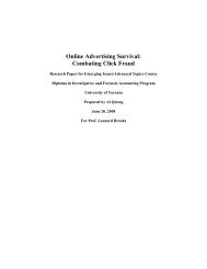
Figure 4. Antigen binding site of Fab-YSd1 and the binding interface with hDR5-ECD. (a) Stereo view of the<br />
superimposition of the variable domains of Fab-YSd1 and humanized Fab-4D5 (1N8Z). 46 The Fab variable domains are<br />
represented as a cartoon. CDR-H3 of Fab-YSd1 is colored yellow; the rest of the Fab-YSd1 heavy chain is in blue and the<br />
light chain is in green. Humanized Fab-4D5 heavy and light chains are colored cyan and red, respectively. (b) Stereo view<br />
of CDR loops that contain residues in contact with the hDR5-ECD (closer than 4.5 Å). The CDR loops are labeled and are<br />
shown as transparent cartoons. CDR side-chains that make contact with the hDR5-ECD are rendered as sticks and are<br />
colored as follows: tyrosine, blue; serine, green; all other amino acids, white. A fragment of the hDR5-ECD is shown as a<br />
molecular surface; residues in contact with the heavy or light chains are colored yellow or red, respectively, and residues<br />
in contact with both are colored orange. (c) The amino acid composition of the interface between Fab-YSd1 and the<br />
hDR5-ECD. Each circle represents the surface area on Fab-YSd1 or the hDR5-ECD that becomes buried upon complex<br />
formation. The colors indicate the proportion of the buried surface area contributed <strong>by</strong> each amino acid type, and all<br />
amino acid types that contribute more than 1% of the total buried surface area on each molecule are shown. (d) Contacts<br />
between CDR-H3 and the hDR5-ECD. The hDR5-ECD is shown as a molecular surface and residues that contact CDR-<br />
H3 are colored pink. The CDR-H3 main-chain is depicted as a yellow cartoon; tyrosine and serine side-chains are<br />
rendered as sticks and colored blue or green, respectively.
1160 <strong>Molecular</strong> <strong>Recognition</strong> <strong>by</strong> a <strong>Binary</strong> <strong>Code</strong><br />
The K d values were determined from the ratios of k on and<br />
k off .<br />
Immunohistochemistry<br />
Human A673 cells expressing murine VEGF-GFP were<br />
stained and imaged, as described. 42 In the plus VEGF<br />
panel, Fab-YSv1 was pre-incubated for five minutes with<br />
a fivefold excess of recombinant hVEGF before being<br />
incubated with the cells.<br />
Immunoprecipitation<br />
A673 cells were metabolically labeled and immunoprecipitations<br />
were performed from the media, as<br />
described, 23 using 15 mg of anti-GFP polyclonal antibody<br />
(Clontech), Fab-YSv1 or monoclonal antibody A4.6.1. The<br />
immune complexes were eluted <strong>by</strong> boiling and resolved<br />
<strong>by</strong> SDS-PAGE on a 14% (w/v) acrylamide gel under<br />
reducing conditions. The gel was dried and then exposed<br />
to a phosphoimager plate overnight.<br />
Crystallization, structure determination and<br />
refinement<br />
DNA coding for the hDR5-ECD (residues 1–130) with<br />
an N-terminal His-tag was cloned into the baculovirus<br />
transfer vector pAcGP67-B (Pharmingen). Following<br />
transfection and viral amplification in Sf9 cells, protein<br />
was expressed and secreted from Hi5 cells grown at 27 8C.<br />
The culture medium was supplemented with 50 mM Tris<br />
(pH 8.0), 1 mM NiCl 2 , 5 mM CaCl 2 , and 1 mM phenylmethylsulfonyl<br />
fluoride. The pH was adjusted to pH 7.6<br />
and the medium was filtered prior to loading on a Ni-<br />
NTA column (Qiagen). After elution with imidazole, the<br />
His-tag was removed <strong>by</strong> digestion with thrombin<br />
followed <strong>by</strong> purification on a Superdex-75 sizing column.<br />
Fab-YSd1 was expressed in E. coli and purified over a<br />
Protein G column. 40 Fab-containing fractions were further<br />
purified <strong>by</strong> passage over a S-Sepharose column, followed<br />
<strong>by</strong> a Superdex-200 sizing column.<br />
Fab–receptor complex was made <strong>by</strong> mixing Fab-YSd1<br />
with an excess of the hDR5-ECD domain, followed <strong>by</strong><br />
purification over a Superdex-200 sizing column. The<br />
complex-containing fractions were pooled and concentrated<br />
to 11 mg/ml in 150 mM NaCl, 20 mM bis-Tris<br />
(pH 6.5) and used for crystallization trials. Crystals grew<br />
at 19 8C from hanging drops containing an equal volume<br />
of protein and well solution consisting of 20% polyethylene<br />
glycol (PEG) 8000, 0.2 M magnesium acetate,<br />
0.1 M sodium cacodylate (pH 6.2–6.6). Crystals were<br />
transferred to mother liquor containing the well solution<br />
with 20% PEG 300, prior to cryo-cooling <strong>by</strong> immersion in<br />
liquid nitrogen. The resulting crystals were visibly nonsingle<br />
and it was necessary to screen many crystals to find<br />
one that was mostly single.<br />
A 3.35 Å data set was collected at beamline 19BM at the<br />
APS. Data processing was done with HKL. 43 Calculation<br />
of the Matthew’s coefficient indicated that the asymmetric<br />
Figure 5. Comparison of the interactions of hDR5 with<br />
Fab-YSd1 and Apo2L/TRAIL. (a) Mapping of the<br />
structural binding epitopes of hDR5 for Fab-YSd1 or<br />
Apo2L/TRAIL. The hDR5-ECD is shown as a molecular<br />
surface. The unshared portions of the epitopes for<br />
binding to Fab-YSd1 or Apo2L/TRAIL are colored yellow<br />
or blue, respectively, and the shared portion is in green.<br />
(b) Comparison of Tyr56 of Fab-YSd1 and Tyr216 of<br />
Apo2L/TRAIL. The hDR5-ECD is shown as a molecular<br />
surface and colored as in (a). Tyr56 of Fab-YSd1 and<br />
Tyr216 of Apo2L/TRAIL are rendered as sticks and<br />
colored cyan or pink, respectively, as are the a-carbon<br />
traces of the surrounding loops. Structures were drawn<br />
with PyMOL (DeLano Scientific, San Carlos, CA).
<strong>Molecular</strong> <strong>Recognition</strong> <strong>by</strong> a <strong>Binary</strong> <strong>Code</strong> 1161<br />
unit of the crystals contained two Fab:antigen complexes.<br />
The structure was solved in space group P3 2 21 <strong>by</strong><br />
molecular replacement with the program Phaser 44 using<br />
the structures of two Fab-4D5 variants as search models. 25<br />
The resulting maps showed clear density for the hDR5-<br />
ECD, which was placed manually, as well as for the Fab<br />
CDRs. Refinement was performed with the program<br />
REFMAC5 45 and included TLS refinement and NCS<br />
restraints imposed separately on the variable and<br />
constant domains of Fab-YSd1, as well as on the hDR5-<br />
ECD.<br />
Protein Data Bank accession numbers<br />
The coordinates and structure factors for the Fab-<br />
YSd1:hDR5-ECD complex have been deposited in the<br />
RCSB Protein Data Bank (PDB:1ZA3).<br />
Acknowledgements<br />
We are grateful to the Genentech Fermentation,<br />
DNA Synthesis and DNA Sequencing groups. We<br />
thank M. Franklin, A. Cochran, A. de Vos and<br />
H. Lowman for helpful discussions and H. Darbon<br />
for his support. We also thank Stephan L. Ginell,<br />
PhD of beamline 19BM at the APS (Advanced<br />
Photon Source). Use of the Argonne National<br />
Laboratory Structural Biology Center beamlines at<br />
the Advanced Photon Source, was supported <strong>by</strong> the<br />
US Department of Energy, Office of Energy<br />
Research, under contract no. W-31-109-ENG-38.<br />
References<br />
1. Kabat, E. A., Wu, T. T., Redi-Miller, M., Perry, H. M. &<br />
Gottesman, K. S. (1987). Sequences of Proteins of<br />
Immunological Interest (4th edit.), National Institutes<br />
of Health, Bethesda, MD.<br />
2. Berek, C., Griffiths, G. M. & Milstein, C. (1985).<br />
<strong>Molecular</strong> events during maturation of the immune<br />
response to oxazolone. Nature, 316, 412–418.<br />
3. Amit, A. G., Mariuzza, R. A., Phillips, S. E. & Poljak,<br />
R. J. (1986). Three-dimensional structure of an antigen–antibody<br />
complex at 2.8 Å resolution. Science,<br />
233, 747–753.<br />
4. Jones, P. T., Dear, P. H., Foote, J., Neuberger, M. S. &<br />
Winter, G. (1986). Replacing the complementaritydetermining<br />
regions in a human antibody with those<br />
from a mouse. Nature, 321, 522–525.<br />
5. Zemlin, M., Klinger, M., Link, J., Zemlin, C., Bauer, K.,<br />
Engler, J. A. et al. (2003). Expressed murine and<br />
human CDR-H3 intervals of equal length exhibit<br />
distinct repertoires that differ in their amino acid<br />
composition and predicted range of structures. J. Mol.<br />
Biol. 334, 733–749.<br />
6. Kabat, E. A., Wu, T. T. & Bilofsky, H. (1977). Unusual<br />
distributions of amino acids in complementaritydetermining<br />
(hypervariable) segments of heavy and<br />
light chains of immunoglobulins and their possible<br />
roles in specificity of antibody-combining sites. J. Biol.<br />
Chem. 252, 6609–6616.<br />
7. Padlan, E. A. (1990). On the nature of antibody<br />
combining sites: unusual structural features that may<br />
confer on these sites an enhanced capacity for binding<br />
ligands. Proteins: Struct. Funct. Genet. 7, 112–124.<br />
8. Lea, S. & Stuart, D. (1995). Analysis of antigenic<br />
surfaces of proteins. FASEB J. 9, 87–93.<br />
9. Ivanov, I., Link, J., Ippolito, G. C. & Schroeder, H. W.,<br />
Jr (2002). Constraints on the hydropathicity and<br />
sequence composition of HCDR3 across evolution.<br />
In The Antibodies (Zanetti, M. & Capra, J., eds), pp. 43–<br />
67, Taylor & Francis, London.<br />
10. Collis, A. V., Brouwer, A. P. & Martin, A. C. (2003).<br />
Analysis of the antigen combining site: correlations<br />
between length and sequence composition of the<br />
hypervariable loops and the nature of the antigen.<br />
J. Mol. Biol. 325, 337–354.<br />
11. Lo Conte, L., Chothia, C. & Janin, J. (1999). The atomic<br />
structure of protein–protein recognition sites. J. Mol.<br />
Biol. 285, 2177–2198.<br />
12. Mian, I. S., Bradwell, A. R. & Olson, A. J. (1991).<br />
Structure, function and properties of antibody binding<br />
sites. J. Mol. Biol. 217, 133–151.<br />
13. Padlan, E. A. (1994). Anatomy of the antibody<br />
molecule. Mol. Immunol. 31, 169–217.<br />
14. Davies, D. R. & Cohen, G. H. (1996). Interactions of<br />
protein antigens with antibodies. Proc. Natl Acad. Sci.<br />
USA, 93, 7–12.<br />
15. Padlan, E. A. (1997). Does base composition help<br />
predispose the complementarity-determining regions<br />
of antibodies to hypermutation? Mol. Immunol. 34,<br />
765–770.<br />
16. Tonegawa, S. (1983). Somatic generation of antibody<br />
diversity. Nature, 302, 575–581.<br />
17. Wilson, P. C., Wilson, K., Liu, Y. J., Banchereau, J.,<br />
Pascual, V. & Capra, J. D. (2000). Receptor revision of<br />
immunoglobulin heavy chain variable region genes in<br />
normal human B lymphocytes. J. Expt. Med. 191,<br />
1881–1894.<br />
18. Bassing, C. H., Swat, W. & Alt, F. W. (2002). The<br />
mechanism and regulation of chromosomal V(D)J<br />
recombination. Cell, 109, S45–S55.<br />
19. Villar, H. O. & Kauvar, L. M. (1994). Amino acid<br />
preferences at protein binding sites. FEBS Letters, 349,<br />
125–130.<br />
20. Fellouse, F. A., Wiesmann, C. & Sidhu, S. S. (2005).<br />
Synthetic antibodies from a four amino acid code: a<br />
dominant role for tyrosine in antigen recognition.<br />
Proc. Natl Acad. Sci. USA, 101, 12467–12472.<br />
21. Ferrara, N. (2001). Role of vascular endothelial growth<br />
factor in regulation of physiological angiogenesis.<br />
Am. J. Physiol. Cell Physiol. 280, C1358–C1366.<br />
22. Folkman, J. (1995). Angiogenesis in cancer, vascular,<br />
rheumatoid and other disease. Nature Med. 1, 27–31.<br />
23. Kim, K. J., Li, B., Houck, K., Winer, J. & Ferrara, N.<br />
(1992). The vascular endothelial growth factor proteins:<br />
identification of biologically relevant regions <strong>by</strong><br />
neutralizing monoclonal antibodies. Growth Factors, 7,<br />
53–64.<br />
24. Ashkenazi, A. & Dixit, V. M. (1998). Death receptors:<br />
signaling and modulation. Science, 281, 1305–1308.<br />
25. Eigenbrot, C., Randal, M., Presta, L., Carter, P. &<br />
Kossiakoff, A. A. (1993). X-ray structures of the<br />
antigen-binding domains from three variants of<br />
humanized anti-p185HER2 antibody 4D5 and<br />
comparison with molecular modeling. J. Mol. Biol.<br />
229, 969–995.<br />
26. Saphire, E. O., Parren, P. W. H. I., Pantophlet, R.,<br />
Zwick, M. B., Morris, G. M., Rudd, P. M. et al. (2001).
1162 <strong>Molecular</strong> <strong>Recognition</strong> <strong>by</strong> a <strong>Binary</strong> <strong>Code</strong><br />
Crystal structure of a neutralizing human IgG against<br />
HIV-1: a template for vaccine design. Science, 293,<br />
1155–1159.<br />
27. Zwick, M. B., Komori, H. K., Stanfield, R. L., Church,<br />
S., Wang, M., Parren, P. W. H. I. et al. (2004). The long<br />
third complementarity-determining region of the<br />
heavy chain is important in the activity of the broadly<br />
neutralizing anti-human immunodeficiency virus<br />
type 1 antibody 2F5. J. Virol. 78, 3155–3161.<br />
28. Darbha, R., Phogat, S., Labrijn, A. F., Shu, Y., Gu, Y.,<br />
Andrykovitch, M. et al. (2004). Crystal structure of the<br />
broadly cross-reactive HIV-1-neutralizing Fab X5 and<br />
fine mapping of its epitope. Biochemistry, 43,<br />
1410–1417.<br />
29. Hymowitz, S. G., Christinger, H. W., Fuh, G., Ultsch,<br />
M., O’Connell, M., Kelley, R. F. et al. (1999). Triggering<br />
cell death: the crystal structure of Apo2L/TRAIL in a<br />
complex with death receptor 5. Mol. Cell, 4, 563–571.<br />
30. Hymowitz, S. G., O’Connell, M. P., Ultsch, M. H.,<br />
Hurst, A., Totpal, K., Ashkenazi, A. et al. (2000). A<br />
unique zinc-binding site revealed <strong>by</strong> a high-resolution<br />
X-ray structure of homotrimeric Apo2L/TRAIL.<br />
Biochemistry, 39, 633–640.<br />
31. Banner, D. W., D’Arcy, A., Janes, W., Gentz, R.,<br />
Schoenfeld, H. J., Broger, C. et al. (1993). Crystal<br />
structure of the soluble human 55 kd TNF receptorhuman<br />
TNF beta complex: implications for TNF<br />
receptor activation. Cell, 73, 431–445.<br />
32. Silverstein, A. M. (2003). Splitting the difference: the<br />
germline-somatic mutation debate on generating<br />
antibody diversity. Nature Immunol. 4, 829–833.<br />
33. Al-Lazikani, B., Lesk, A. M. & Chothia, C. (1997).<br />
Standard conformations for the canonical structures<br />
of immunoglobulins. J. Mol. Biol. 273, 927–948.<br />
34. Chothia, C., Lesk, A. M., Gherardi, E., Tomlinson,<br />
I. M., Walter, G., Marks, J. D. et al. (1992). Structural<br />
repertoire of the human VH segments. J. Mol. Biol.<br />
227, 799–817.<br />
35. Schaffitzel, C., Hanes, J., Jermutus, L. & Pluckthun, A.<br />
(1999). Ribosome display: an in vitro method for<br />
selection and evolution of antibodies from libraries.<br />
J. Immunol. Methods, 231, 119–135.<br />
36. Roberts, R. W. & Szostak, J. W. (1997). RNA-peptide<br />
fusions for the in vitro selection of peptides and<br />
proteins. Proc. Natl Acad. Sci. USA, 94, 12297–12302.<br />
37. Sidhu, S. S., Lowman, H. B., Cunningham, B. C. &<br />
Wells, J. A. (2000). Phage display for selection of novel<br />
binding peptides. Methods Enzymol. 328, 333–363.<br />
38. Deshayes, K., Schaffer, M. L., Skelton, N. J.,<br />
Nakamura, G. R., Kadkhodayan, S. & Sidhu, S. S.<br />
(2002). Rapid identification of small binding motifs<br />
with high-throughput phage display: discovery of<br />
peptidic antagonists of IGF-1 function. Chem. Biol. 9,<br />
495–505.<br />
39. Lee, C. V., Liang, W.-C., Dennis, M. S., Eigenbrot, C.,<br />
Sidhu, S. S. & Fuh, G. (2004). High-affinity human<br />
antibodies from phage-displayed synthetic Fab<br />
libraries with a single framework scaffold. J. Mol.<br />
Biol. 340, 1073–1093.<br />
40. Muller, Y. A., Chen, Y., Christinger, H. W., Li, B.,<br />
Cunningham, B. C., Lowman, H. B. & de Vos, A. M.<br />
(1998). VEGF and the Fab fragment of a humanized<br />
neutralizing antibody: crystal structure of the complex<br />
at 2.4 Å resolution and mutational analysis of the<br />
interface. Structure, 6, 1153–1167.<br />
41. Chen, Y., Wiesmann, C., Fuh, G., Li, B., Christinger,<br />
H. W., McKay, P. et al. (1999). Selection and analysis of<br />
an optimized anti-VEGF antibody: crystal structure of<br />
an affinity-matured Fab in complex with antigen.<br />
J. Mol. Biol. 293, 865–881.<br />
42. Peden, A. A., Oorschot, V., Hesser, B. A., Austin, C. D.,<br />
Scheller, R. H. & Klumperman, J. (2004). Localization<br />
of the AP-3 adaptor complex defines a novel<br />
endosomal exit site for lysosomal membrane proteins.<br />
J. Cell Biol. 164, 1065–1076.<br />
43. Otwinowski, Z. & Minor, W. (1997). Processing of<br />
X-ray diffraction data collected in oscillation mode.<br />
Macromol. Crystallog. Pt A, 276, 307–326.<br />
44. Storoni, L. C., McCoy, A. J. & Read, R. J. (2004).<br />
Likelihood-enhanced fast rotation functions. Acta<br />
Crystallog. sect. D, 60, 432–438.<br />
45. CCP4. (1994). The CCP4 suite: programs for protein<br />
crystallography. Acta Crystallog. sect. D, 50, 760–763.<br />
46. Cho, H.-S., Mason, K., Ramyar, K. X., Stanley, A. M.,<br />
Gabelli, S. B., Denney, D. W. J. & Leahy, D. J. (2003).<br />
Structure of the extracellular region of HER2 alone<br />
and in complex with the Herceptin Fab. Nature, 421,<br />
756–760.<br />
Edited <strong>by</strong> I. Wilson<br />
(Received 22 December 2004; received in revised form 10 March 2005; accepted 14 March 2005)