MAKETA 5/2 po
MAKETA 5/2 po
MAKETA 5/2 po

You also want an ePaper? Increase the reach of your titles
YUMPU automatically turns print PDFs into web optimized ePapers that Google loves.
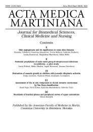
ISSN 1335-8421 Acta Med Mart 2005, 5(2)<br />
ACTA MEDICA<br />
MARTINIANA<br />
Journal for Biomedical Sciences,<br />
Clinical Medicine and Nursing<br />
Contents<br />
3<br />
Assessment of the meconium removal in surfactant vs. saline-lavaged rabbits with<br />
meconium aspiration<br />
Daniela Mokrá, Anna Drgová, Andrea Čalkovská<br />
9<br />
Visualisation of cardiovascular dysregulation in young patients<br />
with type 1 diabetes mellitus by Poincaré plot<br />
Michal Javorka, Jana Javorková, Ingrid Tonhajzerová, Kamil Javorka<br />
17<br />
Stimulation of distal esophagus has no influence on the cough reflex in awake guinea<br />
pigs with experimental allergic rhinitis<br />
Jana Plevková, Mariana Brozmanová, Silvia Varechová, Miloš Tatár<br />
24<br />
Cognitive evoked <strong>po</strong>tentials in patients following concussion<br />
Štefan Sivák, Dušan Trstenský, Egon Kurča, Viera Cisáriková, Ema Kantorová,<br />
Daniela Šútorová<br />
30<br />
Blood serum aluminium content in general <strong>po</strong>pulation compared to dialysed patients<br />
Jela Valachová, Renáta Mikulková, Jana Buchancová, Soňa Funiaková, Monika Jačmeníková<br />
Published by the Jessenius Faculty of Medicine in Martin,<br />
Comenius University in Bratislava, Slovakia
A C T A M E D I C A M A R T I N I A N A 2 0 0 5 5/2<br />
E d i t o r - i n - C h i e f :<br />
Javorka, K., Martin, Slovakia<br />
I n t e r n a t i o n a l E d i t o r i a l B o a r d :<br />
Belej, K., Martin, Slovakia<br />
Buchanec, J., Martin, Slovakia<br />
Honzíková, N., Brno, Czech Republic<br />
Kliment, J., Martin, Slovakia<br />
Lehotský, J., Martin, Slovakia<br />
Lichnovský, V., Olomouc, Czech Republic<br />
Mareš, J., Praha, Czech Republic<br />
Plank, L., Martin, Slovakia<br />
Stránsky, A., Martin, Slovakia<br />
Tatár, M., Martin, Slovakia<br />
Żwirska-Korczala, K., Zabrze-Katowice, Poland<br />
E d i t o r i a l O f f i c e :<br />
Acta Medica Martiniana<br />
Jessenius Faculty of Medicine, Comenius University<br />
(Dept. of Physiology)<br />
Malá Hora 4<br />
037 54 Martin<br />
Slovakia<br />
Instructions for authors: http:|www.jfmed.uniba.sk (Acta Medica Martiniana)<br />
T l a č :<br />
P+M Turany<br />
© Jessenius Faculty of Medicine, Comenius University, Martin, Slovakia, 2005
A C T A M E D I C A M A R T I N I A N A 2 0 0 5 5/2 3<br />
ASSESSMENT OF THE MECONIUM REMOVAL IN SURFACTANT VS.<br />
SALINE-LAVAGED RABBITS WITH MECONIUM ASPIRATION<br />
DANIELA MOKRÁ 1 , ANNA DRGOVÁ 2 , ANDREA CALKOVSKÁ 1<br />
Department of Physiology 1 and Department of Medical Biochemistry 2 , Comenius University,<br />
Jessenius Faculty of Medicine, Martin, Slovakia<br />
A b s t r a c t<br />
Background and aim: Objective quantification of the amount of removed meconium is an im<strong>po</strong>rtant indicator of efficacy<br />
of approaches <strong>po</strong>tentially improving meconium clearance from the lungs. The goal of this study was to assess the<br />
amount of removed meconium by surfactant vs. saline lavage in rabbits with meconium aspiration by spectrophotometry<br />
and meconium-crit methods.<br />
Methods: Adult rabbits were anesthetized, tracheotomized, paralyzed and conventionally ventilated. Human meconium<br />
(25 mg/ml, 4 ml/kg b.w.) was instilled pro<strong>po</strong>rtionally into the right and left lungs. When respiratory failure developed,<br />
surfactant (Surf, n=7) or saline (Sal, n=7) lavage at a dose of 10 ml/kg b.w. in 3 <strong>po</strong>rtions was performed. Lavage<br />
fluid was suctioned 1 and 5 min after the administration and meconium content in the lavage fluid was evaluated by<br />
spectrophotometry and meconium-crit methods.<br />
Results: Animals of both groups were given comparable amount of meconium. Surfactant lung lavage removed significantly<br />
more meconium when evaluated by meconium-crit method (P0.05).<br />
Conclusions: The discrepancy between the results estimated by spectrophotometry and meconium-crit methods<br />
resulted from the different qualities evaluated by these two methods. Moreover, since water-soluble parts of meconium<br />
were effectively removed by both saline and surfactant lavage, surfactant improved the clearance of cholesterol-soluble<br />
fraction of meconium, too.<br />
K e y w o r d s : meconium, lung lavage, rabbit<br />
INTRODUCTION<br />
Despite combined therapy, meconium aspiration syndrome (MAS) is one of the most severe<br />
diseases in the term newborns. Aspiration of meconium causes obstruction of the airways, inactivation<br />
of pulmonary surfactant, inflammation and pulmonary vasoconstriction, that often leads<br />
to the respiratory failure.<br />
Chemically, meconium can be extracted to the water-soluble (gastrointestinal enzymes, bile<br />
acids, bilirubin etc.) and chloroform-soluble (free fatty acids, cholesterol, neutral lipids etc.) fractions<br />
(1). Due to the high concentration of muco<strong>po</strong>lysaccharides, meconium is extremely adhesive<br />
to the airway walls, and clearing procedures such as airway suctioning and physiotherapy<br />
may be ineffective.<br />
Lung lavage with diluted exogenous surfactant increased the meconium clearance from the<br />
lungs in several experimental studies (2-6). Improvement in oxygenation and survival after the<br />
surfactant lavage was found also in the newborns with MAS (7-9).<br />
Although standardization of the lavage procedure in the terms of the number of lavages, volume<br />
of the administered fluid, and concentration and type of surfactant until now have not<br />
inferred to the final conclusion, amount of the removed meconium became an im<strong>po</strong>rtant indicator<br />
of efficacy of approaches <strong>po</strong>tentially improving meconium clearance from the lungs.<br />
Whereas the previous use of the methods objectively quantifying the meconium content in the<br />
recovered lavage fluid led to the controversial results, the goal of this study was to assess the<br />
amount of removed meconium by surfactant vs. saline lavage in rabbits with meconium aspiration<br />
by spectrophotometry and meconium-crit methods.<br />
Address for corres<strong>po</strong>ndence:<br />
Daniela Mokrá, MD, PhD, Department of Physiology, Comenius University, Jessenius Faculty of Medicine,<br />
Malá Hora 4, 037 54 Martin, Slovakia<br />
tel: +421-43-4131426, fax: +421-43-4222260, e-mail: sevecova@jfmed.uniba.sk
4<br />
A C T A M E D I C A M A R T I N I A N A 2 0 0 5 5/2<br />
METHODS<br />
Meconium<br />
Human meconium was collected from 20 healthy newborns, lyophilized, <strong>po</strong>oled and stored in<br />
–20 °C. Before use, meconium was suspended in 0.9 % NaCl at a concentration of 25 mg/ml.<br />
Surfactant<br />
Modified <strong>po</strong>rcine surfactant (Curosurf, Chiesi Pharmaceutici, Italy) was diluted in saline to<br />
a phospholipid concentration of 10 mg/ml.<br />
Design of experiments<br />
Experiments were carried out in concordance with the basic ethical norms and NIH Publication<br />
No. 86-23, revised 1985. Rabbits of the mean body weight (b.w.) of 2.0 kg were anesthetized<br />
with intramuscular ketamine (Narkamon, S<strong>po</strong>fa, Czech Republic) at a dose of 20 mg/kg b.w. and<br />
xylazine (Rometar, S<strong>po</strong>fa, Czech Republic) at a dose of 5 mg/kg b.w., followed by intravenous ketamine<br />
at a dose of 20 mg/kg b.w./hour. Animals were then tracheotomized, paralyzed with<br />
pipecuronium bromide (Arduan, Gedeon Richter, Hungary) at a dose of 0.3 mg/kg b.w./30 min<br />
i.v. and ventilated conventionally by pressure-controlled ventilator Beat-2 (Chirana, Slovakia)<br />
with frequency of 30/min. Meconium at a dose of 4 ml/kg b.w. was instilled pro<strong>po</strong>rtionally into<br />
the right and left lungs. Additional dose of meconium (25 mg/ml, 1 ml/kg b.w.) was given to animals<br />
not showing evidence of respiratory failure, defined as >30 % decrease in lung-thorax compliance<br />
and PaO 2<br />
A C T A M E D I C A M A R T I N I A N A 2 0 0 5 5/2 5<br />
ing to the calibration curve and expressed as a percentage of the total amount administered to<br />
an individual animal.<br />
Estimation of meconium removal by meconium-crit method<br />
Basic suspension of 3.0 g of meconium in 100 ml of normal saline was diluted to seven different<br />
concentrations (30, 20, 15, 10, 6, 3, and 2 mg/ml). Ten samples of each suspension were<br />
taken into the standard microhematocrit glass tubes and centrifuged at 10 000 rpm for 5 minutes<br />
(11). The meconium-crit was then measured directly as the hematocrit, i.e. as a ratio of the<br />
solid content to the total volume, and mean values were calculated. Calibration curve showing<br />
the relationship between the concentration of meconium and meconium-crit value was constructed<br />
(Fig. 2). In experiments, three samples of the lavage fluid were taken into the microhematocrit<br />
glass tubes and centrifuged at 10 000 rpm for 5 min. Percentage of solid content was<br />
read. Average value of removed meconium was expressed as a percentage of the total amount<br />
administered to an individual animal.<br />
Fig. 2. Calibration curve for meconium-crit method.<br />
STATISTICS<br />
Between-group differences were analysed using Mann-Whitney’s test. A P-value0.05). Animals of both groups were given comparable amount of<br />
meconium – 100 mg/kg b.w. (25 mg/ml, 4 ml/kg), i.e. total mean value 110.7±13.4 mg of meconium<br />
per kg b.w.<br />
As shown by spectrophotometry, amount of meconium removed by saline lavage was<br />
12.7±2.0 mg/kg b.w., i.e. 11.0±1.1 % of administered meconium, while by surfactant lung lavage<br />
it was 15.1±3.1 mg/kg b.w., i.e. 13.6±2.5 % of administered meconium. By meconium-crit<br />
method, saline lavage removed 7.9±2.8 mg/kg b.w. of meconium, i.e. 5.4±2.2 % of instilled meconium,<br />
and surfactant lavage removed 19.1±9.7 mg/kg b.w., i.e. 11.1±4.5 % of administered<br />
meconium. Statistical between-group comparison revealed that amount of meconium removed<br />
by surfactant lung lavage compared to saline lavage was higher, when evaluated by meconiumcrit<br />
method than by spectrophotometry (Fig. 3, Fig. 4).
6<br />
A C T A M E D I C A M A R T I N I A N A 2 0 0 5 5/2<br />
Fig. 3. Meconium removal (mg/kg b.w.) evaluated by spectrophotometry and meconium-crit methods in<br />
saline- (Sal) and surfactant-lavaged (Surf) groups.<br />
Fig. 4. Meconium removal (%) evaluated by spectrophotometry and meconium-crit methods in saline-<br />
(Sal) and surfactant-lavaged (Surf) groups.<br />
DISCUSSION<br />
In the previous experimental studies, meconium clearance from the lungs by surfactant lung<br />
lavage was evaluated either by meconium-crit method or by spectrophotometry. However, while<br />
Ohama and Ogawa (2) and Lam et al. (3) using meconium-crit method found significantly more<br />
meconium removed by surfactant lavage than saline lavage, Cochrane et al. (10) using spectrophotometry<br />
found comparable amount of meconium removed by saline and surfactant lavage.<br />
Discrepancy between the results obtained by these two methods yielded with effort to evaluate<br />
meconium removal by surfactant vs. saline lung lavage in a rabbit model of meconium aspiration<br />
syndrome using both methods.<br />
In our study, spectrophotometry and meconium-crit methods showed higher meconium clearance<br />
by diluted exogenous surfactant than by saline lavage. However, the between-group differences<br />
were more pronounced, when evaluated by meconium-crit method. Our results are thus<br />
comparable with the results of the authors mentioned above. Meconium removal by surfactant<br />
lavage evaluated by meconium-crit method was significantly higher compared to saline lavage,
A C T A M E D I C A M A R T I N I A N A 2 0 0 5 5/2 7<br />
in concordance with Ohama and Ogawa (2) and Lam et al. (3). On the other side, in meconium<br />
removal evaluated by spectrophotometry there was no significant difference between the groups,<br />
comparably to Cochrane et al. (10). We sup<strong>po</strong>se that this finding results from the different qualities<br />
estimated by spectrophotometry and meconium-crit methods. While meconium-crit method<br />
is based on the measurement of total solid content in the lavage fluid, spectrophotometric<br />
method measures the optical density of greenish-coloured meconium in the fluid.<br />
Rather high meconium removal evaluated by spectrophotometry, but relatively low by meconium-crit<br />
method indicates that saline lavage removes especially pigmented parts of meconium<br />
from the lungs, which are soluble in water such as gastrointestinal enzymes, bile acids and<br />
bilirubin. On the other side, exogenous surfactant acts as a detergent lowering the surface tension<br />
of highly tenacious meconium. Therefore, we found higher removal of meconium pigments,<br />
but particularly high clearance of those parts of meconium, which are hardly cleared by saline<br />
– muco<strong>po</strong>lysaccharides, free fatty acids, triglycerides, cholesterol etc., estimated by meconiumcrit<br />
method.<br />
Dargaville et al. (4) recently evaluated clearance of both meconium pigments and meconium<br />
solids in the piglet model of MAS, however, with different methods. Dry weight of the purified<br />
meconium pellet was determined after repeated centrifugation of the lavage fluid and dessication,<br />
and meconium pigment concentration in the supernatant was analysed by fluorescent<br />
spectrophotometry. Similarly to our results, the percentual removal of pigments by both saline<br />
and surfactant lavage was higher than the removal of meconium solids despite another type and<br />
concentration of the used surfactant and larger aliquot volume improving distribution of the<br />
lavage fluid throughout the lungs.<br />
According to the presented results, we can conclude that discrepancy between the results<br />
obtained by spectrophotometry and meconium-crit methods is caused by the different qualities<br />
measured by these two methods. While saline lavage removes mostly water-soluble pigmented<br />
parts of meconium, exogenous surfactant improves the clearance of both water-soluble and cholesterol-soluble<br />
fractions of meconium lowering the surface tension of tenacious substances<br />
present in meconium.<br />
ACKNOWLEDGEMENT<br />
We thank to Chiesi Pharmaceutici SpA, Italy for supplying Curosurf and S. Svorková, D. Kulišková,<br />
I. Štritz and J. Benčatová for technical assistance. The study was sup<strong>po</strong>rted by Grant VEGA No.<br />
1/2306/05.<br />
REFERENCES<br />
1. Moses D, Holm BA, Spitale P, Liu M, Enhorning G. Inhibition of pulmonary surfactant function by meconium. Am<br />
J Obstet Gynecol 1991; 164: 477-481.<br />
2. Ohama Y, Ogawa Y. Treatment of meconium aspiration syndrome with surfactant lavage in an experimental rabbit<br />
model. Pediatr Pulmonol 1999; 28: 18-23.<br />
3. Lam BCC, Yeung CY, Fu KH, Wong KY, Chan FL, Tsoi NS. Surfactant tracheobronchial lavage for the management<br />
of a rabbit model of meconium aspiration syndrome. Biol Neonate 2000; 78: 129-138.<br />
4. Dargaville PA, Mills JF, Headley BM, Chan Y, Coleman L, Loughnan PM, Morley CJ. Therapeutic lung lavage in the<br />
piglet model of meconium aspiration syndrome. Am J Respir Crit Care Med 2003; 168: 456-463.<br />
5. Ševecová D, Čalkovská A, Drgová A, Javorka K. Surfactant lung lavage in rabbits with meconium aspiration – a pilot<br />
study. Acta Med Mart 2002; 2 (2): 9-14.<br />
6. Sevecova-Mokra D, Calkovska A, Drgova A, Javorka M, Javorka K. Treatment of experimental meconium aspiration<br />
syndrome with surfactant lung lavage and conventional vs. asymmetric high-frequency jet ventilation. Pediatr Pulmonol<br />
2004; 38 (4): 285-291.<br />
7. Lam BCC, Yeung CY. Surfactant lavage for meconium aspiration syndrome: A pilot study. Pediatrics 1999; 103:<br />
1014-1018.<br />
8. Wiswell TE, Knight GR, Finer NN, Donn SM, Desai H, Walsh WF, Sekar KC, Bernstein G, Keszler M, Visser VE, Merrit<br />
TA, Mannino FL, Mastrioianni L, Marcy B, Revak SD, Tsai H, Cochrane CG. A multicenter, randomized, controlled<br />
trial comparing Surfaxin (Lucinactant) lavage with standard care for treatment of meconium aspiration syndrome.<br />
Pediatrics 2002; 109: 1081-1087.
8<br />
A C T A M E D I C A M A R T I N I A N A 2 0 0 5 5/2<br />
9. Chang HY, Hsu CH, Kao HA, Hung HY, Chang JH, Peng CC, Jim WT. Treatment of severe meconium aspiration syndrome<br />
with dilute surfactant lavage. J Formos Med Assoc 2003; 102: 326-330.<br />
10. Cochrane CG, Revak SD, Merritt TA, Schraufstätter IU, Hoch RC, Henderson C, Andersson S, Takamori H, Oades<br />
ZG. Bronchoalveolar lavage with KL4-Surfactant in models of meconium aspiration syndrome. Pediatr Res 1998; 44:<br />
705-715.<br />
11. Weitzner JS, Strassner HT, Rawlins RG, Mack SR, Anderson RA. Objective assessment of meconium content of amniotic<br />
fluid. Obstet Gynecol 1990; 76: 1143-1144.<br />
Received: March, 23, 2005<br />
Accepted: May,10, 2005
A C T A M E D I C A M A R T I N I A N A 2 0 0 5 5/2 9<br />
VISUALISATION OF CARDIOVASCULAR DYSREGULATION IN YOUNG<br />
PATIENTS WITH TYPE 1 DIABETES MELLITUS BY POINCARÉ PLOT<br />
1<br />
MICHAL JAVORKA, 2 JANA JAVORKOVA, 1 INGRID TONHAJZEROVA,<br />
1<br />
KAMIL JAVORKA<br />
1<br />
Department of Physiology, Comenius University, Jessenius Faculty of Medicine,<br />
2<br />
Paediatric Clinic, Comenius University, Jessenius Faculty of Medicine,Faculty Hospital, Martin, Slovakia<br />
A b s t r a c t<br />
The noninvasive assessment of s<strong>po</strong>ntaneous physiological parameters variations can provide valuable information<br />
about control systems involved in their complex regulation. Time series analysis is usually performed in time and frequency<br />
domains – the so called linear methods. Inspired by effort to apply nonlinear time series analysis into cardiovascular<br />
variability signals, the aim of this study was to compare heart rate and blood pressure variabilities (HRV and<br />
BPV) between young patients with type 1 diabetes mellitus (DM) and control subjects using Poincaré plot.<br />
Patients with type 1 DM (10 females, 7 males) aged 12.9 – 31.5 years (mean ± SEM: 22.4 ± 1.0 years) were investigated.<br />
The control group consisted of 17 healthy probands matched for sex and age. The HRV and BPV were analysed<br />
in time domain (mean, standard deviation - SD) and using quantitative analysis of Poincaré plot pattern measures during<br />
supine rest.<br />
In young patients with type 1 DM, significant reduction of all measured Poincaré plot parameters constructed from<br />
R-R intervals was found. However, no significant difference between groups in BPV Poincaré plot measures was observed.<br />
In conclusion, HRV Poincaré plot was able to reveal beat-to-beat HRV abnormalities. Poincaré plot can provide information<br />
<strong>po</strong>tentially usable for diagnosis and prognosis in visually understandable manner. We suggest that parasympathetic<br />
dysfunction in cardiac regulation occurs earlier than dysregulation of sympathetic control of the vessels in young<br />
diabetics.<br />
K e y w o r d s : heart rate variability, blood pressure variability, diabetes mellitus, Poincaré plot<br />
INTRODUCTION<br />
The noninvasive assessment of s<strong>po</strong>ntaneous physiological parameters variations in time can<br />
provide valuable information about control systems involved in their complex regulation. Repeatedly<br />
measured values of any assessed parameter form time series that can be regarded as a signal<br />
encompassing information about structure and status of dynamical system that generates<br />
and modifies given parameter. Time series analysis is able to acquire data about normal dynamical<br />
system as well as system changes during pathological circumstances with clinically im<strong>po</strong>rtant<br />
applications (diagnosis, prognosis) (1, 2).<br />
Time series analysis of physiological parameters is usually performed in time and frequency<br />
domains – the so called linear methods. Time domain parameters based on basic statistical<br />
parameters (e.g. mean value, standard deviation) provide summarized information on short- and<br />
long-term variability in time series, but did not comprise information concerning oscillations`<br />
patterns (3). These parameters are sensitive to artefacts and require gaussian distribution of<br />
measured parameter. Time series analysis in frequency domain (spectral analysis) enables to<br />
quantify cyclic oscillations (e.g. respiratory sinus arrhythmia). The nonperiodic oscillations are<br />
ignored and usually regarded as a noise (1,4).<br />
Cardiovascular control system com<strong>po</strong>nents (baroreceptors, chemoreceptors, sympathetic<br />
and parasympathetic nervous system, cardiac pacemaker, smooth muscles of blood vessels,<br />
etc.) interact in a complex manner. These interactions are usually not linear – output is not pro<strong>po</strong>rtional<br />
to input (e.g. heart rate changes are not directly pro<strong>po</strong>rtional to blood pressure<br />
changes). The nonlinear systems are able to generate very complex signals that cannot be distinguished<br />
from noise using linear time series analysis tools. Therefore, there is an ongoing<br />
Address for corres<strong>po</strong>ndence:<br />
Michal Javorka, MD, PhD, Department of Physiology, JLF UK<br />
Malá Hora 4, 037 54 Martin, Slovakia<br />
Phone:++421 43 41314 26, fax: ++ 421 43 42222 60, e-mail: mjavorka@jfmed.uniba.sk
10<br />
A C T A M E D I C A M A R T I N I A N A 2 0 0 5 5/2<br />
effort to apply nonlinear time series analysis into cardiovascular variability signals (1, 3, 4, 5).<br />
Application of new mathematical tools based on nonlinear dynamics to heart rate and blood<br />
pressure variabilities analysis provides supplementary information about systems involved in<br />
cardiovascular parameters changes. Computing of the commonly used nonlinear parameters<br />
(Lyapunov ex<strong>po</strong>nent, correlation dimension) from the signal requires relatively long and stationary<br />
signals which are difficult to obtain from living animals and humans. The graphical analysis<br />
by Poincaré plot is increasingly used because it can be performed from shorter quasi-stationary<br />
records. Poincaré plot is on the boundary between linear methods of biosignal analysis<br />
and tools based on nonlinear dynamics – the principle of its construction is taken from the nonlinear<br />
dynamics theory, but parameters used for its quantification are essentially linear (6). In<br />
addition, Poincaré plot enables to display information about beat-to-beat variability of heart rate<br />
in compact visual format which is easy to read (4,7).<br />
Autonomic neuropathy and cardiovascular dysregulation are usually regarded as the late<br />
complications of diabetes mellitus (DM). However, autonomic nervous system dysregulation can<br />
be detected by modern sensitive methods even in early phases of DM (8, 9, 10, 11). Early diagnosis<br />
of autonomic neuropathy is im<strong>po</strong>rtant because the mortality of the patients with this complication<br />
is markedly higher (12, 13).<br />
The reduction of s<strong>po</strong>ntaneous heart rate variability (HRV) is regarded as one of the early signs<br />
of cardiac autonomic neuropathy (8, 14). HRV originates predominantly from parasympathetic<br />
nervous traffic oscillations (15) and therefore HRV analysis can provide information about vagal<br />
com<strong>po</strong>nent of the autonomic nervous system (16). On the other side, smooth muscles of the vessels<br />
and hence peripheral vascular resistance are under dominant sympathetic nervous system<br />
control. Therefore, the analysis of blood pressure variability (BPV) can be useful for detection of<br />
sympathetic dysfunction (16, 17, 18).<br />
Relatively few studies were focused on cardiovascular dysregulation in adolescents and young<br />
adults with type 1 DM, although a multicentric study EURODIAB has found the cardiac autonomic<br />
neuropathy in 19% of diabetics in the age group 15-29 years (19). Short-term BPV was<br />
analysed in diabetic patients only in time domain (20, 21, 22) and, to our knowledge, Poincaré<br />
plot of BPV in young diabetics has not been used yet.<br />
The aim of the study was to compare heart rate and blood pressure variabilities between<br />
young patients with type 1 DM and control subjects using Poincaré plot.<br />
METHODS<br />
Subjects<br />
We have investigated 17 young patients with type 1 DM (10 females, 7 males) aged 12.9 – 31.5<br />
years (mean ± SEM: 22.4 ± 1.0 years). The mean duration of DM was 12.4 ± 1.2 years. The control<br />
group consisted of 17 healthy probands matched for sex and age. All subjects were familiarised<br />
with investigation protocol and they gave informed consent. Probands were instructed to<br />
avoid smoking and drinking alcoholic beverages for 24 hours before investigation. Several basic<br />
study groups characteristics are presented in Table 1.<br />
Table 1: Study groups characteristics (control group – C, group of patients with type 1 diabetes mellitus - DM). Values<br />
are presented as mean ± SEM and P-values were obtained using Mann-Whitney U-test. Asterisk indicate significant<br />
(P
A C T A M E D I C A M A R T I N I A N A 2 0 0 5 5/2 11<br />
Protocol<br />
The length of R-R intervals was measured using telemetric system (VariaCardio TF4, Sima<br />
Media, Olomouc, Czech Republic) where ECG signal (sampling freqency 1000 Hz) from thoracic<br />
belt with integrated electrodes was transferred into PC for further analysis. Systolic blood pressure<br />
(SBP) was monitored beat-to-beat using volume-clamp method (23) by Finapres 2300<br />
(Ohmeda, USA). The finger cuff of appropriate size was wrapped around middle phalanx of the<br />
third finger of the left hand. The finger was passively maintained at the heart level to avoid blood<br />
pressure distortion caused by hydrostatic pressure changes. Analog output of the Finapres was<br />
transferred into PC by analog-digital convertor PCL-711 (Advantech Co., Taiwan) with the sampling<br />
frequency of 500 Hz. The SBP values were obtained on-line using specially developed software<br />
and stored in PC for subsequent analysis.<br />
All subjects were investigated in quiet room from 7.30 to 12.00 a.m. The thoracic belt with<br />
ECG electrodes and finger cuff of Finapres device were applied after 10 minutes in sitting <strong>po</strong>sition.<br />
Then, the subject was in supine <strong>po</strong>sition on the bed during next 70 minutes of the continuous<br />
recording of the cardiovascular parameters. We have asked the probands to avoid voluntary<br />
movements and speaking as much as <strong>po</strong>ssible.<br />
Data analysis<br />
HRV and BPV analysis was performed off-line in selected interval (interval started 30 min<br />
after reclining, the length of interval was 600 s) of the records using special software.<br />
In analysed time interval we quantified several basic time domain HRV and BPV parameters:<br />
mean R-R interval duration (mean RR), standard deviation of the R-R intervals (SDRR), mean<br />
SBP and standard deviation of SBP values (SDSBP).<br />
Poincaré plot is the semiquantitative tool for physiological parameters variability analysis<br />
(most commonly used in for HRV analysis) which enable to assess their non-random beat-to-beat<br />
changes. The HRV Poincaré plot is the scatterplot of current R-R interval length against the R-<br />
R interval length immediately preceding it and provides visually understandable information<br />
about both overall and beat-to-beat HRV. If the heart rate rhythm is regular then the <strong>po</strong>ints in<br />
Poincaré plot are located closely around the line of identity (axis of the 1st quadrant) (4, 24).<br />
Analogously, BPV Poincaré plot is the scatterplot where x-coordinate of each <strong>po</strong>int is the current<br />
SBP and y-coordinate is the SBP value during previous heart beat.<br />
Poincaré plots of HRV and BPV were constructed from resampled R-R and SBP time series (at<br />
1 Hz) and quantitatively analysed. Quantitative analysis of the Poincaré plot patterns (Fig. 1) was<br />
performed using self-developed software. The widths of Poincaré plot pattern at 10th, 25th, 50th,<br />
75th and 90th percentiles of R-R intervals (SBP) distribution – W10, W25, W50, W75 and W90 –<br />
were quantified according to Schechtman et al. (1992) and Silke et al.(1999). This approach<br />
enables to quantify beat-to-beat heart rate (and SBP) changes at given “basal” heart rate (SBP)<br />
Fig. 1: Poincaré plot analysis with quantified measures
12<br />
A C T A M E D I C A M A R T I N I A N A 2 0 0 5 5/2<br />
level - common comet-like shape of Poincaré plot pattern is caused by higher beat-to-beat variability<br />
around longer R-R intervals (higher widths at 75th and 90th percentiles). The length of<br />
the Poincaré plot was defined as the difference between 10th and 90th percentiles of R-R intervals<br />
(SBP) distribution.<br />
Statistics<br />
Nonparametric tests were used due to non-gaussian distribution of the HRV nad BPV parameters.<br />
The non-gaussian distribution of the variables was ascertained using the Lilliefors test.<br />
Between-groups comparisons (DM vs control group) were performed using Mann-Whitney U-test.<br />
All inferential statistics were considered significant at P
A C T A M E D I C A M A R T I N I A N A 2 0 0 5 5/2 13<br />
Fig. 3: HRV Poincaré plots illustrate differences in beat-to-beat<br />
heart rate variability between a healthy control subject and<br />
a patient with DM. Marked reduction in all Poincaré plot indices<br />
was found in young DM patients.<br />
Fig. 4: Heart rate variability analysed using Poincaré plot in<br />
control subjects (dark gray) and patients with DM (light gray).<br />
Bars and error lines represent mean and SEM, respectively.<br />
Asterisks indicate significant (p
14<br />
A C T A M E D I C A M A R T I N I A N A 2 0 0 5 5/2<br />
Fig. 6: BPV Poincaré plot parameters in control subjects (dark gray) and<br />
patients with DM (light gray). Bars and error lines represent mean and<br />
SEM, respectively.<br />
DISCUSSION<br />
HRV Poincaré plot changes during various physiological manoeuvres and diseases<br />
Kamen et al. (25) assessed changes of Poincaré plot pattern constructed from R-R intervals<br />
during head-up tilt test and pharmacological interventions. Head-up tilt test and parasympathetic<br />
blockade by atropine were accompanied by reduced Poincaré plot length and width. On<br />
the other hand, transdermal application of sco<strong>po</strong>lamine (parasympathetic activator) increased<br />
Poincaré plot measures. Parasympathetic nervous system influence on Poincaré plot parameters<br />
was analysed also by Tulp<strong>po</strong> et al. (26). Increasing doses of atropine caused predominantly<br />
reduction of pattern width although the decrease in length was also significant. These findings<br />
indicate that Poincaré plot pattern width reflecting beat-to-beat HRV can be regarded as an cardiac<br />
parasympathetic system activity index.<br />
Poincaré plot was able to detect HRV abnormalities also during pathological circumstances. In<br />
children who later succumbed to sudden infant death syndrome (SIDS), significant reduction in Poincaré<br />
plot width on 90 th percentile of R-R interval distribution was found (27). Reduced width and<br />
length of Poincaré plot pattern was found in patients with chronic ischemic heart disease. Potentially<br />
useful for the patients` prognosis can be observation that in <strong>po</strong>st- myocardial infarction patients<br />
with ventricular tachyarrhythmias is reduced length and width of pattern compared to patients without<br />
arrhythmias (28). Epidemiological study proved that Poincaré plot parameters provide independent<br />
prognostic value concerning sudden death in patients with chronic cardiac failure (29).<br />
Heart rate variability in DM patients<br />
The characteristic findings in adult diabetic patients with cardiovascular autonomic neuropathy<br />
are resting tachycardia and mostly reduced HRV, which is the earliest sign of cardiac<br />
autonomic dysfunction (8). In our study we did not find any significant difference in mean heart<br />
rate (represented by its reciprocal value – mean RR) in supine <strong>po</strong>sition between diabetic patients<br />
and control group, but we found reduced overall HRV (reduced SDRR) in young patients with<br />
DM. These findings are in agreement with other studies (13, 14, 20, 30, 31) where nonsignificant<br />
difference in resting mean heart rate was often accompanied by significant differences in conventional<br />
(time and frequency domains) HRV parameters in diabetic patients.<br />
From the Poincaré plot parameters, young diabetic patients had all measures of the pattern<br />
reduced. This was not surprising, beacause the decreased overall HRV (represented by the length<br />
of Poincaré plot pattern) and decreased beat-to-beat HRV (represented by the width) was expected<br />
in DM patients. The studies of Kamen et al.(25), Tulp<strong>po</strong> et al. (26) and others confirmed the<br />
reduction of Poincaré plot pattern measures in diseases and manoeuvres linked with parasympathetic<br />
nervous system activity inhibition. As known before (25, 26, 32), we have also observed<br />
significant <strong>po</strong>sitive correlations between Poincaré plot measures and mean R-R interval (results<br />
not shown). Despite this fact, Poincaré plot can provide supplementary information about beatto-beat<br />
variability at various heart rates which is unavailable by time and frequency domain<br />
analysis (4, 28, 33, 34). These findings indicate dysfunction of the parasympathetic com<strong>po</strong>nent<br />
of autonomic nervous system in DM patients, because the short-term HRV is mostly mediated<br />
by vagal nerve discharge changes (15, 35).
A C T A M E D I C A M A R T I N I A N A 2 0 0 5 5/2 15<br />
Blood pressure variability in DM patients<br />
Direct intraarterial measurement of blood pressure is the most precise method for its continuous<br />
monitoring. However, the usage of this method is markedly limited by its invasiveness<br />
(36). The volume-clamp method is the only alternative for noninvasive beat-to-beat blood pressure<br />
monitoring (23, 37). Although absolute values of the blood pressure obtained using this<br />
method can be distorted (overestimated systolic and underestimated diastolic blood pressures),<br />
volume-clamp method is able to reliably follow s<strong>po</strong>ntaneous blood pressure oscillations (38).<br />
Short term blood pressure changes are mediated mostly by sympathetic nevous system and<br />
therefore the analysis of short term BPV has been taken as more sensitive for detection of sympathetic<br />
dysregulation than HRV analysis (16, 17, 18).<br />
Scaramuzza et al. (10) observed significant reduction in mean peripheral SBP values measured<br />
by Finapres in adolescents with type 1 DM. However, we did not find any significant difference<br />
in mean SBP between young DM patients and control group.<br />
Several authors observed dysfunction of sympathetic control of the vessels in diabetic<br />
patients manifested as an reduction of spectral <strong>po</strong>wer in low frequency band in systolic blood<br />
pressure and skin blood flow signals (12, 39). Mésangeau et al. (40) found reduced standard deviation<br />
of blood pressure signal in animals with induced type 1 DM. In contrast, Chau et al. (20)<br />
did not observe changes in overall BPV in DM patients compared to control group. In our study<br />
no significant changes in overall systolic BPV were found.<br />
We hy<strong>po</strong>thesized that Poincaré plot analysis could be able to detect subtle abnormalities in<br />
beat-to-beat SBP control at various basal SBP levels in young patients with DM. However, no significant<br />
changes in BPV quantified by Poincaré plot pattern measures was observed in our group<br />
of patients compared to control group. We suggest that the significant sympathetic nervous system<br />
dysfunction was not present in our group of young diabetic patients.<br />
In conclusion, Poincaré plot constructed from R-R intervals was able to reveal beat-to-beat<br />
HRV abnormalities. Poincaré plot can provide information <strong>po</strong>tentially usable for diagnosis and<br />
prognosis in visually understandable manner. No significant difference between DM group and<br />
control group was observed in BPV quantified by Poincaré plot and standard deviation of SBP.<br />
We suggest that parasympathetic dysfunction in cardiac chronotropic regulation occurs earlier<br />
than dysregulation of sympathetic control of the vessels in young diabetics.<br />
Acknowledgements<br />
This study was sup<strong>po</strong>rted by VEGA grant N. 1/2305/05.<br />
REFERENCES<br />
1. Kantz H, Schreiber T. Nonlinear time series analysis. Cambridge UK: Cambridge University Press; 1997.<br />
2. ftp://amath.colorado.edu/pub/dynamics/papers/sci.nonlinearFAQ.rtf<br />
3. Hanratty CG, Silke B, Riddell JG. Evaluation of the effect on heart rate variability of a ß2 – adrenoceptor agonist and<br />
antagonist using non-linear scatterplot and sequence methods. Br J Clin Pharmacol 1999; 47: 157-166.<br />
4. Kamen PW, Tonkin AM. Application of the Poincaré plot to heart rate variability: a new measure of functional status<br />
in heart failure. Aust N Z J Med 1995; 25: 18-26.<br />
5. Kaplan D, Glass L. Understanding nonlinear dynamics. New York: Springer Verlag; 1995.<br />
6. Brennan M, Palaniswami M, Kamen P. Do existing measures of Poincaré plot geometry reflect nonlinear features of<br />
heart rate variability? IEEE Trans Biomed Eng 2001; 48: 1342-1347.<br />
7. Keeley EC, Lange RA, Hillis LD, Joglar JA, Page RL. Correlation between time-domain measures of heart rate variability<br />
and scatterplots in patients with healed myocardial infarcts and the influence of metoprolol. Am J Cardiol<br />
1997; 79: 412-414.<br />
8. Ziegler D. Diabetic cardiovascular autonomic neuropathy: prognosis, diagnosis and treatment. Diabetes Metab Rev<br />
1994; 10: 339-383.<br />
9. Spallone V, Uccioli L, Menzinger G. Diabetic autonomic neuropathy. Diabetes Metab Rev 1995; 11: 227-257.<br />
10. Scaramuzza A, Salvucci F, Leuzzi S, Radaelli A, d`Annunzio G, Fratino P, Lorini R, Bernardi L. Cardiovascular autonomic<br />
testing in adolescents with type I (insulin-dependent) diabetes mellitus: an 18-month follow-up study. Clin Sci<br />
1998; 94: 615-621.<br />
11. Sima AAF. Does insulin play a role in cardiovascular autonomic regulation ? Diabetes Care 2000; 23: 724-725.<br />
12. Spallone V, Menzinger G. Diagnosis of cardiovascular autonomic neuropathy in diabetes. Diabetes 1997; 46: S67-S76.
16<br />
A C T A M E D I C A M A R T I N I A N A 2 0 0 5 5/2<br />
13. Osterhues H-H, Großmann G, Kochs M, Hombach V. Heart-rate variability for discrimination of different types of<br />
neuropathy in patients with insulin-dependent diabetes mellitus. J Endocrinol Invest 1998; 21: 24-30.<br />
14. Rollins MD, Jenkins JG, Carson DJ, McGlure BG, Mitchell RH, Imam SZ. Power spectral analysis of the electrocardiogram<br />
in diabetic children. Diabetologia 1992; 35: 452-455.<br />
15. Eckberg DL. Physiological basis for human autonomic rhythms. Ann Med 2000; 32: 341-349.<br />
16. Takalo R, Korhonen I, Turjanmaa V, Majahalme S, Tuomisto M, Uusitalo A. Short-term variability of blood pressure<br />
and heart rate in borderline and mildly hypertensive subjects. Hypertension 1994; 23: 18-24.<br />
17. Cottin F, Papelier Y, Escourrou P. Effects of exercise load and breathing frequency on heart rate and blood pressure<br />
variability during dynamic exercise. Int J S<strong>po</strong>rts Med 1999; 20: 232-238.<br />
18. Laitinen T, Hartikainen J, Niskanen L, Geelen G, Länsimies E. Sympathovagal balance is major determinant of shortterm<br />
blood pressure variability in healthy subjects. Am J Physiol 1999; 276: H1245-H1252.<br />
19. Donaghue KC. Autonomic neuropathy: diagnosis and impact on health in adolescents with diabetes. Horm Res 1998;<br />
50: 33-37.<br />
20. Chau NP, Mestivier D, Chanudet X, Bauduceau B, Gautier D, Larroque P. Use of runs test to assess cardiovascular<br />
autonomic function in diabetic subjects. Diabetes Care 1994; 17: 146-148.<br />
21. Pecis M, Azevedo MJ, Moraes RS, Ferlin EL, Gross JL. Autonomic dysfunction and urinary albumin excretion rate<br />
are associated with an abnormal blood pressure pattern in normotensive normoalbuminuric type 1 diabetic patients.<br />
Diabetes Care 2000; 23: 989-993.<br />
22. Watkins LL, Surwit RS, Grossman P, Sherwood A. Is there a glycemic threshold for impaired autonomic control ?<br />
Diabetes Care 2000; 23: 826-830.<br />
23. Peňáz J. Photo-electric measurement of blood pressure, volume and flow on the finger. Digest 10th Int Conf Med Biol<br />
Eng, Dresden 1973: 104.<br />
24. Woo MA, Stevenson WG, Moser DK, Trelease RB, Harper RM. Patterns of beat-to-beat heart rate variability in<br />
advanced heart failure. Am Heart J 1992; 123(3): 704-710.<br />
25. Kamen PW, Krum H, Tonkin AM. Poincaré plot of heart rate variability allows quantitative display of parasympathetic<br />
nervous activity in humans. Clin Sci 1996; 91: 201-208.<br />
26. Tulp<strong>po</strong> MP, Mäkikallio TH, Takala TES, Seppänen T, Huikuri HV. Quantitative beat-to-beat analysis of heart rate<br />
dynamics during exercise. Am J Physiol 1996; 271: H244-H252.<br />
27. Schechtman VL, Raetz SL, Harper K, Garfinkel A, Wilson AJ, Southall DP, Harper RM. Dynamic analysis of cardiac<br />
R-R intervals in normal infants and in infants who subsequently succumbed to the sudden infant death syndrome.<br />
Pediatr Res 1992; 31: 606-612.<br />
28. Mäkikallio T. Analysis of heart rate dynamics by methods derived from nonlinear mathematics: Clinical applicability<br />
and prognostic significance. Oulun yliopiston kirjasto 2000: (URL: http://hercules.oulu.fi/isbn9514250133/html).<br />
29. Brouwer J, van Veldhuisen DJ, Man in’t Veld AJ, Haaksma J, Dijk WA, Visser KR, Boomsma F, Dunselman PH. Prognostic<br />
value of heart rate variability during long-term follow-up in patients with mild to moderate heart failure. The<br />
Dutch Ibopamine Multicentre Trial Study Group. J Am Coll Cardiol 1996; 28: 1183-1189.<br />
30. Yamasaki Y, Ueda N, Kishimoto M, Tani A, Ishida Y, Kawamori R, Kamada T. Assessment of early stage autonomic<br />
nerve dysfunction in diabetic subjects – application of <strong>po</strong>wer spectral analysis of heart rate variability. Diabetes Res<br />
1991; 17: 73-80.<br />
31. Javorka K, Javorková J, Petrášková M, Tonhajzerová I, Buchanec J, Chromá O. Heart rate variability and cardiovascular<br />
tests in young patients with diabetes mellitus type 1. J Pediatr Endocrinol Metab 1999; 12: 423-431.<br />
32. Copie X, Pousset F, Lechat P, Jaillon P, Guize L, Le Heuzey J-Y. Cardiac Insufficiency Bisoprolol Study Investigators.<br />
Effects of b-blockade on heart rate variability in advanced heart failure: Analysis of scatterplots of R-R intervals at<br />
selected heart rates. Am Heart J 1996; 132: 369-375.<br />
33. Bergfeldt L, Haga Y. Power spectral and Poincaré plot characteristics in sinus node dysfunction. J Appl Physiol 2003;<br />
94: 2217-2224.<br />
34. Javorka M, Žila I, Balhárek T, Javorka K. On- and off-res<strong>po</strong>nses of heart rate to exercise – relations to heart rate<br />
variability. Clin Physiol & Func Im 2003; 23: 1-8.<br />
35. Task Force of the European Society of Cardiology and the North American Society of Pacing and Electrophysiology.<br />
Heart rate variability. Standards of measurement, physiological interpretation and clinical use. Circulation 1996; 93:<br />
1043-1065.<br />
36. Porter KB, O`Brien WF, Kiefert V, Knuppel RA. Finapres: a noninvasive device to monitor blood pressure. Obstet<br />
Gynecol 1991; 78: 430-433.<br />
37. Virolainen J. Use of non-invasive finger blood pressure monitoring in the estimation of aortic pressure at rest and<br />
during the Mueller manoeuvre. Clin Physiol 1992; 12: 619-628.<br />
38. Castiglioni P, Parati G, Omboni S, Mancia G, Imholz BPM, Wesseling K, Di Rienzo M. Broad-band spectral analysis<br />
of 24h continuous finger blood pressure: comparison with intra-arterial recordings. Clin Sci 1999; 97: 129-139.<br />
39. Bernardi L, Rossi M, Leuzzi S, Mevio E, Fornasari G, Calciati A, Orlandi C, Fratino P. Reduction of 0.1 Hz microcirculatory<br />
fluctuations as evidence of sympathetic dysfunction in insulin-dependent diabetes. Cardiovasc Res 1997;<br />
34: 185-191.<br />
40. Mésangeau D, Laude D, Elghozi J-L. Early detection of cardiovascular autonomic neuropathy in diabetic pigs using<br />
blood pressure and heart rate variability. Cardiovasc Res 2000; 45: 889-899.<br />
Received: March, 23, 2005<br />
Accepted: May, 11, 2005
A C T A M E D I C A M A R T I N I A N A 2 0 0 5 5/2 17<br />
STIMULATION OF DISTAL ESOPHAGUS HAS NO INFLUENCE ON THE<br />
COUGH REFLEX IN AWAKE GUINEA PIGS WITH EXPERIMENTAL<br />
ALLERGIC RHINITIS<br />
JANA PLEVKOVÁ, MARIANA BROZMANOVÁ, SILVIA VARECHOVÁ, MILOŠ TATÁR<br />
Department of Pathophysiology, Comenius University, Jessenius Faculty of Medicine in Martin, Slovakia<br />
A b s t r a c t<br />
Intranasal stimulation with capsaicin and experimentally induced rhinitis enhances cough res<strong>po</strong>nse probably via<br />
central plasticity of the nucleus of the solitary tract neurons due to strong afferent inputs from the nose (1). Isolated<br />
intraesophageal administration of capsaicin did not induce any changes in the cough res<strong>po</strong>nse (2).<br />
This study was designed to test whether combinations of afferent inputs from the nose and esophagus could enhance<br />
the intensity of citric acid (CA) induced cough in guinea pigs.<br />
16 male TRIK strain guinea pigs were sensitised by ovalbumin. 21 days after sensitization was confirmed by skin<br />
tests. Experimental allergic rhinitis was induced in these animals by intranasal ovalbumin challenge. After development<br />
of nasal symptoms the animals of experimental group (E) were challenges with intraesophageal (IE) capsaicin (1000 μM,<br />
250 μl). Controls received IE saline. Cough was induced during IE stimulation both in E and C groups by citric acid and<br />
compared to baseline cough res<strong>po</strong>nse tested in the beginning of the study. Number of coughs was analysed from pneumotachographic<br />
records of airflow changes typical for cough.<br />
The number of CA induced coughs in C was increased after the induction of rhinitis and IE administration of saline<br />
[4 (1.5-5) vs 6 (3.5-8), p < 0.05]. Similar result was obtained for group of E animals with rhinitis IE challenged with capsaicin<br />
[3(2-4) vs 5 (5-6.5), p < 0.05]. This increase of the number of coughs is due to rhinitis. We did not find any differences<br />
in the cough res<strong>po</strong>nse that could be ascribed to IE administration of saline or capsaicin (p = 0.26).<br />
Conclusion: Intraesophageal administration of capsaicin did not affect CA induced cough in guinea pigs suffering<br />
from allergic rhinitis.<br />
K e y w o r d s : cough plasticity, allergic rhinitis, intraesophageal capsaicin challenge, guinea pig cough models<br />
INTRODUCTION<br />
In 1977, Irwin and coworkers (3) published a comprehensive review pro<strong>po</strong>sing that the cause<br />
of cough could be determined if a systematic evaluation was performed of the anatomic locations<br />
where cough receptors reside in the afferent limb of the cough reflex. The afferent limb includes<br />
the vagus nerve, which also innervates the esophagus. Using this protocol, Irwin and coworkers<br />
(3) re<strong>po</strong>rted a prospective trial in which the cause of cough was found in all subjects and specific<br />
therapy toward the cause resulted in cough resolution in 98% of subjects. Gastroesophageal<br />
reflux was the etiology of cough in 10% of their <strong>po</strong>pulation when the diagnosis was made by history,<br />
endoscopy, or barium esophagogram (4).<br />
The utility of the anatomic diagnostic protocol for chronic persistent cough and the im<strong>po</strong>rtance<br />
of GERD as a cause of cough is proved (5, 6, 7).<br />
There are two pro<strong>po</strong>sed mechanisms of GERD-associated cough: (A) acid in the distal esophagus<br />
stimulating an esophageal-tracheobronchial cough reflex, and (B) microaspiration or<br />
macroaspiration of esophageal contents into the larynx and tracheobronchial tree. A number of<br />
studies have been done to examine <strong>po</strong>tential mechanisms affecting cough reflex in patients suffering<br />
from pathological gastroesophageal reflux (8, 9, 10).<br />
In addition to the discussed <strong>po</strong>ints of pathogenesis of chronic cough in patients with GERD,<br />
there may be at least one im<strong>po</strong>rtant mechanism involved. It is sup<strong>po</strong>sed that activity of central<br />
cough pattern generator (CPG) is under plasticity of numerous stimuli, conducted to the brainstem<br />
via afferent nerve connections of these afferents with neuronal circuits (network) res<strong>po</strong>nsible<br />
for cough. Afferent stimuli originated in the nasal mucosa in patients suffering from rhinitis<br />
Address for corres<strong>po</strong>ndence:<br />
Jana Plevková MD, PhD,Department of Pathophysiology,<br />
Jessenius Faculty of Medicine, Commenius University, Sklabinska str. 26, 037 53 MARTIN,<br />
Phone: 4238 213, 0904 828 142, e-mail: plevkova@jfmed.uniba.sk
18<br />
A C T A M E D I C A M A R T I N I A N A 2 0 0 5 5/2<br />
and those originated in esophagus in patients with GERD and subsequent convergence of these<br />
afferent inputs in the nucleus of the solitary tract (nTs) could be res<strong>po</strong>nsible for cough in this<br />
group of patients, although the pathological process is localized outside the respiratory tract<br />
area, from which the cough could be clearly elicited (11).<br />
The simplest hy<strong>po</strong>thesis explaining the cough enhancement by reflux is that the intense activation<br />
of sensory nerves in esophagus either directly triggers cough and/or sensitizes the cough<br />
reflex. These changes would result in the inappropriately active cough reflex leading to chronic<br />
coughing.<br />
In our previous study we approached this hy<strong>po</strong>thesis in the guinea pig cough models. Citric<br />
acid–induced cough was elicited during stimulation of the esophageal afferent nerves in the<br />
intact esophagus or esophagus with injured mucosa. We found that in either model the transient<br />
introduction of the nociceptive sensory nerve activator capsaicin into the esophagus did not trigger<br />
cough or affected cough induced by citric acid. Our results suggest that acute localized stimulation<br />
of esophageal mucosal nerves is not sufficient to trigger and/or enhance cough in guinea<br />
pigs.<br />
There is also suggestion that stimulation of afferent nerve endings in injured esophageal<br />
mucosa is able to change the cough sensitivity only in patients with some subclinical pathological<br />
changes in the airways (12). For that reason we decided to assess the effect of intraesophageal<br />
administration of capsaicin in supramaximal concentration on citric- acid induced<br />
cough in guinea pigs with experimentally induced allergic disorder of the respiratory system.<br />
Aim: The aim of the study was to assess the effect of intraesophageal administration of capsaicin<br />
(noxious stimulus activating nerve endings of C – fibres and sub<strong>po</strong>pulation of Aδ nerve<br />
fibres) on citric acid induced cough in animals with experimental allergic rhinitis after passive<br />
sensitisation with ovalbumin and repeated sequence of active sensitisations with intranasal ovalbumin<br />
challenges, as well.<br />
METHODS<br />
Animals: All experiments were approved by Jessenius Faculty of Medicine Ethical Committee<br />
and followed the criteria of welfare of experimental animals, as well.<br />
Animals (guinea pigs n = 16) (body weight 350-450 g) were housed in an approved animal<br />
house, maintained at room temperature 21-22 O C, humidity 60-70%, ventilation, 12-h light-dark<br />
cycle and free access to water and standard animal food. Male TRIK strain guinea pigs were<br />
obtained from the Department of Experimental Pharmacology, Slovak Academy of Science (Dobra<br />
Voda, Slovak Republic) and used after at least 1week adaptation period in the animal house.<br />
Guinea pigs were adapted to experimental conditions two times, by inhalation of nebulized saline<br />
in the plethysmographic box.<br />
Ovalbumin sensitization: All animals (n = 16) were passively sensitized with ovalbumin (OA)<br />
(10 μg, Sigma) administered intraperitoneally together with aluminum hydroxide (100 mg) in<br />
saline (1 ml i.p) (13). Twenty-one days after successful sensitization was confirmed by skin prick<br />
test (intradermal injection of ovalbumin 25 μl of 200 μg.ml -1 ) on the skin of the back. Only sensitised<br />
animals with marked erythema and oedema were involved in the study. Animals were<br />
used for experiments 7 days later.<br />
Model of allergic rhinitis: Sensitized animals were used to develop a model of allergic rhinitis<br />
by repeated intranasal instillation of 0.015 ml of 0.5% OA into both nares via a thin catheter.<br />
These animals were repeatedly intranasally challenged with OA at 7-day intervals for 6 weeks.<br />
These challenges of antigen were followed by allergic nasal res<strong>po</strong>nse (sneezing, nasal discharge<br />
and worsening of nasal breathing) and so this model of experimental allergic rhinitis was taken<br />
as confirmed.<br />
Intraesophageal administration of stimulating substances: Animals (n=16) were placed<br />
into a plastic cylinder (a part of the body chamber of the plethysmographic box equipment (double<br />
chamber plethysmograph type 855, Hugo Sachs Elektronik, Germany). This allowed immobilization<br />
of animals.
A C T A M E D I C A M A R T I N I A N A 2 0 0 5 5/2 19<br />
After that, using a mouth opener device, thin <strong>po</strong>rtex catheter (external diameter 0.3 cm) with<br />
conducting wire was introduced into the esophagus of the animal. The catheter was tipped with<br />
cotton tam<strong>po</strong>n and its end was <strong>po</strong>sitioned in the middle part of esophagus.<br />
Capsaicin (Sigma, 1000 μM, 0.2 ml) was applied into the catheter in a manner that allowed<br />
absorption of the solutions used into the cotton tam<strong>po</strong>n. Conducting wire was then introduced<br />
into the catheter and cotton tam<strong>po</strong>n was disengaged from its tip by means of careful moving of<br />
the conducting wire. Then the catheter was pulled out from esophagus and mouth.<br />
Induction of coughing: Awake guinea pigs were placed into the plastic cylinder (a part of<br />
plethysmographic box equipment) allowing immobilization of the animal. Then the animals were<br />
placed individually in a bodyplethysmograph (type 855, Hugo Sachs Electronic, Germany) (Fig.1)<br />
consisting of a head chamber and a body chamber. The opening between the head chamber and<br />
body chamber was equipped with a plastic collar lining around animal neck to prevent communication<br />
between the chambers. Appropriate collar size was chosen for each animal to prevent<br />
neck compression, which could cause airway obstruction and/or mechanical stimulation of the<br />
upper trachea and larynx.<br />
Fig. 1: Double chamber bodyplethysmograph (type 855, Hugo Sachs<br />
Elektronik, Germany) which is used in our department for induction of<br />
cough by administration of tussive aerosols into the front chamber of the<br />
plethysmograph.<br />
To ex<strong>po</strong>se an animal to aerosol, the head chamber was connected to a nebulizer (Pari Provokation<br />
Test I, Menzel, Germany, manufacturer’s specification: output 5 l.min -1 , particle mass<br />
median aerodynamic diameter 1.2 μm). A suction device adjusted to the same input 5 l. min -1<br />
was connected to the head chamber to maintain constant airflow through the chamber during<br />
aerosol administration. Respiratory changes in the airflow were measured using pneumotachograph<br />
(Godart, Germany) with Fleisch head (No. 1, Gould Godart Statham BV) connected to the<br />
head chamber and recorded directly with pen recorder (Multiscriptor Hellige, Germany).<br />
Respiratory sounds including cough and sneezing were recorded with a microphone placed in<br />
the roof of the head chamber and connected to a preamplifier and loudspeaker. Pneumotachograph<br />
and tape recorder outputs were simultaneously recorded on a PC for off- line analysis.<br />
Cough challenge was performed using inhalation of citric acid (CA) (Lachema, 0.3 M) for 1<br />
minute. Cough was detected from the expiratory change of airflow interrupting basic respiratory<br />
pattern accompanied by a cough sound during 1 min ex<strong>po</strong>sure to CA and subsequent 2 minutes<br />
period.<br />
Statistical analysis: Data for number of the cough efforts are expressed as a median and<br />
interquartil range. Statistical analysis of the samples was performed using a Kruskal – Wallis<br />
test. A probability value of P< 0.05 was considered as significant.
20<br />
A C T A M E D I C A M A R T I N I A N A 2 0 0 5 5/2<br />
Experimental protocol<br />
In all animals (n = 16) the control cough res<strong>po</strong>nse to 0.3 M citric acid was determined.<br />
According reactivity the animals were divided into controls and experimental group.<br />
After 7 days interval the animals of both groups were challenged with intranasal ovalbumin<br />
to induce rhinitis.<br />
When the clear clinical signs of rhinitis were present (sneezing, nasal rubbing, crackles or discharge)<br />
controls were intraesophageally challenged with saline and experimental animals were<br />
intraesophageally challenged with supramaximal concentration of capsaicin.<br />
The cough res<strong>po</strong>nse to citric acid was determined just after the intraesophageal administration<br />
of capsaicin or saline.<br />
These values (number of coughs) were compared to basal values obtained just in the beginning<br />
of the experimental protocol.<br />
RESULTS<br />
Model of allergic rhinitis<br />
Clinical symptoms of rhinitis have occurred in all sensitized animals that were challenged<br />
intranasally with ovalbumin within 15 minutes after the challenge. These nasal symptoms<br />
involved sneezing, nasal discharge, nasal rubbing and nasal crackles. Some of the animals<br />
showed signs of laboured breathing - paradoxical movements of abdominal wall during inspiration<br />
(due to congestion of nasal mucosa).<br />
Intraesophageal challenges<br />
Intraesophageal challenges with both saline and capsaicin were well tolerated by animals. We<br />
did not notice any signs of esophageal irritation due to tam<strong>po</strong>n (it means excessive salivation,<br />
vomitus or abnormal behaviour of the animals after the challenge). The animals had no problems<br />
with deglutition of standard food and water after the procedure.<br />
Citric acid-induced cough<br />
We have found that the number of cough efforts induced by inhalation of citric acid in controls<br />
was increased after the induction of rhinitis and intraesophageal administration of saline (med<br />
± interquartil range) [4 (1.5-5) vs 6 (3.5-8), p = 0.05]. Very similar result was obtained in the group<br />
of experimental animals with rhinitis, which were intraesophageally challenged with supramaximal<br />
concentration of capsaicin (med ± interquartil range) [3 (2-4) vs 5 (5-6.5), p= 0.034].<br />
This mentioned increase of the number of cough efforts is due to rhinitis, but on the other<br />
hand, we did not find any differences in the cough res<strong>po</strong>nse that could be ascribed to intraesophageal<br />
administration of saline or capsaicin (p = 0.26) (Fig.2).<br />
Fig. 2:<br />
Changes of citric acid – induced cough<br />
intensity (expressed as a median and<br />
interquartil range of number of coughs)<br />
in guinea pigs with experimental allergic<br />
rhinitis during stimulation of<br />
esophageal mucosa. The increase of<br />
cough res<strong>po</strong>nse in both groups is due to<br />
rhinitis, but there was found no difference<br />
in cough res<strong>po</strong>nse that could be<br />
ascribed to esophageal administration<br />
of capsaicin (rhinitis + IE saline vs<br />
rhinitis + IE capsaicin)<br />
* p< 0.05, # = non significant difference.
A C T A M E D I C A M A R T I N I A N A 2 0 0 5 5/2 21<br />
DISCUSSION<br />
In our previous study we showed that the transient introduction of capsaicin into intact<br />
esophagus or esophagus injured by acid or alkaline pretreatment did not trigger cough or affect<br />
the citric acid-induced cough in the awake guinea pigs models. This esophageal stimulation does<br />
not influence either the specific airway resistance measured by Pennock`s method (2). These data<br />
indicate that acute localized stimulation of esophageal mucosal nerves is not sufficient to trigger<br />
and/or enhance cough in naive animals.<br />
Our present study was based on the suggestion that stimulation of vagal afferents in the<br />
esophagus in patients suffering both from the GERD and the chronic cough could be sufficient<br />
to influence the reflex arc of coughing only in those patients with some subclinical changes in<br />
the airways (12). This suggestion could be also one of the <strong>po</strong>ssible explanations of our previous<br />
negative results in animals with injured esophagi, but intact respiratory system (2).<br />
A model of allergic rhinitis in guinea pigs sensitized to ovalbumin was chosen for this pur<strong>po</strong>se<br />
due to previous good experiences with this model as suitable for enhancement of citric acidinduced<br />
cough in guinea pigs. In this case the inflammatory process was located in the nasal<br />
cavity and probably close proximity of nasopharynx, however, it is well known that inflammatory<br />
process from the nasal cavity could be easily spread into the whole respiratory system (the<br />
larynx and the lower airways – the sites from which the cough could be elicited) via airways (<strong>po</strong>st<br />
nasal dripping, microaspiration) (14) or via systemic circulation and subsequent homing of<br />
immune cells (15). We did not have a convincing evidence for the presence of inflammatory<br />
changes in the lower airways in guinea pigs suffering from experimentally induced allergic rhinitis.<br />
This caveat could be used for the interpretation of the negative results from these studies.<br />
We have found that cough res<strong>po</strong>nse in animals is enhanced, however, this increase of the<br />
cough intensity is due to rhinitis. This result is well known and was published before (14). But<br />
we did not find any differences in the cough res<strong>po</strong>nse, which could be ascribed to intraesophageal<br />
administration of capsaicin.<br />
Capsaicin, one of the most frequently used stimulants for sensory afferents, was chosen for<br />
esophageal vagal nerve endings. This substance acts via vanilloid receptor (TRVR1) on the nerve<br />
terminals. Morphological and neurophysiological studies indicate that only a pro<strong>po</strong>rtion of the<br />
sensory nerve terminals in healthy esophageal mucosa can be accessed by diffusion form the<br />
esophageal lumen (16). To offset the barrier effect we introduced large concentration of li<strong>po</strong>filic<br />
capsaicin (1000 μM, ≅ 1000 times the maximal effective concentration on the guinea pig TRPV1<br />
receptor).<br />
Our results indicate that this afferent input from the distal esophagus conducted to the<br />
nucleus of the solitary tract (nTs) was not sufficient to enhance cough in the guinea pig. NTs is<br />
the site of central projections of vagal afferents (including putative cough-mediating afferents) it<br />
is believed that esophageal afferents affect the activity of cough pathways neurons (11). Indeed,<br />
a mechanism of interaction between disparate afferent inputs (termed convergence) has been<br />
pro<strong>po</strong>sed in regulation of the bronchial tone (18, 19) and other C-fibres-mediated reflexes (20).<br />
However, although convergence would provide the simplest mechanism, the interaction between<br />
esophageal afferent input and cough pathways can take place at other central levels.<br />
Another very interesting question is the chemosensitivity of the vagal afferent in the esophagus.<br />
Although chemo sensitivity of the esophageal vagal afferent was demonstrated in the<br />
guinea pigs, it was also found that in the dog, the esophagus appears to have scant vagal innervations,<br />
with a pre<strong>po</strong>nderance of SARs having a <strong>po</strong>ssible role in the mechanisms of deglutition.<br />
Moreover, there is a <strong>po</strong>or res<strong>po</strong>nse to acid solutions and other irritants. These receptors have different<br />
properties than have vagal airway receptors do not appear to play an im<strong>po</strong>rtant role of the<br />
esophagus in res<strong>po</strong>nse to chemical irritants (21).<br />
In this relation, very im<strong>po</strong>rtant finding is that whereas the majority of cough associated with<br />
esophageal diseases is due to reflux, there is some recent experience suggesting that other<br />
esophageal diseases such as dysmotility and esophageal spasm may be equally im<strong>po</strong>rtant in the<br />
etiology of chronic cough rising from the upper gastrointestinal tract (21). It was suggested that
22<br />
A C T A M E D I C A M A R T I N I A N A 2 0 0 5 5/2<br />
in these patients, cough may arise directly from receptors in muscle or in the submucsa stimulated<br />
by muscle contraction. There is also another factor that should be taken into consideration.<br />
Although ineffective esophageal motility (lack of mechanical protection against irritating<br />
substances and impaired trans<strong>po</strong>rt of saliva) could be a pathogenetic factor in developing inflammatory<br />
changes in the esophagus, inflammatory changes in the esophageal wall could result into<br />
formation of esophageal strictures and dysmotility, as well (22).<br />
The mechanisms res<strong>po</strong>nsible for coughing in patients with GERD are still not completely<br />
understood, but the close proximity of the trachea and the esophagus with the afferent nerve<br />
fibres of the vagus running between the two organs give the op<strong>po</strong>rtunity for multiple mechanisms<br />
to be relevant in the production of cough from esophageal disease (23).<br />
Our results indicate that the acute guinea pig model is not suitable for studying the mechanisms<br />
of reflux–related chronic cough. The acute model probably cannot adequately mimic the<br />
changes occurring in esophagus and or neural pathways in patients with reflux– related chronic<br />
cough. The chronic ex<strong>po</strong>sure of esophagus to noxious com<strong>po</strong>nents in refluxed material leads<br />
to functional and morphological changes in the esophageal sensory nerves. Indeed, the nerve<br />
density and as well as neuropeptide and nociceptive receptors expression by neural tissue is<br />
increased in the esophagi of patients with GER (24). In addition, chronic stimulation of<br />
esophageal sensory nerves may lead to changes in the properties and connectivity of central com<strong>po</strong>nents<br />
of esophageal sensory pathways. For example, the expression of neurotransmitter substance<br />
P is regularly increased in sensory nerves supplying the injured tissues. Recent papers<br />
suggest that substance P acts as a volume neurotransmitter in the sites of central projections of<br />
vagal sensory nerves (i.e. nucleus of the solitary tract) and this effect may substantiate the interactions<br />
between esophageal and bronchopulmonary (including cough or bronchoconstriction)<br />
pathways (12). In any event, these plastic changes typically develop over long periods of time and<br />
are therefore inherently absent in the acute models (15). To further explore this question<br />
a chronic model of esophageal injury is needed.<br />
Acknowledgment:<br />
This study was sup<strong>po</strong>rted by VEGA Grant 1/2273/05.<br />
REFERENCES<br />
1. Plevkova J, Kollarik M, Brozmanova M, Revallo M, Varechova S, Tatar M: Modulation of experimentally induced<br />
cough by stimulation of nasal mucosa. Respir Physiol & Neurobiol 142, 2004, 225-235.<br />
2. Kollarik M, Plevkova J, Brozmanova M, Revallo M, Varechova S, Bartos V, Plank L, Tatar M: The effect of chemical<br />
stimulation of esophageal mucosa on citric acid induced cough and specific airway resistance in guinea pig models.<br />
Bratisl Med J, 2005, Bratisl Lek Listy 2005, 106(3) 101 –106.<br />
3. Irwin RS, Rosen MJ, Braman SS. Cough: a comprehensive review. Arch Intern Med 1977; 137:1186-91.<br />
4. Poe RH, Harder RV, Israel RH, et al. Chronic persistent cough: experience in diagnosis and outcome using an<br />
anatomic diagnostic protocol. Chest 1989; 95:723-28.<br />
5. Zabert G, Zabert E, Pelaez V, et al. Chronic cough: usefulness of the anatomic approach for diagnosis and treatment<br />
[abstract]. Chest 1993; 104:72S<br />
6. Villanova CA, Palombini BC, Pereira EA, et al. Post-nasal drip syndrome as a cause of chronic cough: its place among<br />
other conditions [abstract]. Am J Respir Crit Care Med 1996; 153:A517<br />
7. Irwin RS, Zawacki JK, Curley FJ, et al. Chronic cough as the sole presenting manifestation of gastroesophageal<br />
reflux. Am Rev Respir Dis 1989; 140:1294-1300<br />
8. Ing AJ, Ngu MC, Breslin ABX. Chronic persistent cough and clearance of esophageal acid. Chest 1992; 102:1668-<br />
71.<br />
9. Ing AJ, Ngu MC, Breslin ABX. Pathogenesis of chronic persistent cough associated with gastroesophageal reflux. Am<br />
J Respir Crit Care Med 1994; 149:160-67.<br />
10. Ing AJ, Ngu MC, Breslin ABX. Chronic persistent cough and gastroesophageal reflux. Thorax 1991; 46:479-83<br />
11. Mazzone SB, Canning BJ, Widdicombe JG. Sensory pathways for the cough reflex. In: Cough: Causes, mechanisms<br />
and therapy (Chung F, Widdicombe JG, Boushey HA, eds), pp 161-172. Oxford: Blackwell Publishing, 2003.<br />
12. Benini L, Ferrari M, Sembenini C, Olivieri M, Micciolo R, Zuccali V, Bulghon GM, Fiorino F, Ederle A, Lo Cascio V,<br />
Vantini I: Cough threshold in reflux esophagitis: influence of acid and of laryngeal esophageal damage. Gut 2000,<br />
46: 762-767.
A C T A M E D I C A M A R T I N I A N A 2 0 0 5 5/2 23<br />
13. Underwood S, Foster M, Raeburn D, Bottoms S, Karlsson JA. Time course of antigen – induced airway inflammation<br />
in the guinea- pig and its relationship to airway hyper res<strong>po</strong>nsiveness. Eur Respir J 1995, 8: 2104-2113.<br />
14. Plevkova J, Brozmanova M, Tatar M: The effect of intensified nasal breathing on cough reflex intensity in guinea pigs<br />
with ovalbumin induced rhinitis. Acta Med Martiniana, 4 (2), 2004, 3-11.<br />
15. Magnan A, Romanet S, Varvloet D: Rhinitis, nasosinusal <strong>po</strong>ly<strong>po</strong>sis and asthma: clinical aspects. In: The nose and<br />
lung diseases. Eds. Wallaert B. Eur Respir Monograph 2001, 6:101-114.<br />
16. Tatar M, Karcolova D, Pecova R, Kollarik M, Plevkova J, Brozmanova M: Experimental modulation of the cough reflex.<br />
Eur Respir Rev 2002, 85: 264-269.<br />
17. Blackshaw LA, Page AJ, Partosoedarso ER. Acute effects of capsaicin on gastrointestinal vagal afferents. Neuroscience<br />
2000, 96:407-416.<br />
18. Mazzone SB, Canning BJ. Synergistic interactions between airway afferent nerve subtypes mediating reflex bronchospasm<br />
in guinea pigs. Am J Physiol Regul Integr Comp Physiol, 2002, 283, 86-98.<br />
19. Mazzone SB, Canning BJ: Plasticity of the cough reflex. Eur Respir Rev 2002a, 12:236-242.<br />
20. Bonham AC, Chen CY, Mutoh T, Joad JP. Lung C – fibre CNS reflex: Role in the respiratory consequences of extended<br />
environmental tobacco smoke ex<strong>po</strong>sure in young guinea pigs. Environ Health Persp, 2001,4 109: 573-578.<br />
21. Sekizawa S, Ishikawa T, SanĘt Ambrogio SB, SanĘt Ambrogio G: Vagal esophageal receptors in anaesthetized dogs:<br />
mechanical and chemical res<strong>po</strong>nsiveness. J Appl Physiol 1999, 86:1234-1235.<br />
22. Kastelik JA, Azis I, Thomsopson R: Gastroesophageal dysmotility as a cause of chronic persistent cough. Am J Respir<br />
Crit Care Med 2001, 163: A60<br />
23. Simren M, Silny J, Holloway R, Tack J, Janssen J, Sifrim D. Relevance of ineffective oesophageal motility during<br />
esophageal acid clearance. GUT ONLINE, http://gut.jmjjournals.com<br />
24. Morice AH: Epidemiology of cough. Pulmonary Pharmacology and Therapeutics 2002, 15: 253-259.<br />
Received: February, 9, 2005<br />
Accepted: May, 2, 2005
24<br />
A C T A M E D I C A M A R T I N I A N A 2 0 0 5 5/2<br />
COGNITIVE EVOKED POTENTIALS IN PATIENTS FOLLOWING CONCUSSION<br />
ŠTEFAN SIVÁK 1 , DUŠAN TRSTENSKÝ, EGON KURČA, VIERA CISÁRIKOVÁ 2 ,<br />
EMA KANTOROVÁ 1 , DANIELA ŠÚTOROVÁ 1<br />
1<br />
Clinic of Neurology, Comenius University, 2 Clinic of Radiodiagnostics,<br />
Jessenius Faculty of Medicine and Faculty Hospital, Martin, Slovakia<br />
A b s t r a c t<br />
Background and pur<strong>po</strong>se: Concussion is one of the most common neurological diagnosis that can lead to detecting<br />
the damage to cellular systems of the brain and long-term <strong>po</strong>stconcussion symptoms. The aim of this study was to neuropsychologically<br />
and neurophysiologically examine the patients in acute period following concussion and to compare<br />
the results with the control group of volunteers.<br />
Methods: Eleven patients and eleven controls were included in the study. They were neuropsychologically tested and<br />
underwent two standard auditory oddball ERP paradigms within 2 - 4 days after concussion.<br />
Results: We found significant differences in all measured scores of reaction time, attentional and memory functions.<br />
There were no significant differences in measured variables of N2 and P3 wave.<br />
Conclusion: Standard auditory oddball paradigm was insensitive to identify <strong>po</strong>ssible physiologic changes that are<br />
associated with cognitive, emotional and behavioral signs and symptoms soon after mild closed head injuries. In the<br />
future we plan to increase the number of patients and reexamine them in 3 to 6 months.<br />
K e y w o r d s : concussion, mild traumatic brain injury, event-related <strong>po</strong>tentials, cognitive evoked <strong>po</strong>tentials, P300<br />
INTRODUCTION<br />
Concussion accounts for 70-80% of all head injuries and it is one of the most common neurological<br />
diagnosis. In the year 2003 the incidence of hospital-treated patients was about<br />
250/100 000 in Slovakia and 310/100 000 in the Czech Republic (1,2). It was the 7-th most frequent<br />
reason for hospitalisation in the Slovak Republic. Concussions are mostly caused by<br />
motor-vehicle collisions and falls. The risk is approximately 2 times higher for males and higher<br />
in teenage and young adults (3).<br />
Although concussions were considered as a tem<strong>po</strong>rary disruption of neurological functions<br />
without long-term effects, it is now understood that concussions may result in various transient<br />
and permanent damage to cellular systems of the brain that can lead to long-term <strong>po</strong>stconcussion<br />
symptoms (4,5). Epidemiological studies have re<strong>po</strong>rted that 1 year after injury 15-50% of<br />
the patients declare <strong>po</strong>stconcussive symptoms (6,7).<br />
Standard methods used to assess the damage related to concussion are neurological examination,<br />
neuropsychological testing and structural imaging (CT, MR) with little emphasis on neurophysiological<br />
methods. In our study patients and controls were examined by two paradigms of<br />
event-related <strong>po</strong>tentials (ERPs).<br />
Cognitive ERPs belong to a wider group of neurophysiological methods. ERPs differ from<br />
evoked <strong>po</strong>tentials while they are associated with cognitive processes such as attention, memory,<br />
executive functions and anticipation that follow processing information in primary sensory systems.<br />
They reveal activity of inner neuronal circles in the brain during performing specific tasks.<br />
Two com<strong>po</strong>nents of the cognitive ERPs that deserve special attention with respect to mild brain<br />
trauma are the N200 (N2) and P300 (P3) res<strong>po</strong>nses. The generators of these waveforms have been<br />
attributed to medial tem<strong>po</strong>ral structures, hip<strong>po</strong>campus, thalamus, parietal lobe, tem<strong>po</strong>ro-parietal<br />
junction, frontal lobe (8). Given that concussions are often accompanied by difficulties in<br />
processing information, ERPs may be suited to detect such impairment particularly well.<br />
METHODS<br />
The aim of this study was to examine the patients in acute stage following concussion and to<br />
compare the results with control group of volunteers.<br />
Address for corres<strong>po</strong>ndence:<br />
Štefan Sivák, MD, Clinic of Neurology, Jessenius Faculty of Medicine, Faculty Hospital,<br />
Kollárova Str.N.2, 036 01 Martin, Slovakia
A C T A M E D I C A M A R T I N I A N A 2 0 0 5 5/2 25<br />
Subjects: The group of patients consisted of n=11 subjects (7 men; M=31.45 years; SD=<br />
11.69) who were suffered by concussion. Control group included n=11 volunteers (7 men;<br />
M=30.55 years; SD=12.2). Both groups did not differ in terms of age, sex and the level of education<br />
(Tab.1). All patients and controls were neurologically and neuropsychologically tested and<br />
underwent ERP, MRI and EEG examination within 2-4 days after the accident. The severity of<br />
the symptoms was assessed by Postconcussion Symptoms Scale (PCSS) (9) rating of 19 the most<br />
common <strong>po</strong>stconcussion symptoms. The severity of each symptom was rated on a scale ranging<br />
from 0 (none) to 6 (severe).<br />
Table 1. Population<br />
Patients<br />
Controls<br />
Difference /p/<br />
Male/Female<br />
7/4<br />
7/4<br />
Age ( Mean ± SD ) years x<br />
30.09 ± 11.12<br />
29.18 ± 13.3<br />
0.58<br />
Education ( Mean ± SD ) years<br />
13.18 ± 1.94<br />
14.55 ± 2.38<br />
0.18<br />
Inclusion criteria: In our study concussion was defined as any blow to the head which<br />
results in tem<strong>po</strong>rary impairment of neurological functions such as unconsciousness up to 30<br />
mins, confusion, disorientation, anterograde or retrograde amnesia not longer than 24 hours.<br />
Later on it can be accompanied by other <strong>po</strong>stconcussive signs and symptoms such as headache,<br />
nausea, vomiting, drowsiness, dizziness, emotional and cognitive changes.<br />
Exclusion criteria : We excluded subjects with history of chronic alcohol or drug abuse, previous<br />
traumatic brain injury, pre-existing neurological disorder (e.g. stroke, epilepsia, multiple<br />
sclerosis), psychiatric illnessess, arterial hypertension, diabetes mellitus and those under 18<br />
years of age. Exclusion criteria were designed to minimise misleading effects of external influences<br />
on neuropsychological test scores, ERPs and structural brain imaging.<br />
Neuropsychological testing: Neuropsychological testing used a battery of standard tests<br />
designed to identify abnormalities in reaction time, attention and memory. Memory was assessed<br />
by subtests of Wechsler Memory Scale, 3rd edition (10)- Logical Memory I&II, Faces I&II, Verbal<br />
Paired Associates I&II, Word list I&II, Letter-Number Sequencing. For our study we defined Score<br />
of Working Memory and Attention as the score of Letter-Number Sequencing subtest, Score of<br />
Immediate Memory as the sum of the scores of Logical Memory I, Faces I, Verbal Paired Associates<br />
I and Word List I subtests. Score of Delayed Memory is defined as the sum of scores of Logical<br />
Memory II, Faces II, Verbal Paired Associates II, Word List II subtests and Score of Reaction<br />
Time as the score of Disjunctive Reaction Time test (11).<br />
Cognitive Event Related Potentials (ERP): Two standard auditory oddball paradigms were<br />
used with a series of tones containing 40 target and 160 non-target tones. A high tone of 2000<br />
Hz served as a target stimulus and a low tone of 1000 Hz was the non-target stimulus. Each<br />
burst was presented at an intensity of 70 dB. The stimulus duration was 100 ms for both tones<br />
with an inter- stimulus interval of 1.25 s. During the task subjects were comfortably seated in<br />
a armchair. ERP res<strong>po</strong>nses were recorded from three midline scalp electrodes Fz, Cz, Pz applied<br />
according to the International 10-20 System. A reference electrode was attached to the left mastoid<br />
area and the ground electrode to the forehead. In the first paradigm the subject’s instruction<br />
was as follows: „Try to relax and close your eyes. Count the number of higher tones. When<br />
the recording is finished I will ask you their number. “ In the second paradigm the subjects<br />
pressed the button on each target tone and counted their number as well. We measured N2 and<br />
P3 latencies and amplitudes (baseline to peak), N2/P3 (peak to peak) amplitude in each paradigm<br />
and reaction time of pressing the button in second paradigm. Each paradigm was performed<br />
twice and mean values were calculated from both repetitions.<br />
Statistics: We assumed that the data were not normally distributed and therefore the nonparametric<br />
Mann-Whitney U test was used for comparison between patiens and control groups<br />
in neuropsychological and ERPs variables. The significance level was set to alpha= 0.05. We used<br />
statistical program Past (website: ).
26<br />
A C T A M E D I C A M A R T I N I A N A 2 0 0 5 5/2<br />
RESULTS<br />
PCCS and Neuropsychological testing (Table 2):<br />
Patients declare more subjective complaints in PCCS than healthy volunteers in control group<br />
(p=0.03). Neuropsychological testing revealed signifficant differences in all measured scores of<br />
attentional and memory functions (A+WM: p=0.009; IM: p=0.009; DM:p=0.003). Differences in<br />
reaction time from both tasks (Table 4) were significant.<br />
ERPs:<br />
There were no significant differences in measured variables of N2 and P3 wave (Table 3,4).<br />
Table 2. PCCS and Neuropsychological testing<br />
Score<br />
Patients<br />
( Mean ± SD )<br />
Controls<br />
( Mean ± SD )<br />
Difference<br />
p<br />
PCCS<br />
26.2 ± 18.78<br />
9.5 ± 6.36<br />
0.03<br />
Attention and working memory<br />
10.09 ± 2.74<br />
13.2 ± 1.23<br />
0.009<br />
Immediate memory<br />
130.27 ± 12.19<br />
151.1 ± 17.64<br />
0.009<br />
Delayed memory<br />
140.82 ± 18.28<br />
165.6 ± 10.25<br />
0.003<br />
Reaction time<br />
31.9 ± 13.0<br />
48.67 ± 10.24<br />
0.015<br />
Table 3. ERP – first paradigm<br />
Patients<br />
( Mean ± SD )<br />
Controls<br />
( Mean ± SD )<br />
Difference<br />
p<br />
N200 lat (ms)<br />
289.26 ± 32.81<br />
274.25 ± 29.91<br />
0.28<br />
N200 ampl (μV)<br />
10.78 ± 3.89<br />
10.88 ± 3.11<br />
0.97<br />
P300 lat (ms)<br />
396.82 ± 26.87<br />
402.92 ± 21.59<br />
0.46<br />
P300 ampl (μV)<br />
8.01 ± 3.05<br />
8.38 ± 2.37<br />
0.65<br />
Ampl N2/P3 (μV)<br />
18.78 ± 5.53<br />
19.26 ± 4.54<br />
1<br />
Table 4. ERP – second paradigm<br />
Patients<br />
( Mean ± SD )<br />
Controls<br />
( Mean ± SD )<br />
Difference<br />
p<br />
N200 lat (ms)<br />
278.17 ± 21.97<br />
274.43 ± 26.34<br />
0.55<br />
N200 ampl (μV)<br />
9.09 ± 3.52<br />
9.64 ± 3.5<br />
0.46<br />
P300 lat (ms)<br />
390.91 ± 29.64<br />
406.9 ± 29.19<br />
0.23<br />
P300 ampl (μV)<br />
7.01 ± 3.67<br />
7.81 ± 2.08<br />
0.65<br />
Ampl N2/P3 (μV)<br />
16.1 ± 5.25<br />
17.45 ± 3.99<br />
0.60<br />
Reaction time (ms)<br />
385.82 ± 90.9<br />
289.9 ± 52.24<br />
0.01<br />
DISCUSSION<br />
Traditionally, early ERP com<strong>po</strong>nents, such as N100 and P200 waves have been considered to<br />
be a manifestation of primarily sensory processing in auditory cortex or auditory association
A C T A M E D I C A M A R T I N I A N A 2 0 0 5 5/2 27<br />
areas, whereas later ERP com<strong>po</strong>nents including N2 and P3 waves have been associated with cognitive<br />
processes. Shape of N2 wave appears to depend on retaining and comparing memory<br />
traces of stimuli, a process that leads to categorization of the current stimulus and selection of<br />
the appropriate res<strong>po</strong>nse. Longer latency and smaller amplitude are associated with fewer cognitive<br />
resources involved in stimulus discrimination (12). P3 latency is normally considered to<br />
reflect speed of stimulus evaluation and categorization. P3 amplitude is a manifestation of attentional<br />
allocation to a stimulus, subjective significance and stimulus probability (13). N2-P3<br />
<strong>po</strong>tential have been used to assess the severity of severe closed head injuries, as well as the<br />
recovery period (14,15,16,17). In our study of patients with mild brain injury we found no significant<br />
differences in N2 and P3 latencies and amplitudes, despite the <strong>po</strong>sitive neuropsychological<br />
findings. Prolongation of patients’ reaction times (RT) both in Disjunctive Reaction Time<br />
test and in the second paradigm did not produce a prolongation of N2-P3 latencies. The reason<br />
for this is, that stimulus evaluation time which contribute to N2-P3 latency was not affected and<br />
RT prolongation was due to slowness of res<strong>po</strong>nse execution time (18). It has been pro<strong>po</strong>sed, that<br />
patients who sustained a head injury are unsure about res<strong>po</strong>nding. We sup<strong>po</strong>se, that the<br />
insignificant reduction of mean P3 latency in the patients group is probably due to a small size<br />
of the sample. Moreover, neuropsychologically proven impairment of attention has no correlation<br />
in the attenuation of the P3 amplitude. The P3 amplitude is usually reduced when the processing<br />
required for the stimulus evaluation is more complex due to difficult task (19) or limited<br />
capacity of attentional resources directed to the stimulus evaluation (20,21). Both standard paradigms<br />
we used, were not difficult enough to exhaust attentional capacity and therefore did not<br />
lead to a significant amplitude reduction. And for this reason more complex auditory oddball paradigms<br />
such as Dichotic Listening task (22), Three-tone task (23), Duration or Duration with<br />
Distraction tasks (19,22,24) and also Speech Evoked task (25), Contingent Negative Variation<br />
paradigm (19,24) seem to be more suitable to reveal an attentional limitation.<br />
There are inconsistencies in results between studies that use standard auditory oddball paradigm<br />
soon after the concussive injury. Our results are similar to those of Werner and Vanderzant<br />
(26). They found, that standard paradigm was insensitive in quantifying the <strong>po</strong>ssible<br />
physiologic changes that are associated with cognitive, emotional and behavioral symptoms after<br />
mild closed head injuries. On the other hand Pratap-Chand et al. (27) studied a group of 20 controls<br />
and 20 patients with acute mild head injury and found significant abnormalities of P300<br />
latency and amplitude. This discrepancy with our results can be due to fact that variables such<br />
as age and level of education were probably not fully considered. Two other studies (28,29) have<br />
demonstrated significant prolongation of the P3 latency, when measured in the immediate <strong>po</strong>stconcussion<br />
period. In this period patients were still confused and had problems with encoding<br />
information to memory (which result in <strong>po</strong>straumatic amnesia-PTA). During the period of recovery<br />
from PTA, P3 latency decreased. However, all our patients were already oriented at the time<br />
of ERP examination. We sup<strong>po</strong>se that difference of the <strong>po</strong>stinjury time evaluation is the main<br />
reason for differences between the results in our studies.<br />
We realize that a small number of patients in our case-control study, is the main weak <strong>po</strong>int<br />
of it. We plan to test more subjects and then evaluate differences in ERP variables of patients<br />
with and without MR abnormalities.<br />
There are a few studies of standard auditory ERPs, in which patients were not examinated<br />
soon after head injury, but several months and years after. They reveal inconsistent results. In<br />
studies of Sangal and Sangal (30), Gaetz and Weinberg (8) no significant differences were found<br />
in N2/P3 parameters. In another study, P3 latency and amplitude abnormalities were found in<br />
a group of mild to moderate head injury subjects (31). Another two studies re<strong>po</strong>rted a reduction<br />
in P3 amplitude in patients several years after mild head injury (19,24). For better comparisons<br />
with these studies, all our patients and controls will be tested once more in 3 to 6 months time.<br />
If we find any abnormalities in parameters N2/P3, despite their absence soon after the injury,<br />
we sup<strong>po</strong>se, they could be caused by subtle organic changes, that can develop during the weeks<br />
following the concussion (4,5,32).<br />
We found that standard auditory oddball ERP paradigm was insensitive to identify <strong>po</strong>ssible<br />
physiological changes that are associated with cognitive, emotional and behavioral signs and
28<br />
A C T A M E D I C A M A R T I N I A N A 2 0 0 5 5/2<br />
symptoms soon after mild closed head injuries. In the future we plan to increase the number of<br />
patients and reexamine them in 3 to 6 months.<br />
REFERENCES<br />
1. Statistic information. The Institute of Health Information and Statistics of the Slovak Republic, 2004a. [Cit. 2004-7-<br />
24]. Web site: <br />
2. Statistic information. The Institute of Health Information and Statistics of the Czech Republic, 2004b. [Cit. 2004-8-<br />
9]. Web site: <br />
3. Cassidy JD, Carroll LJ, Peloso PM, Borg J, von Holst H, Holm L, Kraus J, Coronado VG. Incidence, risk factors and<br />
prevention of mild traumatic brain injury: results of the WHO Collaborating Centre Task Force on Mild Traumatic<br />
Brain Injury. J Rehabil Med 2004 Feb;(43 Suppl):28-60.<br />
4. King NS. Post-concussion syndrome: clarity amid the controversy?. Br J Psychiatry 2003 Oct;183:276-8.<br />
5. Hofman PA, Stapert SZ, van Kroonenburgh MJ, Jolles J, de Kruijk J, Wilmink JT. MR imaging, single-photon emission<br />
CT, and neurocognitive performance after mild traumatic brain injury. AJNR Am J Neuroradiol. 2001<br />
Mar;22(3):441-9.<br />
6. Alexander MP. Mild traumatic brain injury: pathophysiology, natural history, and clinical management. Neurology<br />
1995 Jul;45(7):1253-60.<br />
7. Thornhill S, Teasdale GM, Murray GD, McEwen J, Roy CW, Penny KI. Disability in young people and adults one year<br />
after head injury: prospective cohort study. BMJ 2000 Jun 17;320(7250):1631-5.<br />
8. Gaetz M, Weinberg H. Electrophysiological indices of persistent <strong>po</strong>st-concussion symptoms. Brain Inj. 2000<br />
Sep;14(9):815-32.<br />
9. Aubry M, Cantu R, Dvorak J, Graf-Baumann T, Johnston K, Kelly J, Lovell M, McCrory P, Meeuwisse W, Schamasch<br />
P; Concussion in S<strong>po</strong>rt Group. Summary and agreement statement of the First International Conference on Concussion<br />
in S<strong>po</strong>rt, Vienna 2001. Recommendations for the improvement of safety and health of athletes who may suffer<br />
concussive injuries. Br J S<strong>po</strong>rts Med. 2002 Feb;36(1):6-10.<br />
10. Wechsler D. Wechsler Memory Scale- Third Edition WMS-III. Bratislava: Psychodiagnostika, a.s.; 1999<br />
11. Vonkomer J. Disjunkčný reakčný čas. Bratislava: Psychodiagnostika, s. r.o.; 1992.<br />
12. Duncan CC, Kosmidis MH, Mirsky AF. Event-related <strong>po</strong>tential assessment of information processing after closed<br />
head injury. Psychophysiology. 2003 Jan;40(1):45-59.<br />
13. Lavoie ME, Dupuis F, Johnston KM, Leclerc S, Lassonde M. Visual p300 effects beyond symptoms in concussed college<br />
athletes. J Clin Exp Neuropsychol. 2004 Feb;26(1):55-73.<br />
14. Keren O, Ben-Dror S, Stern MJ, Goldberg G, Groswasser Z. Event-related <strong>po</strong>tentials as an index of cognitive function<br />
during recovery from severe closed head injury.J Head Trauma Rehabil. 1998 Jun;13(3):15-30.<br />
15. Rappa<strong>po</strong>rt M, McCandless KL, Pond W, Krafft MC. Passive P300 res<strong>po</strong>nse in traumatic brain injury patients. J Neuropsychiatry<br />
Clin Neurosci. 1991 Spring;3(2):180-5.<br />
16. Olbrich HM, Nau HE, Lodemann E, Zerbin D, Schmit-Neuerburg KP. Evoked <strong>po</strong>tential assessment of mental function<br />
during recovery from severe head injury. Surg Neurol. 1986 Aug;26(2):112-8.<br />
17. Yingling CD, Hosobuchi Y, Harrington M. P300 as a predictor of recovery from coma. Lancet. 1990 Oct<br />
6;336(8719):873<br />
18. Deacon D, Campbell KB. Decision-making following closed-head injury: can res<strong>po</strong>nse speed be retrained? J Clin Exp<br />
Neuropsychol. 1991 Sep;13(5):639-51.<br />
19. Segalowitz SJ, Bernstein DM, Lawson S. P300 event-related <strong>po</strong>tential decrements in well-functioning university students<br />
with mild head injury. Brain Cogn. 2001 Apr;45(3):342-56.<br />
20. Naatanen R. Mismatch negativity outside strong attentional focus: a commentary on Woldorff et al. (1991). Psychophysiology.<br />
1991 Jul;28(4):478-84.<br />
21. Picton TW. The P300 wave of the human event-related <strong>po</strong>tential. J Clin Neurophysiol. 1992 Oct;9(4):456-79.<br />
22. Solbakk AK, Reinvang I, Nielsen C, Sundet K. ERP indicators of disturbed attention in mild closed head injury:<br />
a frontal lobe syndrome? Psychophysiology. 1999 Nov;36(6):802-17.<br />
23. Solbakk AK, Reinvang I, Nielsen CS. ERP indices of resource allocation difficulties in mild head injury. J Clin Exp<br />
Neuropsychol. 2000 Dec;22(6):743-60.<br />
24. Bernstein DM. Information processing difficulty long after self-re<strong>po</strong>rted concussion. J Int Neuropsychol Soc. 2002<br />
Jul;8(5):673-82.<br />
25. Lew HL, Slimp J, Price R, Massagli TL, Robinson LR. Comparison of speech-evoked v tone-evoked P300 res<strong>po</strong>nse:<br />
implications for predicting outcomes in patients with traumatic brain injury. Am J Phys Med Rehabil. 1999 Jul-<br />
Aug;78(4):367-71.<br />
26. Werner RA, Vanderzant CW. Multimodality evoked <strong>po</strong>tential testing in acute mild closed head injury. Arch Phys Med<br />
Rehabil. 1991 Jan;72(1):31-4.<br />
27. Pratap-Chand R, Sinniah M, Salem FA. Cognitive evoked <strong>po</strong>tential (P300): a metric for cerebral concussion. Acta Neurol<br />
Scand. 1988 Sep;78(3):185-9.<br />
28. Onofrj M, Curatola L, Malatesta G, Bazzano S, Colamartino P, Fulgente T. Reduction of P3 latency during outcome<br />
from <strong>po</strong>st-traumatic amnesia. Acta Neurol Scand. 1991 May;83(5):273-9.
A C T A M E D I C A M A R T I N I A N A 2 0 0 5 5/2 29<br />
29. Papanicolaou AC, Levin HS, Eisenberg HM, Moore BD, Goethe KE, High WM Jr. Evoked <strong>po</strong>tential correlates of <strong>po</strong>sttraumatic<br />
amnesia after closed head injury. Neurosurgery. 1984 Jun;14(6):676-8.<br />
30. Sangal RB, Sangal JM. Closed head injury patients with mild cognitive complaints without neurological or psychiatric<br />
findings have abnormal visual P300 latencies.Biol Psychiatry. 1996 Feb 15;39(4):305-7.<br />
31. Reinvang I, Nordby H, Nielsen CS. Information processing deficits in head injury assessed with ERPs reflecting early<br />
and late processing stages. Neuropsychologia. 2000;38(7):995-1005.<br />
32. Kellerová V, Štefan J. Difúzní axonální <strong>po</strong>ranění I. Čes a Slov. Neurol. Neurochir 2003, 66/99, No.3, p. 152-160<br />
Received: February, 16, 2005<br />
Accepted: May, 13, 2005
30<br />
A C T A M E D I C A M A R T I N I A N A 2 0 0 5 5/2<br />
BLOOD SERUM ALUMINIUM CONTENT IN GENERAL POPULATION<br />
COMPARED TO DIALYSED PATIENTS<br />
JELA VALACHOVÁ 1 , RENÁTA MIKULKOVÁ 1 , JANA BUCHANCOVÁ 1 , SOŇA FUNIAKOVÁ 2 ,<br />
MONIKA JAČMENÍKOVÁ 3<br />
1<br />
Clinic of Occupational Medicine and Toxicology, Comenius University, Jessenius Faculty of Medicine and Faculty<br />
Hospital, Martin, 2 Nephro - dialysis centre, Martin, 3 Clinic of Internal Medicine I., Comenius University,<br />
Jessenius Faculty of Medicine and Faculty Hospital, Martin, Slovakia<br />
A b s t r a c t<br />
Objective: The goal of this work was to examine the concentrations of aluminium (Al) in the blood serum of children<br />
and adults (who during their life were not professionally ex<strong>po</strong>sed to aluminium) in Martin Region Slovakia, to find<br />
out the average concentrations of aluminium in the serum of general <strong>po</strong>pulation of this region. These values were compared<br />
to the concentrations of Al in the serum of adult patients of Nephro - Dialysis Centre of Martin suffering from chronic<br />
renal insufficiency.<br />
Methods: The authors examinated blood sera of 27 adults and 24 children and 53 blood sera of the dialysed patients<br />
(DP). The used analytical method was the atomic absorption spectroscopy in graphite furnace (GFAAS) with detection<br />
limit of samples 0.1 - 5 μmol.l -1 and temperature 2500 °C.<br />
Results: The values of serum aluminium of adults were 0.521 ± 0.05 μmol.l -1 (x ± SE ) and of children were 0.320<br />
± 0.04 μmol.l-1 (x ± SE). The measured concentrations of Al in the blood serum of dialysed patients were 0.530<br />
± 0.03 μmol.l -1 in year 2002 and 0.916 ± 0.08 μmol.l -1 in 2004. In comparison to the control group of adults the values<br />
of aluminium in the serum of the dialysed patients in 2002 were not higher. However, the examination of the same dialysed<br />
patients two years after (in 2004) revealed a significant increase of serum aluminium.<br />
Conclusion: Although the concentrations of serum aluminium were safe, the regular monitoring should be maintained<br />
also in the future.<br />
K e y w o r d s : aluminium, non-occupational ex<strong>po</strong>sure, dialysed patients, serum<br />
INTRODUCTION<br />
Aluminium (Al) is the most abundant metal and the third most abundant element, after oxygen<br />
(O 2<br />
) and silicon (Si), in the earth’s crust. This element is very reactive (in com<strong>po</strong>unds as Al 3+ )<br />
and it is never found as free metal in the nature. It is found in com<strong>po</strong>unds with other elements,<br />
most frequently with oxygen, silicon, and fluorine. These chemical com<strong>po</strong>unds are commonly<br />
found in soil, rocks and clays. These are the natural forms of aluminium rather than the silvery<br />
metal. The metal is obtained from aluminium-containing minerals, primarily bauxite. Small<br />
amounts of aluminium are even found in water in dissolved or ionic form. The most frequenty<br />
found ionic forms of aluminium are complexes formed with hydroxyl (OH - ) ions (1).<br />
We are most familiar with aluminium in beverage cans, <strong>po</strong>ts and pans, aeroplanes, roofing,<br />
and foil. Aluminium metal is light in weight and silvery-white in appearance. Since pure aluminium<br />
is very soft, aluminium is often mixed with small amounts of other metals. These aluminium<br />
com<strong>po</strong>unds are used in many diverse and im<strong>po</strong>rtant industrial applications. The world<br />
industrial production is about 29 million tons per year mainly for automobile industry. Powdered<br />
aluminium metal is often used in explosives and fireworks. It is also found in the consumer products<br />
such as antacids, astringents, buffered aspirin, food additives, and antiperspirants.<br />
The way of its intake into the food chain is from Al contaminated air or earth’s surface layer<br />
into the raining water, then into drinking water and finally into the food plants by bio concentrate.<br />
The human body is always ex<strong>po</strong>sed to some aluminium either through gastrointestinal<br />
tract, or by breathing air, or sometimes even through skin (1).<br />
Aluminium levels in urban and industrial areas can vary between 0.4 - 10 ng.m -3 . The max-<br />
Address for corres<strong>po</strong>ndence:<br />
Jela Valachová, Clinic of Occupational Medicine and Toxicology, Jessenius Faculty of Medicine, Faculty Hospital Martin,<br />
Kolárova Str. N.2, 036 59 Martin, Slovak<br />
Phone: ++421 43 42 03 768, e-mail: valachova@mfn.sk
A C T A M E D I C A M A R T I N I A N A 2 0 0 5 5/2 31<br />
imum permitted concentration of metal aluminium in working area during occupational ex<strong>po</strong>sure<br />
in Slovakia is 1.5 mg.m -3 (2). The amount of aluminium people inhale by breathing during<br />
the day is much lower than that they consume in food. They may breathe in higher levels of aluminium<br />
in the form of dust if they live in areas where the air is dusty, where aluminium is mined<br />
or processed into aluminium metal (1).<br />
People generally consume very little aluminium from drinking water (3). Drinking water is<br />
sometimes treated with aluminium salts and generally the levels of aluminium in water are<br />
increasing. The maximum permitted concentration of aluminium in drinking water in Slovakia<br />
is 0.200 mg.l -1 (4). People are ex<strong>po</strong>sed to aluminium also through some cosmetics and pharmaceuticals.<br />
The amount of aluminium ingested in antacids on the base of Al is as much as 200<br />
milligram per tablet. Aluminium occurs naturally in many different kinds of food. Eating large<br />
amounts of processed food containing aluminium additives, cooking acid food in aluminium<br />
<strong>po</strong>ts, or taking aluminium-containing drugs are the most common ways how families may be<br />
ex<strong>po</strong>sed to high levels of aluminium. But, on the other hand, it has been proved that aluminium<br />
cooking utensils, aluminium foil, antiperspirants, antacids, and other aluminium products are<br />
generally safe. The human body contains small amount of aluminium, but its biological significance<br />
has not been clarified yet. Aluminium is regarded as a non-toxic element for the current<br />
<strong>po</strong>pulation. Despite of that the acid rains and the use of aluminium in industry can cause a dramatic<br />
increase of the amount of aluminium in the ecosystem. Therefore, the level of aluminium<br />
in <strong>po</strong>pulation will be increasing.<br />
The daily intake of Al is 2 mg for babies and 7 - 14 mg for adults (5). The gastrointestinal tract<br />
is relatively impermeable to aluminium. The normal absorption is only about 2%. Al is absorbed<br />
by a mechanism related to calcium. Gastric acidity and oral citrate favour absorption, H 2<br />
-blockers<br />
reduce absorption. Transferrin is the primary protein binder and an aluminium carrier in the<br />
plasma, where 80% is protein bound and 20% is free or com<strong>po</strong>und in small molecules. Most aluminium<br />
leaves body quickly in faeces. The small amount of aluminium that does enter the bloodstream<br />
leaves in the urine. Very little amount can enter body through the skin and lungs (3). Al<br />
is cumulated mainly in the brain and bones and during professional ex<strong>po</strong>sition largely in lungs.<br />
The patients with chronic renal failure are ex<strong>po</strong>sed to more aluminium than the healthy <strong>po</strong>pulation.<br />
Their daily intake of Al is not only from food and drinking water but also from Alantacids<br />
such as phosphate binders and Al-containing dialysis fluids. At the beginning of the<br />
1980s dialysed patients (DP) were heavily overloaded with Al (6). At present time the mentioned<br />
factors have been eliminated: elimination of Al content in dialysis concentrate by producer,<br />
purification of water for preparing dialysis solution, application of antacids on the base of calcium<br />
carbonate, not on the base of Al. The removal of Al by dialysis is not easy because almost 85<br />
– 90% is protein-bound aluminium, thus only a small amount of Al (10 - 15%) is diffusible and<br />
ultrafiltratable (6). Elevated aluminium levels have been the cause of various disorders, including<br />
dialysis encephalopathy or dementia, Al-induced bone disease and microcytic anaemia (7).<br />
Dialysis dementia can arise after three to seven years of haemodialysis treatment. Speech disorders<br />
precede dementia and convulsions. The death in this case has been re<strong>po</strong>rted as sudden cardiac<br />
arrest usually associated with acute pulmonary oedema (8). Aluminium contributes to various<br />
forms of renal osteodystrophy, because absorbed Al quickly leaves the serum and accumulates<br />
in the bone, where it is protected from excretion (9).<br />
Several deaths have been re<strong>po</strong>rted in 1960s after occupational ex<strong>po</strong>sure to a finely <strong>po</strong>wdered<br />
metallic aluminium used in paints, explosives, and fireworks (10). It should be noted that<br />
changes in production technology, the use of breathing masks and controls of the dust levels in<br />
factories have resulted in decreased occupational ex<strong>po</strong>sures to finely <strong>po</strong>wdered aluminium. Factory<br />
workers who breathe large amounts of aluminium dust can have lung problems, such as<br />
coughing. Pulmonary fibrosis is the most frequently re<strong>po</strong>rted respiratory effect observed by workers<br />
after long term ex<strong>po</strong>sure to fine aluminium dust, aluminium oxide, or bauxite. However, the<br />
re<strong>po</strong>rts prove the fibrinogen <strong>po</strong>tential of aluminium. In some of the cases, the fibrosis was attributed<br />
to concomitant ex<strong>po</strong>sure to other dusts with content of silicon oxide (11). For example, pulmonary<br />
fibrosis has been observed in a number of bauxite workers. By these workers, it is very
32<br />
A C T A M E D I C A M A R T I N I A N A 2 0 0 5 5/2<br />
likely that there was simultaneous ex<strong>po</strong>sure to silica and that this latter was the causative agent<br />
rather than the aluminium (12, 13).<br />
There is a relationship between Alzheimer’s disease and accumulation of Al in brain tissue.<br />
Though we do not know for certain whether aluminium accumulation is a result of this disease<br />
or its cause (14).<br />
The amount of aluminium can be measured in the blood (serum), urine, dialysis fluid or<br />
cerebrospinal fluid. These measured values are im<strong>po</strong>rtant for the diagnosis of intoxication,<br />
monitoring of aluminium ex<strong>po</strong>sed persons (industry) and for patients with chronic renal<br />
insufficiency .<br />
METHODS<br />
The goal of this study was to detect the concentration of Al in the blood serum. Four groups<br />
of samples were examined: DP 02 - dialysed patients from Martin in 2002 (n =38), DP 04 - dialysed<br />
patients from Martin in 2004 (n =36), CA - control adults (n =27) and CCH - control children<br />
(n =24). The samples of serum in volume 1 ml were prepared from 5 ml of native blood. All<br />
samples were stored in refrigerator at 5 °C and analysed within three days. The analytical<br />
method which was used was atomic absorption spectroscopy in graphite furnace (GFAAS) with<br />
detection limit of sample 0.1 - 5 μmol.l -1 and the atomise temperature 2500 °C. The atomic<br />
absorption spectrophotometer AAS Varian Spectr. AA 30 P was used with graphite furnace GTA<br />
- 96. All measurements were performed at the Clinic of Occupational Medicine and Toxicology,<br />
Faculty Hospital Martin.<br />
Results of the examinations were processed by mathematical and statistical methods. Arithmetic<br />
mean values (x), standard error of the arithmetic mean (±SE) and standard deviations<br />
(±SD) were calculated. Groups were compared by using Student’s t-test. The data were processed<br />
by means of the Stargraphics programme.<br />
A group of 24 children living in Martin was examined. This group was used as a control<br />
group. Serum samples contained of 0.5 ml of serum which remained from other examinations.<br />
We sup<strong>po</strong>sed that the concentration of Al in body and afterwards in the blood increases by age,<br />
therefore we expected the concentration to be low.<br />
RESULTS<br />
All results are presented in tables and figures. Table 1 shows the basic characteristics of the<br />
examined groups - the number of people in the group and their mean age. Figure 1 shows the<br />
concentrations of Al in the serum of the studied groups from Martin.<br />
Table 1 Basic characteristics of the examination groups - dialysis patients in years 2002 and 2004, the control groups<br />
of adults and children: the number of examinated, the mean age and age range<br />
DP-02<br />
Dialysed<br />
patients<br />
2002<br />
DP-04<br />
Dialysed<br />
patients<br />
2004<br />
CA<br />
Control<br />
adults<br />
CCH<br />
Control<br />
children<br />
N<br />
38<br />
36<br />
27<br />
24<br />
mean age in years<br />
59.01<br />
60.95<br />
36.21<br />
5.78<br />
± SD<br />
15.09<br />
15.66<br />
11.20<br />
2.77<br />
± SE<br />
2.45<br />
2.61<br />
2.16<br />
0.57<br />
Minimal<br />
20<br />
22<br />
20<br />
1<br />
Maximal<br />
88<br />
90<br />
66<br />
10
A C T A M E D I C A M A R T I N I A N A 2 0 0 5 5/2 33<br />
μmol.l -1 1.000<br />
0.916 ± 0.08<br />
0.800<br />
0.600<br />
0.400<br />
0.530 ± 0.03 0.521 ± 0.13<br />
0.320 ± 0.04<br />
0.200<br />
0.000<br />
dialysis 2002 dialysis 2004 control control adults<br />
children<br />
n=38 n=36 n=24 n=27<br />
Fig.1 The average concentration of Al ( - x - ± SE μmol.l -1 ) in the serum of the examined groups: dialysed patients in years<br />
2002 and 2004, adults and children from Martin<br />
The children had low concentrations of Al in the serum ( - x - ± SE, 0.320 ± 0.04 μmol.l -1 ). We<br />
tried to prove that the content of Al in the blood serum increases by age. Correlation coefficient<br />
for relation between age and concentration of Al in the serum was + 0.411 for the control group<br />
of adults (CA) and + 0.083 for the control of children (CCH) - the non significant correlation coefficients.<br />
Therefore, these measurements do not confirm relation between Al concentration in the<br />
serum and the age, but it is necessary to examine a larger group of people in the future. In comparison<br />
to the control group of adults, dialysed patients did not have increased concentrations<br />
of serum aluminium in 2002. These two groups of DP ( DP - 02 and DP - 04) were not compared,<br />
because the groups did not have the same members.<br />
Seventy percent of the group members DP-02 and DP-04 ( n=21) consisted of the same people.<br />
We compared their concentrations after two years and we could see the increased values of Al concentrations<br />
in the serum of dialysis patients after two years of dialysis programme (Table. 2, Fig.<br />
2). The values of concentration of Al in the serum were significantly increased, p
34<br />
A C T A M E D I C A M A R T I N I A N A 2 0 0 5 5/2<br />
μmol.l -1 ***<br />
0.977 ± 0.08<br />
1.0<br />
0.8<br />
0.509 ± 0.05<br />
0.6<br />
0.4<br />
0.2<br />
0<br />
year 2002 year 2004<br />
Fig. 2. The average concentration of Al ( - x - ± SE μmol.l -1 ) in the serum of dialysis patients from Martin, years 2002 and 2004, n=21,<br />
*** p
A C T A M E D I C A M A R T I N I A N A 2 0 0 5 5/2 35<br />
patients < 60 μg.l -1 ( = 2.224 μmol.l -1 ) and <strong>po</strong>ints out the value > 200 μg.l -1 ( = 7.413 μmol.l -1 )<br />
where an urgent action is required, because high risk of toxicity exists in all cases (15).<br />
Aluminium balance in haemodialysis depends mainly on the gradient of diffusible aluminium,<br />
on the type of dialysis membranes, on their surface and thickness and also on many other<br />
factors, such as the pH of the dialysate. Among all these factors, undoubtedly the most im<strong>po</strong>rtant<br />
is the concentration of aluminium in dialysis fluids (18). Maximum allowed concentration<br />
for dialysis fluid is 30 μg.l -1 ( = 1.112 μmol.l -1 ) - CEC recommendation (15).<br />
The increased of values Al in serum of DP (groups DP MT - 02 vs DP MT - 04) after two years<br />
of haemodialysis is the evidence that the regular annual monitoring of the serum concentration<br />
of Al of dialysed patients remains necessary.<br />
REFERENCES<br />
1. [ cit. 29. 11. 2004]<br />
2. Nariadenie vlády SR č. 45/2002 Z. z. o ochrane zdravia pri práci s chemickými faktormi.<br />
3. Yokel RA, McNamara PJ. Aluminium toxokinetics: an updated minireview. Pharmacol Toxicol 2001; 4: 159 - 167.<br />
4. Zbierka zákonov č. 151/2004 Vyhláška MZ SR z 26. januára 2004 o <strong>po</strong>žiadavkách na pitnú vodu a kontrolu kvality<br />
pitnej vody.<br />
5. Bougle D, Bureau F, Voirin J et al. Aluminium levels in term and premature infants on enteral nutrition. Trace Elem<br />
Med 1991; 8: 172 - 174.<br />
6. Fernandéz-Martin JL, Canteros A, Serrano M, González-Carcedo A et al. Prevention of aluminium ex<strong>po</strong>sure through<br />
dialysis fluids. Analysis of changes in the last 8 years. Nefrol Dial Transplant 1998; 13: 82 - 87.<br />
7. Alfrey AC. Aluminium metabolism and toxicity in uremia. J of the University of Occupational and Environmental<br />
Health 1987; 9: 123 - 132.<br />
8. Rossoff IS. Aluminium. p. 54 – 57, In: Rossoff IS. Encyclopedia of Clincal Toxicology. A CRC Press company New<br />
York; 2002: p. 1234; ISBN 1-84214-101-5.<br />
9. Diaz Lopez JB, Jorgetti V et al. Epidemiology of renal osteodystrophy in Iberoamerica. Nefrol Dial Transplant 1998;<br />
13: 41 - 45.<br />
10. Mitchell J, Manning GB, Molyneux M et al. Pulmonary fibrosis in workers ex<strong>po</strong>sed to finely <strong>po</strong>wdered aluminum. Br<br />
J Ind Med 1961; 18: 10-20.<br />
11. Buchancová J. Ostatné pneumokoniózy. p. 594 - 598, in: Buchancová J, Klimentová G, Šulcová M, Fabiánová E et<br />
al. Pracovné lekárstvo a toxikológia. Osveta Martin; 2003: p. 1133; ISBN 80 - 8063 - 113 - 2.<br />
12. Hosovski E, Vidakovic A, Hosovski M. Dermal and bronchial res<strong>po</strong>nsiveness of aluminum smelter workers. J Occup<br />
Health 1998; 40: 44 - 49.<br />
13. Bast-Pettersen R, Skaug V, Ellingsen D, Thomassen Y. Neurobehavioral performance in aluminum welders. Am J Ind<br />
Med 2000; 37: 184 -192.<br />
14. Yokel RA. Brain uptake, retention, and efflux of aluminum and manganese. Environ Health Perspect 2002; 110: 699<br />
- 704.<br />
15. [ cit. 10. 02. 2005]<br />
16. Duriš I, Hulín I, Bernadič M. Princípy internej medicíny. SAP Bratislava; 2001: p. 2951.<br />
17. Sulkova S. Aluminiová osteopatie - zkušenosti s léčbou. Vnitřní Lék 1993; 39: 459-463.<br />
18. Cannata-Andia JB, Fernandéz-Martin JL. The clinical impact of aluminium overload in renal failure. Nefrol Dial<br />
Transplant 2002; 17: 9 - 12.<br />
Received: February, 11, 2005<br />
Accepted: May, 5, 2005