Orthodontic Diagnosis (Chephalomerty) - Pharos University in ...
Orthodontic Diagnosis (Chephalomerty) - Pharos University in ...
Orthodontic Diagnosis (Chephalomerty) - Pharos University in ...

Create successful ePaper yourself
Turn your PDF publications into a flip-book with our unique Google optimized e-Paper software.
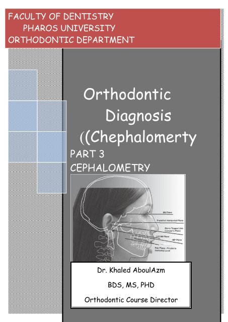
FACULTY OF DENTISTRY<br />
PHAROS UNIVERSITY<br />
ORTHODONTIC DEPARTMENT<br />
<strong>Orthodontic</strong><br />
<strong>Diagnosis</strong><br />
)(<strong>Chephalomerty</strong><br />
PART 3<br />
CEPHALOMETRY<br />
Enjy<br />
Dr. Khaled AboulAzm<br />
BDS, MS, PHD<br />
<strong>Orthodontic</strong> Course Director<br />
Dr.Khaled AboulAzm 2010
<strong>Orthodontic</strong> <strong>Diagnosis</strong> (<strong>Chephalomerty</strong>)<br />
Dr.Khaled AboulAzm<br />
2010<br />
Cephalometric radiography is a standardized<br />
method of production of skull radiographs,<br />
which are useful <strong>in</strong> mak<strong>in</strong>g measurements of<br />
the cranium and the orofacial complex.<br />
The radiograph thus obta<strong>in</strong>ed is called a<br />
cephalogram.<br />
Posterior-Anterior (Frontal view)<br />
Assessment of transverse conditions<br />
(assymetry)<br />
WORD DERIVATION<br />
• Cephalo=Head<br />
• Metric=Measurement<br />
Cephalometrics <strong>in</strong>troduced for use by<br />
orthodontists by Dr. Broadbent <strong>in</strong> 1931<br />
Orig<strong>in</strong>al purpose: Research on growth<br />
patterns of the craniofacial complex. Soon<br />
thereafter, it was shown that they could<br />
also be used to evaluate dentofacial<br />
proportions and clarify the anatomic basis<br />
for a malocclusion<br />
Purpose of Cephalometrics<br />
Views<br />
Lateral (Profile view)<br />
Assessment of A-P & Vertical conditions<br />
(prognatism, vertical growth)<br />
A. An aid to diagnosis<br />
<strong>Diagnosis</strong> of craniofacial deformity help <strong>in</strong><br />
identify<strong>in</strong>g, locat<strong>in</strong>g and quantify<strong>in</strong>g the<br />
nature of the problem, the most important<br />
result be<strong>in</strong>g a differentiation between<br />
skeletal and dental malrelationships.<br />
B. Treatment plann<strong>in</strong>g<br />
By help<strong>in</strong>g <strong>in</strong> diagnosis and prediction of<br />
craniofacial morphology and future growth,<br />
cephalometries help <strong>in</strong> develop<strong>in</strong>g a clear<br />
treatment plan. Even prior to start<strong>in</strong>g<br />
orthodontic treatment an orthodontist can<br />
predict the f<strong>in</strong>al position of each tooth<br />
with<strong>in</strong> a given patient's craniofacial I<br />
skeleton to achieve aesthetic and more<br />
stable results.<br />
2
<strong>Orthodontic</strong> <strong>Diagnosis</strong> (<strong>Chephalomerty</strong>)<br />
Dr.Khaled AboulAzm<br />
2010<br />
C. Evaluation of treated cases<br />
Serial cephalograms permit the orthodontist<br />
to evaluate and assess the progress of<br />
treatment and also helps <strong>in</strong> guid<strong>in</strong>g any<br />
desired change.<br />
D. Study of craniofacial growth<br />
Serial cephalogram studies have helped <strong>in</strong><br />
provid<strong>in</strong>g <strong>in</strong>formation regard<strong>in</strong>g<br />
• The various growth patterns.<br />
• The formation of standards, aga<strong>in</strong>st which<br />
other cephalograms can be compare<br />
E. Medico-legal documentation<br />
OBTAINING THE CEPHALOGRAM<br />
Cephalometric apparatus consists of head<br />
hold<strong>in</strong>g device (cephalostat). An x-ray<br />
source and a cassette holder.<br />
3
<strong>Orthodontic</strong> <strong>Diagnosis</strong> (<strong>Chephalomerty</strong>)<br />
Dr.Khaled AboulAzm<br />
2010<br />
What Are We Try<strong>in</strong>g to<br />
Accomplish?<br />
F. F<strong>in</strong>d out skeletal classification<br />
Antroposterior<br />
Is the patient class I,II,III skeletal<br />
Vertical<br />
Does the patient have a skeletal open bite<br />
growth pattern, or a deep bite growth<br />
pattern, or a normal growth<br />
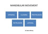
G. F<strong>in</strong>d out angulation of <strong>in</strong>cisors<br />
Are the maxillary/mandibular<br />
<strong>in</strong>cisors procl<strong>in</strong>ed, retrocl<strong>in</strong>ed or<br />
normal<br />
H. Consider soft tissue<br />
Facial profile<br />
Are the lips protrusive, retrusive,<br />
or normal<br />
COMMONLY USED<br />
CEPHALOMETRIC LANDMARKS<br />
• Nasion (N): junction of nasal and frontal<br />
bones<br />
Orbitale (O): lowest po<strong>in</strong>t on lower marg<strong>in</strong><br />
of orbit<br />
Po<strong>in</strong>t A (Subsp<strong>in</strong>ale) : most concave portion<br />
of premaxilla<br />
Po<strong>in</strong>t B (Supramentale) : most concave<br />
portion of symphysis<br />
Pogonion (Pog): most anterior portion of<br />
symphysis<br />
Menton (M): lowermost portion of<br />
symphysis<br />
Gnathion (Gn): most anterior <strong>in</strong>ferior<br />
portion of symphysis<br />
Gonion (Go): most <strong>in</strong>ferior posterior portion<br />
of the angle of the mandible<br />
COMMONLY USED PLANES IN<br />
CEPHALOMETRICS<br />
Frankfort Horizontal Plane (FH)<br />
• Extends from the upper border of the<br />
external auditory canal to the upper<br />
border of the lower orbital rim<br />
• Porion (Po): superior po<strong>in</strong>t of external<br />
auditory meatus<br />
Sella Turcica (S): midpo<strong>in</strong>t of the pituitary<br />
fossa<br />
4
<strong>Orthodontic</strong> <strong>Diagnosis</strong> (<strong>Chephalomerty</strong>)<br />
Dr.Khaled AboulAzm<br />
2010<br />
Porion-Orbitale<br />
Mandibular Plane<br />
L<strong>in</strong>e drawn from Gnathion (Gn) to<br />
Gonion (Go)<br />
Sella-Nasion Plane (SN)<br />
5
<strong>Orthodontic</strong> <strong>Diagnosis</strong> (<strong>Chephalomerty</strong>)<br />
Dr.Khaled AboulAzm<br />
2010<br />
Cephalometric Analysis<br />
I.Skeletal relation<br />
A. Antroposterior relation<br />
1. SNA<br />
Evaluates A-P position of the maxilla<br />
relative to the cranial base<br />
Norm=82 degrees<br />
• >82 deg=maxillary protrusion<br />
• 79 deg=mandibular protrusion<br />
• 3 deg=skeletal Cl II tendency<br />
Therefore the outcomes of the skeletal<br />
antroposterior analysis are to evaluate<br />
skeletal patterns (bone-to-bone relations)<br />
• Does the patient have a Class I, Class<br />
II or Class III skeletal pattern?<br />
• Is the problem due to a prognathic<br />
maxilla, a retrognathic maxilla, a<br />
prognathic mandible, a retrognathic<br />
mandible or a comb<strong>in</strong>ation of these?<br />
•
<strong>Orthodontic</strong> <strong>Diagnosis</strong> (<strong>Chephalomerty</strong>)<br />
Dr.Khaled AboulAzm<br />
2010<br />
• Mean 61°<br />
B. Vertical relation<br />
1. FrankFurt Mandibular plane angle (FMA)<br />
• Angle formed by mandibular plane and<br />
Frankfurt plane<br />
• Increase angle –vertical growth<br />
pattern( long face tendency)<br />
• Decreased angle –horizontal growth<br />
pattern( deep bite)<br />
• (Avrage 25°)<br />
• Increase FMA-high anlgle case<br />
(vertical growers)<br />
2. Y(growth) Axis<br />
• Anlgle by Y axis( S-Gn) and FH plane<br />
• Indicate the type of growth of the<br />
mandible<br />
7
<strong>Orthodontic</strong> <strong>Diagnosis</strong> (<strong>Chephalomerty</strong>)<br />
Dr.Khaled AboulAzm<br />
2010<br />
Dental analysis<br />
1. Upper <strong>in</strong>cisor ( 1:SN )<br />
• Increased –procl<strong>in</strong>ed upper <strong>in</strong>cisors<br />
• Decresed –retrocl<strong>in</strong>ed upper <strong>in</strong>cisors<br />
2. lower <strong>in</strong>cisor( 1:SN )<br />
• Increased –procl<strong>in</strong>ed lower <strong>in</strong>cisors<br />
• Decresed –retrocl<strong>in</strong>ed lower <strong>in</strong>cisors<br />
3. Inter<strong>in</strong>cisal angle<br />
Soft tissue analysis<br />
Esthetic Plane (E-plane)<br />
• Tip of nose to tip of soft tissue ch<strong>in</strong><br />
• In balance face<br />
• Lower lip should fall 0-4 mm beh<strong>in</strong>d<br />
l<strong>in</strong>e<br />
• Upper lip a little further posterior to<br />
the l<strong>in</strong>e.<br />
• Angle formed by the long axis of<br />
upper and lower <strong>in</strong>cisor<br />
• Increased –retruded <strong>in</strong>cisors<br />
• decreased –protruded <strong>in</strong>cisors<br />
8