Hemolytic Disease of the Newborn
Hemolytic Disease of the Newborn
Hemolytic Disease of the Newborn

Create successful ePaper yourself
Turn your PDF publications into a flip-book with our unique Google optimized e-Paper software.
Intensive Care Nursery House Staff Manual<br />
<strong>Hemolytic</strong> <strong>Disease</strong> <strong>of</strong> <strong>the</strong> <strong>Newborn</strong><br />
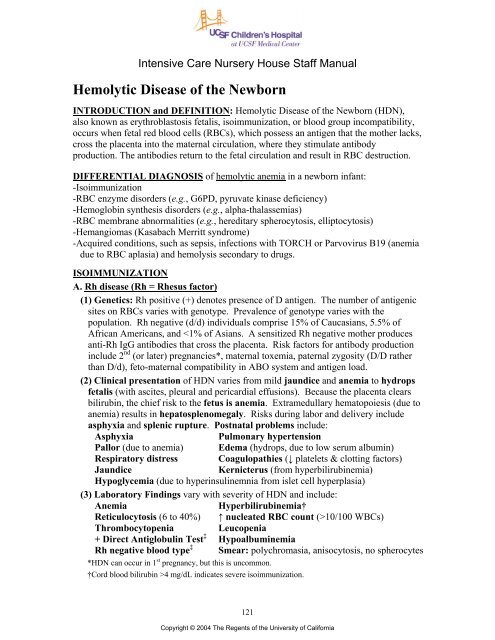
INTRODUCTION and DEFINITION: <strong>Hemolytic</strong> <strong>Disease</strong> <strong>of</strong> <strong>the</strong> <strong>Newborn</strong> (HDN),<br />
also known as erythroblastosis fetalis, isoimmunization, or blood group incompatibility,<br />
occurs when fetal red blood cells (RBCs), which possess an antigen that <strong>the</strong> mo<strong>the</strong>r lacks,<br />
cross <strong>the</strong> placenta into <strong>the</strong> maternal circulation, where <strong>the</strong>y stimulate antibody<br />
production. The antibodies return to <strong>the</strong> fetal circulation and result in RBC destruction.<br />
DIFFERENTIAL DIAGNOSIS <strong>of</strong> hemolytic anemia in a newborn infant:<br />
-Isoimmunization<br />
-RBC enzyme disorders (e.g., G6PD, pyruvate kinase deficiency)<br />
-Hemoglobin syn<strong>the</strong>sis disorders (e.g., alpha-thalassemias)<br />
-RBC membrane abnormalities (e.g., hereditary spherocytosis, elliptocytosis)<br />
-Hemangiomas (Kasabach Merritt syndrome)<br />
-Acquired conditions, such as sepsis, infections with TORCH or Parvovirus B19 (anemia<br />
due to RBC aplasia) and hemolysis secondary to drugs.<br />
ISOIMMUNIZATION<br />
A. Rh disease (Rh = Rhesus factor)<br />
(1) Genetics: Rh positive (+) denotes presence <strong>of</strong> D antigen. The number <strong>of</strong> antigenic<br />
sites on RBCs varies with genotype. Prevalence <strong>of</strong> genotype varies with <strong>the</strong><br />
population. Rh negative (d/d) individuals comprise 15% <strong>of</strong> Caucasians, 5.5% <strong>of</strong><br />
African Americans, and 10/100 WBCs)<br />
Thrombocytopenia Leucopenia<br />
+ Direct Antiglobulin Test ‡ Hypoalbuminemia<br />
Rh negative blood type ‡ Smear: polychromasia, anisocytosis, no spherocytes<br />
*HDN can occur in 1 st pregnancy, but this is uncommon.<br />
†Cord blood bilirubin >4 mg/dL indicates severe isoimmunization.<br />
121<br />
Copyright © 2004 The Regents <strong>of</strong> <strong>the</strong> University <strong>of</strong> California
<strong>Hemolytic</strong> <strong>Disease</strong> <strong>of</strong> <strong>the</strong> <strong>Newborn</strong><br />
‡ With severe HDN, high quantities <strong>of</strong> antibody may block Rh antigen site resulting in an Rh+ infant<br />
typing as Rh- and having a negative Direct Antiglobulin test.<br />
(4) Intra-uterine Transfusion (IUT): When iso-immunization is severe, IUTs are<br />
given to <strong>the</strong> fetus to prevent hydrops fetalis and fetal death. After multiple IUTs,<br />
most <strong>of</strong> <strong>the</strong> baby’s blood will be Rh negative donor blood. Therefore, <strong>the</strong> Direct<br />
Antiglobulin test will be negative, but <strong>the</strong> Indirect Antiglobulin Test will be positive.<br />
After IUTs, <strong>the</strong> cord bilirubin is not an accurate indicator <strong>of</strong> rate <strong>of</strong> hemolysis or <strong>of</strong><br />
<strong>the</strong> likelihood <strong>of</strong> <strong>the</strong> need for post-natal exchange transfusion.<br />
B. Minor Blood Group Incompatibility is uncommon, occurs in ~0.8% <strong>of</strong> pregnant<br />
women and usually with E, c, Kell, Kidd or Duffy. Clinical presentation is similar to<br />
Rh disease. Anti-Kell disease may be severe due to hemolysis or erythroid<br />
suppression. Lewis antigen stimulates only IgM production, so maternal antibody<br />
screen may be positive, but fetus is not affected.<br />
C. ABO Incompatibility<br />
(1) Genetics: With maternal blood types A and B, isoimmunization does not occur<br />
because <strong>the</strong> naturally occurring antibodies (anti-A and -B) are IgM, not IgG. In type<br />
O mo<strong>the</strong>rs, <strong>the</strong> antibodies are predominantly IgG, cross <strong>the</strong> placenta and can cause<br />
hemolysis in <strong>the</strong> fetus. The association <strong>of</strong> a type A or B fetus with a type O mo<strong>the</strong>r<br />
occurs in ~15% <strong>of</strong> pregnancies. However, HDN occurs in only 3%, is severe in only<br />
1%, and
<strong>Hemolytic</strong> <strong>Disease</strong> <strong>of</strong> <strong>the</strong> <strong>Newborn</strong><br />
-If <strong>the</strong> infant is hydropic, intubate immediately and begin assisted ventilation with<br />
oxygen. If ventilation is difficult, drain pleural and ascitic fluid; during<br />
paracentesis, take care to avoid puncturing <strong>the</strong> enlarged liver and spleen.<br />
-Insert umbilical arterial (UAC) and venous ca<strong>the</strong>ters (UVC) (See section on<br />
Umbilical Ca<strong>the</strong>ters, P. 25) and immediately measure blood pressures, arterial<br />
pH and blood gas tensions, hematocrit (Hct) and blood sugar.<br />
-Correct metabolic acidosis with alkali, but only if giving assisted ventilation (see<br />
section on Acid Base Balance, P. 62).<br />
-Correct anemia, which is essential for effective resuscitation.<br />
•If arterial blood pressure is low, give simple transfusion <strong>of</strong> packed RBCs<br />
(e.g., for Hct <strong>of</strong> 30%, push 10 mL/kg over 5 min; for Hct <strong>of</strong> 20%, push 10<br />
mL/kg over 5 min, <strong>the</strong>n repeat).<br />
•Do not infuse packed RBCs or blood through UAC because <strong>of</strong> risk <strong>of</strong><br />
damage to spinal cord from emboli.<br />
•With normal blood pressure, elevated central venous pressure, metabolic<br />
acidosis or hydrops, correct anemia by partial ExTx (exchange 20 mL/kg,<br />
<strong>the</strong>n repeat hematocrit).<br />
-Measure blood sugar frequently and correct hypoglycemia.<br />
-Follow platelet counts; consider platelet transfusion for counts 20 mg/dL in a term infant with HDN).<br />
Kernicterus occurs at lower levels <strong>of</strong> bilirubin in <strong>the</strong> presence <strong>of</strong> acidosis,<br />
hypoalbuminemia, prematurity and certain drugs (e.g., sulfonamides).<br />
-Measure bilirubin in cord blood and at least q4h for <strong>the</strong> first 12 to 24h. Plot<br />
bilirubin concentrations over time. A graph for plotting bilirubin over time is<br />
available at <strong>the</strong> unit clerk’s desk.<br />
-Begin photo<strong>the</strong>rapy shortly after birth. Although photo<strong>the</strong>rapy may not eliminate<br />
<strong>the</strong> need for ExTx, it may delay ExTx and decrease <strong>the</strong> number required.<br />
-Use ExTx for hyperbilirubinemia not controlled by photo<strong>the</strong>rapy (see P. 42).<br />
•Indications depend upon absolute serum concentration <strong>of</strong> bilirubin, <strong>the</strong> rate <strong>of</strong><br />
rise <strong>of</strong> bilirubin, gestational age, albumin concentration and acid-base status. In<br />
general, perform ExTx for cord bilirubin >5 mg/dL, for a rate <strong>of</strong> rise <strong>of</strong> bilirubin<br />
>0.7 mg/h, and to prevent bilirubin >20 mg/dL in a term infant, and lower levels<br />
in preterm infants (e.g., maintain serum bilirubin
<strong>Hemolytic</strong> <strong>Disease</strong> <strong>of</strong> <strong>the</strong> <strong>Newborn</strong><br />
infant’s blood volume (e.g., 5 mL/kg). The blood should be warmed and <strong>the</strong><br />
bag agitated every few minutes (to prevent settling <strong>of</strong> <strong>the</strong> RBCs).<br />
•Complications <strong>of</strong> ExTx:<br />
-Hypocalcemia due to Ca ++ binding by citrate. Give Ca-gluconate 100 mg<br />
after every 100 mL <strong>of</strong> blood exchanged.<br />
-Hypoglycemia, particularly after <strong>the</strong> ExTx, due to dextrose load from anticoagulant<br />
<strong>of</strong> donor blood and hyperinsulinism in HDN.<br />
-Thrombocytopenia and granulocytopenia due to washout with <strong>the</strong> ExTx.<br />
-Hyperkalemia, especially with older units <strong>of</strong> blood.<br />
-Hypo<strong>the</strong>rmia, associated with inadequate warming <strong>of</strong> blood.<br />
OUTCOME:<br />
(A) Late anemia: Antibodies persist for weeks, cause continued hemolysis and can cause<br />
anemia as late as age 6 months (especially in infants who had received IUTs). After<br />
discharge, follow Hct weekly. Erythropoietin treatment will help prevent severe<br />
anemia and fur<strong>the</strong>r transfusions (see P. 111).<br />
(B) Neurological prognosis is good. Commonest problem is sensorineural hearing loss.<br />
124<br />
Copyright © 2004 The Regents <strong>of</strong> <strong>the</strong> University <strong>of</strong> California