HEPATITS C HCV IgG - ELISA kits - Rapid tests
HEPATITS C HCV IgG - ELISA kits - Rapid tests
HEPATITS C HCV IgG - ELISA kits - Rapid tests

You also want an ePaper? Increase the reach of your titles
YUMPU automatically turns print PDFs into web optimized ePapers that Google loves.
DIAGNOSTIC AUTOMATION, INC.<br />
23961 Craftsman Road, Suite D/E/F, Calabasas, CA 91302<br />
Tel: (818) 591-3030 Fax: (818) 591-8383<br />
onestep@rapidtest.com<br />
technicalsupport@rapidtest.com<br />
www.rapidtest.com<br />
See external label 2°C-8°C Σ=96 <strong>tests</strong> Cat # 1709-12<br />
<strong>HEPATITS</strong> C<br />
<strong>HCV</strong> <strong>IgG</strong><br />
<strong>IgG</strong> ANTIBODIES TO HEPATITIS C VIRUS<br />
(<strong>HCV</strong> <strong>IgG</strong>) <strong>ELISA</strong> KIT<br />
Two-Step Incubation, Indirect Principle<br />
Cat # 1709-12<br />
INTENDED USE<br />
This kit is an enzyme-linked immunosorbent assay for qualitative detection of <strong>IgG</strong>-class antibodies to<br />
hepatitis C virus in human serum or plasma. It is intended for clinical diagnosing and management of<br />
patients related to infection with hepatitis C virus and for serological investigation studies of <strong>HCV</strong><br />
antibody prevalence.<br />
SUMMARY<br />
Hepatitis C virus (<strong>HCV</strong>) is an envelope, single stranded positive sense RNA (9.5 kb) virus belonging to<br />
the family of Flaviviridae. <strong>HCV</strong> is now recognized as the major cause for transfusion associated non-A,<br />
non-B hepatitis. The disease is characterized with acute and chronic form although more than 50% of<br />
the infected individuals develop severe, life threatening chronic hepatitis with liver cirrhosis and<br />
hepatocellular carcinomas. Sixty to seventy percent of <strong>HCV</strong> infected individuals have no noticeable<br />
symptoms. Jaundice (yellowing of skin and whites of the eyes) may be present in 20 – 30% of <strong>HCV</strong><br />
infected individuals, while another 10 – 20% may exhibit non-specific symptoms (e.g., anorexia,<br />
malaise, or abdominal pain). When symptoms are present, the average time period after exposure is 6<br />
to 7 weeks. The average time period from exposure to seroconversion is 8 to 9 weeks.<br />
The serological detection of <strong>HCV</strong> <strong>IgG</strong> antibodies is important clinical marker for diagnosis and<br />
management of <strong>HCV</strong> infected individuals and serological studies. Usually the antibody is detectable for<br />
life.<br />
DAI Code # 12 1
This <strong>HCV</strong> <strong>IgG</strong> <strong>ELISA</strong> belongs to the third generation <strong>HCV</strong> assays which have improved sensitivity and<br />
shorten the time between infection with <strong>HCV</strong> and the appearance of detectable antibodies (window<br />
period) to 60 days.<br />
Fig. 1 The serological and viral responses during <strong>HCV</strong> infection<br />
PRINCIPLE OF THE ASSAY<br />
This kit employs solid phase, indirect <strong>ELISA</strong> method for detection of <strong>IgG</strong>-class antibodies to <strong>HCV</strong> in<br />
two-step incubation procedure. Polystyrene microwell strips are pre-coated with recombinant, highly<br />
immunoreactive antigens corresponding to the core and non-structural regions of <strong>HCV</strong> (third generation<br />
<strong>HCV</strong> <strong>ELISA</strong>). During the first incubation step, <strong>HCV</strong> <strong>IgG</strong> specific antibodies, if present, will be bound to<br />
the solid phase pre-coated <strong>HCV</strong> antigens. The wells are washed to remove unbound serum proteins,<br />
and than, rabbit anti-human <strong>IgG</strong> antibodies (anti-<strong>IgG</strong>) conjugated to horseradish peroxidase (HRP-<br />
Conjugate) are added. During the second incubation step, these HRP-conjugated antibodies will be<br />
bound to any antigen-antibody complexes previously formed and the unbound HRP-conjugate is then<br />
removed by washing. Chromogen solutions containing Tetramethylbenzidine (TMB) and urea peroxide<br />
are added to the wells and in presence of the antigen-antibody-anti-<strong>IgG</strong> (HRP) immunocomplex, the<br />
colorless Chromogens are hydrolyzed by the bound HRP-conjugate to a blue-colored product. The blue<br />
color turns yellow after stopping the reaction with sulfuric acid. The amount of color intensity can be<br />
measured and it is proportional to the amount of antibody captured in the wells, and to the amount of<br />
antibody in the sample respectively. Wells containing samples negative for <strong>HCV</strong> <strong>IgG</strong> remain colorless.<br />
Assay principle scheme: Indirect <strong>ELISA</strong><br />
Ag(p)+Ab(s)→[Ag(p)–Ab(<strong>IgG</strong>)+ENZ]→[Ag(p)–Ab(<strong>IgG</strong>)–ENZ]→blue→yellow (+)<br />
Ag(p)+ → [Ag(p) +ENZ]→ [Ag(p) ] → no color (-)<br />
Incubation1 Incubation 2 Immobilized Complex Coloring Results<br />
30min. 30min. 15min<br />
DAI Code # 12 2
Ag(p)–pre-coated <strong>HCV</strong> antigens(core,NS3/4,NS5);<br />
Ab(<strong>IgG</strong>)–<strong>HCV</strong> antibodies in sample (<strong>IgG</strong>);<br />
ENZ–HRP conjugated rabbit anti-human <strong>IgG</strong>;<br />
COMPONENTS<br />
96 Tests:<br />
• MICROWELL PLATE 1plate<br />
Blank microwell strips fixed on white strip holder.<br />
The plate is sealed in aluminium pouch with desiccant.<br />
8×12/12×8-well strips per plate.<br />
Each well contains recombinant <strong>HCV</strong> antigens. The<br />
microwell strips can be broken to be used separately.<br />
Place unused wells or strips in the plastic sealable storage bag<br />
together with the desiccant and return to 2~8°C.<br />
• NEGATIVE CONTROL 1vial<br />
Blue-colored liquid filled in a vial with green screw cap<br />
0.2ml per vial.<br />
Protein-stabilized buffer tested non-reactive for <strong>HCV</strong> <strong>IgG</strong> antibodies.<br />
Preservatives: 0.1% ProClin 300.<br />
Ready to use as supplied.<br />
Once open, stable for one month at 2-8°C.<br />
• POSITIVE CONTROL SERUM 1vial<br />
Red-colored liquid filled in a vial with red screw cap<br />
0.2ml per vial.<br />
<strong>HCV</strong> <strong>IgG</strong> antibodies diluted in protein-stabilized buffer<br />
Preservatives: 0.1% ProClin 300.<br />
Ready to use as supplied.<br />
Once open, stable for one month at 2-8°C.<br />
• SPECIMEN DILUENT 1vial<br />
Blue-colored liquid filled in a white vial with blue screw cap.<br />
13ml per vial.<br />
Protein-stabilized buffer, casein, and sucrose solution.<br />
Ready to use as supplied.<br />
Once open, stable for one month at 2-8°C.<br />
• HRP-CONJUGATE REAGENT 1vial<br />
Red-colored liquid filled in a white vial with red screw cap.<br />
13ml per vial.<br />
Horseradish peroxidase-conjugated rabbit anti-human <strong>IgG</strong> antibodies.<br />
Ready to use as supplied.<br />
Once open, stable for one month at 2-8°C.<br />
• STOCK WASH BUFFER 1bottle<br />
Colorless liquid filled in a clear bottle with white screw cap.<br />
50ml per bottle.<br />
PH 7.4, 20 × PBS (Contains Tween-20 as a detergent)<br />
DILUTE BEFORE USE-The concentrate must be diluted 1 to 20<br />
with distilled/deionized water before use.<br />
Once diluted, stable for one week at room temperature or for two<br />
weeks at 2-8°C.<br />
• CHROMOGEN SOLUTION A 1vial<br />
Colorless liquid filled in a white vial with green screw cap.<br />
DAI Code # 12 3
8ml per vial.<br />
Urea peroxide solution.<br />
Ready to use as supplied.<br />
Once open, stable for one month at 2-8°C.<br />
• CHROMOGEN SOLUTION B 1vial<br />
Colorless liquid filled in a black vial with black screw cap.<br />
8ml per vial.<br />
TMB solution( Tetramethylbenzidine dissolved in citric acid).<br />
Ready to use as supplied.<br />
Once open, stable for one month at 2-8°C.<br />
• STOP SOLUTION 1vial<br />
Colorless liquid filled in a white vial with yellow screw cap.<br />
8ml per vial.<br />
Diluted sulfuric acid solution (2.0M H 2 SO 4 ).<br />
Ready to use as supplied.<br />
• PLASTIC SEALABLE BAG 1unit<br />
For enclosing the strips not in use.<br />
• CARDBOARD PLATE COVER 2sheets<br />
To cover the plates during incubation and prevent evaporation<br />
or contamination of the wells.<br />
• PACKAGE INSERTS 1copy<br />
ADDITIONAL MATERIALS AND INSTRUMENTS REQUIRED BUT<br />
NOT PROVIDED<br />
- Freshly distilled or deionized water.<br />
- Disposable gloves and timer.<br />
- Appropriate waste containers for potentially contaminated materials.<br />
- Disposable V-shaped troughs.<br />
- Dispensing system and/or pipette (single or multichannel), disposable pipette tips.<br />
- Absorbent tissue or clean towel.<br />
- Dry incubator or water bath, 37±0.5°C.<br />
- Microshaker for dissolving and mixing conjugate with samples.<br />
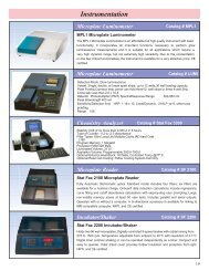
- Microwell plate reader, single wavelength 450nm or dual wavelength 450nm and 630nm.<br />
- Microwell aspiration/wash system.<br />
SPECIMEN COLLECTION, TRANSPORTATION AND STORAGE<br />
1. Sample Collection: Either fresh serum or plasma samples can be used for this assay. Blood<br />
collected by venipuncture should be allowed to clot naturally and completely – the serum/plasma<br />
must be separated from the clot as early as possible as to avoid hemolysis of the RBC. Care should<br />
be taken to ensure that the serum samples are clear and not contaminated by microorganisms. Any<br />
visible particulate matters in the sample should be removed by centrifugation at 3000 RPM for at<br />
least 20 minutes at room temperature, or by filtration on 0.22u filters. Plasma samples collected into<br />
EDTA, sodium citrate or heparin may be tested, but highly lipaemic, icteric, or hemolized samples<br />
should not be used as they could give erroneous results in the assay. Do not heat inactivate<br />
samples. This can cause sample deterioration.<br />
2. Transportation and Storage: Store samples at 2-8°C. Samples not required for assaying within 3<br />
days should be stored frozen (-20°C or lower).Multiple freeze-thaw cycles should be avoided. For<br />
shipment, samples should be packaged and labeled in accordance with the existing local and<br />
international regulations for transport of clinical samples and ethological agents.<br />
DAI Code # 12 4
SPECIAL INSTRUCTIONS FOR WASHING<br />
1. A good washing procedure is essential to obtain correct and precise analytical data.<br />
2. It is therefore recommended to use a good quality <strong>ELISA</strong> microplate washer, maintained at the best<br />
level of washing performances. In general, no less than 5 automatic washing cycles with dispensing<br />
of 350-400µl/well, are sufficient to avoid false positive reactions and high background (all wells turn<br />
yellow).<br />
2. To avoid cross-contaminations of the plate with sample or HRP-conjugate, after incubation do not<br />
discard the content of the wells, but allow the plate washer to aspirate it automatically.<br />
3. Anyway, we recommend calibrating the washing system on the kit itself in order to match the<br />
declared analytical performances. Assure that the microplate washer’s liquid dispensing channels<br />
are not blocked or contaminated, and sufficient volume of Wash buffer is dispensed each time into<br />
the wells.<br />
5. In case of manual washing, we suggest to perform at least 5cycles, dispensing 350-400µl/well and<br />
aspirating the liquid for 5times. If poor results (high background) are observed, increase the<br />
washing cycles or soaking time per well.<br />
6. In any case, the liquid aspirated out the strips should be treated with a sodium hypochlorite<br />
solution(final concentration of 2.5%) for 24 hours, before liquids are disposed in an appropriate way.<br />
The concentrated Washing solution should be diluted 1 to 20 before use. For one plate, mix 50 ml<br />
of the concentrate with 950ml of water for a final volume of 1000ml diluted Wash Buffer. If less than<br />
a whole plate is used, prepare the proportional volume of solution.<br />
STORAGE AND STABILITY<br />
The components of the kit will remain stable through the expiration date indicated on the label and<br />
package when stored between 2-8 °C; do not freeze. To assure maximum performance of this <strong>HCV</strong><br />
<strong>IgG</strong> <strong>ELISA</strong> kit, during storage protect the reagents from contamination with microorganism or<br />
chemicals.<br />
PRECAUTIONS AND SAFETY<br />
This kit is intended FOR IN VITRO USE ONLY<br />
FOR PROFESSIONAL USE ONLY<br />
<strong>ELISA</strong> is a time and temperature sensitive method. To avoid incorrect result, strictly follow the test<br />
procedure steps and do not modify them.<br />
1. Do not exchange reagents from different lots, or use reagents from other commercially available<br />
<strong>kits</strong>. The components of the kit are precisely matched as to achieve optimal performance during<br />
testing.<br />
2. Make sure that all reagents are within the validity indicated on the kit box and are of the same lot.<br />
Never use reagents beyond the expiry date stated on reagents labels or on the kit box.<br />
3. CAUTION - CRITICAL STEP: Allow the reagents and samples to reach room temperature<br />
(18-30°C) before use. Shake reagent gently before, and return to 2-8°C immediately after use.<br />
4. Use only sufficient volume of sample as indicated in the procedure steps. Failure to do so, may<br />
cause in low sensitivity of the assay.<br />
5. Do not touch the bottom exterior of the wells; fingerprints or scratches may interfere with microwell<br />
reading.<br />
DAI Code # 12 5
6. When reading the results, ensure that the plate bottom is dry and there are no air-bubbles inside<br />
the wells.<br />
7. Never allow the microplate wells to dry after the washing step. Immediately proceed to the next<br />
step. Avoid the formation of air-bubbles when adding the reagents.<br />
8. Avoid assay steps long time interruptions. Assure same working conditions for all wells.<br />
9. Calibrate the pipette frequently to assure the accuracy of samples/reagents dispensing. Always use<br />
different disposal pipette tips for each specimen and reagents as to avoid cross-contaminations.<br />
Never pipette solutions by mouth.<br />
10. The use of automatic pipettes is recommended.<br />
11. Assure that the incubation temperature is 37°C inside the incubator.<br />
12. When adding samples, avoid touching the well’s bottom with the pipette tip.<br />
13. When reading the results with a plate reader, it is recommended to determine the absorbance at<br />
450nm or at 450nm with reference at 630nm.<br />
14. All specimens from human origin should be considered as potentially infectious.<br />
15. Materials from human origin may have been used in the kit. These materials have been tested with<br />
<strong>tests</strong> <strong>kits</strong> with accepted performance and found negative for antibodies to HIV ½, <strong>HCV</strong>, TP and<br />
HBsAg. However, there is no analytical method that can assure that infectious agents in the<br />
specimens or reagents are completely absent. Therefore, handle reagents and specimens with<br />
extreme caution as if capable of transmitting infectious diseases. Strict adherence to GLP (Good<br />
Laboratory Practice) regulations can ensure the personal safety. Never eat, drink, smoke, or apply<br />
cosmetics in the assay laboratory.<br />
16. Bovine derived sera may have been used in this kit. Bovine serum albumin (BSA) and fetal calf sera<br />
(FCS) are derived from animals from BSE/TSE free-geographical areas.<br />
17. The pipette tips, vials, strips and sample containers should be collected and autoclaved for 1hour at<br />
121°C or treated with 10% sodium hypochlorite for 30minutes to decontaminate before any further<br />
steps for disposal.<br />
18. The Stop solution (2M H 2 SO 4 ) is a strong acid. Corrosive. Use it with appropriate care. Wipe up<br />
spills immediately or wash with water if come into contact with the skin or eyes. ProClin 300 used<br />
as a preservative can cause sensation of the skin.<br />
19. The enzymatic activity of the HRP-conjugate might be affected from dust, reactive chemical, and<br />
substances like sodium hypochlorite, acids, alkalins etc. Do not perform the assay in the presence<br />
of such substances.<br />
20. Materials Safety Data Sheet (MSDS) available upon request.<br />
21. If using fully automated microplate processing system, during incubation, do not cover the plates<br />
with the plate cover. The tapping out of the remainders inside the plate after washing, can also be<br />
omitted.<br />
ASSAY PROCEDURE<br />
Step1 Reagents preparation: Allow the reagents to reach room temperature (18-30°C) for at least<br />
15-30minutes. Check the Wash Buffer concentrate for the presence of salt crystals. If crystals<br />
have formed in the solution, resolubilize by warming at 37°C until crystals dissolve. Dilute the<br />
stock wash Buffer 1 to 20 with distilled or deionized water. Use only clean vessels to dilute the<br />
Wash Buffer.<br />
Step2 Numbering Wells: Set the strips needed in strip-holder and number sufficient number of wells<br />
including three Negative control (e.g. B1, C1, D1), two Positive control (e.g. E1, F1) and one<br />
Blank (e.g. A1, neither samples nor HRP-Conjugate should be added into the Blank well). If the<br />
results will be determined by using dual wavelength plate reader, the requirement for use of<br />
Blank well could be omitted. Use only number of strips required for the test.<br />
Step3 Adding Diluent: Add 100µl Specimen Diluent into each well except the blank.<br />
Step4 Adding Sample: Add 10µl of Positive control, Negative control, and Specimen into their<br />
respective wells. Note: Use a separate disposal pipette tip for each specimen, Negative<br />
Control and Positive Control as to avoid cross-contamination. Mix by tapping the plate<br />
DAI Code # 12 6
gently.<br />
Step5 Incubating(1): Cover the plate with the plate cover and incubate for 30minutes at 37°C. It is<br />
recommended to use thermostat-controlled water tank to assure the temperature stability and<br />
humidity during the incubation. If dry incubator is used, do not open the door frequently.<br />
Step6 Washing(1): After the end of the incubation, remove and discard the plate cover. Wash each<br />
well 5times with diluted Wash buffer. Each time allow the microwells to soak for 30-60<br />
seconds. After the final washing cycle, turn the strips plate onto blotting paper or clean towel,<br />
and tap it to remove any remainders.<br />
Step7 Adding HRP-Conjugate: Add 100µl HRP-Conjugate to each well except the Blank.<br />
Step8 Incubating HRP-Conjugate(2): Cover the plate with the plate cover and incubate for<br />
30minutes at 37°C.<br />
Step9 Washing(2): After the end of the incubation, remove and discard the plate cover. Wash each<br />
well 5 times with diluted Wash buffer as in Step6.<br />
Step10 Coloring: Dispense 50µl of Chromogen A and 50µl Chromogen B solution into each well<br />
including the Blank and mix by tapping the plate gently. Incubate the plate at 37°C for 15<br />
minutes avoiding light. The enzymatic reaction between the Chromogen A/B solutions<br />
produces blue color in Positive control and <strong>HCV</strong> <strong>IgG</strong> positive sample wells.<br />
Step11 Stopping Reaction: Using a multichannel pipette or manually add 50µl Stop Solution into<br />
each well and mix by tapping the plate gently. Intensive yellow color develops in Positive<br />
control and <strong>HCV</strong> <strong>IgG</strong> positive sample wells.<br />
Step12 Measuring the Absorbance: Calibrate the plate reader with the Blank well and read the<br />
absorbance at 450nm. If a dual filter instrument is used, set the reference wavelength at<br />
630nm. Calculate the Cut-off value and evaluate the results (Note: read the absorbance within<br />
5 minutes after stopping the reaction).<br />
INTERPRETATION OF RESULTS AND QUALITY CONTROLS<br />
Each microplate must be considered separately when calculating and interpreting results of the assay,<br />
regardless of the number of plates concurrently processed. The results are calculated by relating each<br />
sample’s optical density (OD) value to the Cut-off value (C.O.) of the plate. If the Cut-off reading is<br />
based on Single filter plate reader, the results must be calculated by subtracting the Blank well OD<br />
value from the print report values of samples and controls. In case the reading is based on dual filter<br />
plate reader, do not subtract the Blank well OD from the print report values of samples and controls.<br />
1. Calculation of Cut-off value (C.O.) = *Nc + 0.12<br />
*Nc = the mean absorbance value for three negative controls.<br />
Important: If the mean OD value of the negative control is lower than 0.02, take it as 0.02.If<br />
higher than 0.02 see the Quality control range.<br />
Example:<br />
1. Calculation of Nc:<br />
Well No B1 C1 D1<br />
Negative control OD value 0.02 0.012 0.016<br />
Nc=0.016<br />
(The mean value is lower than 0.02, so take it as 0.02).<br />
2. Calculation of Cut-off (C.O.) = 0.02 + 0.12 = 0.140<br />
If one of the Negative control values does not meet the Quality<br />
Control Range specifications, it should be discarded and the mean value is calculated again using the<br />
remaining two values. If more than one negative control OD value does not meet the Quality Control<br />
Range specifications, the test is invalid and must be repeated.<br />
DAI Code # 12 7
2. Quality control range:<br />
The test results are valid if the Quality Control criteria are verified. It is recommended that each<br />
laboratory must establish appropriate quality control system with quality control material similar to or<br />
identical with the patient sample being analyzed.<br />
1. The OD value of the Blank well, which contains only Chromogens and Stop solution, is less than<br />
0.080 at 450 nm.<br />
2. The OD value of the Positive control must be equal to or greater than 0.800 at 450/630nm or at<br />
450nm after blanking.<br />
3. The OD value of the Negative control must be less than 0.100 at 450/630nm or at 450nm after<br />
blanking.<br />
3. Interpretations of the results:<br />
(S = the individual absorbance (OD) of each specimen)<br />
Negative Results (S/C.O.
Reproducibility Within run Between run<br />
Sample No Mean OD CV% Mean OD CV%<br />
Weak positive 10 0.436 9.1% 0.401 9.5%<br />
Moderate positive 10 0.946 7.0% 0.916 7.5%<br />
Strong positive 1 10 1.917 4.4% 1.895 4.2%<br />
Strong positive 2 10 2.372 3.8% 2.309 4.0%<br />
LIMITATIONS<br />
1. Non-repeatable positive result may occur due to the general biological characteristics of <strong>ELISA</strong><br />
method. The assay is designed to achieve very high performances of sensitivity and specificity and<br />
the “indirect model” minimizes the unspecific reactions, which can occur due to interference<br />
between unknown meters in sample and the rabbit anti-human <strong>IgG</strong> used as a conjugate. Antibodies<br />
may be undetectable during the early stages of the disease and in some immunosuppresed<br />
individuals.<br />
2. Positive results must be confirmed with another available method. Any positive result must be<br />
interpreted together with the patient clinical information and other laboratory results.<br />
3. Common sources for mistakes: <strong>kits</strong> beyond the expiry date, bad washing procedures, contaminated<br />
reagents, incorrect assay procedure steps, insufficient aspiration during washing, failure to add<br />
samples or reagents, equipment, timing, volumes, sample nature and quality.<br />
4. The prevalence of the marker will affect the assay’s predictive values.<br />
5. This kit is intended ONLY for testing of individual serum or plasma samples. Do not use it for testing<br />
of cadaver samples, saliva, urine or other body fluids, or pooled (mixed) blood.<br />
6. This is a qualitative assay and the results cannot be use to measure antibodies concentrations.<br />
VALIDITY<br />
Please do not use this kit beyond the expiration indicated on the kit box and reagent labels。<br />
REFERENCES:<br />
1. Alter HJ. (1978) You will wonder where the yellow went: A 15-year retrospective of posttransfusion<br />
hepatitis. In: Moore SB, ed. Transfusion-Transmitted Viral Diseases. Alington, VA. Am. Assoc. Blood<br />
Banks, pp. 53-38.<br />
2. Alter HJ., Purcell RH, Holland PV, et al. (1978) Transmissible agent in non-A, non-B hepatitis.<br />
Lancet I: 459-463.<br />
3. Choo Q-L,Weiner AJ, Overby LR, Kuo G, Houghton M. (1990) Hepatitis C Virus: the major causative<br />
agent of viral non-A, non-B hepatitis. Br Med Bull 46: 423-441.<br />
4. Engvall E, Perlmann P. (1971) Enzyme linked immunosorbent assay (<strong>ELISA</strong>): qualitative assay of<br />
<strong>IgG</strong>. Immunochemistry 8:871-874.<br />
5. Esaban JI, Viladomiu L, Gonzalez A, et al,. (1989) Hepatitis C virus antibodies among risk groups in<br />
Spain. Lancet ii: 294-297.<br />
6. I. Saito et al., (1990) Hepatitis C virus infection is associated with the development of hepatocellular<br />
carcinoma. Oroc. Natl. Acad. Sci., 87: 6547-65749.<br />
7. Kuhnl P, Seidi S, Stangel W et al. (1989) Antibodies to hepatitis C in German blood donors. Lancet<br />
ii: 324.<br />
8. M.R. Wick, S. Moore, H.F. Taswell (1985) Non-A, Non-B hepatitis associated with blood transfusion.<br />
DAI Code # 12 9
Transfusion, 25: 93-101.<br />
9. Van der Poel CM, Reesink HW, Schaasberg W, et al. (1990) Infectivity of blood seropositive for<br />
hepatitis C virus antibodies. Lancet 335: 558-560.<br />
SUMMARY OF THE ASSAY PROCEDURE:<br />
Add sample diluent 100µl<br />
Add sample 10µl<br />
Incubate<br />
30minutes<br />
Wash<br />
5times<br />
Add HRP-Conjugate 100µl<br />
Incubate<br />
30minutes<br />
Wash<br />
5times<br />
Coloring<br />
50µl A + 50µl B<br />
Incubate<br />
15minutes<br />
Stop the reaction<br />
50µl stop solution<br />
Read the absorbance 450nm or 450/630 nm<br />
SUMMARY OF THE MAJOR COMPONENTS OF THE KIT:<br />
Microwell plate<br />
One/ 96 wells<br />
Negative/Postive control One each/ 0.2ml<br />
Sample Diluent<br />
One/ 13ml<br />
HRP-Conjugate<br />
One/ 13ml<br />
Wash Buffer<br />
One/ 50ml<br />
Chromogen A/B/Stop Solution One each/ 8ml<br />
Note: the components of individual <strong>kits</strong> are not interchangeable<br />
Date Adopted Reference No.<br />
2008-01-05 DA-<strong>HCV</strong> <strong>IgG</strong>-2008<br />
DIAGNOSTIC AUTOMATION, INC.<br />
23961 Craftsman Road, Suite D/E/F, Calabasas, CA 91302<br />
Tel: (818) 591-3030 Fax: (818) 591-8383<br />
ISO 13485-2003<br />
Revision Date: 6/18/08<br />
DAI Code # 12 10