Guide to Converting ELISA to DELFIA - PerkinElmer
Guide to Converting ELISA to DELFIA - PerkinElmer
Guide to Converting ELISA to DELFIA - PerkinElmer

Create successful ePaper yourself
Turn your PDF publications into a flip-book with our unique Google optimized e-Paper software.
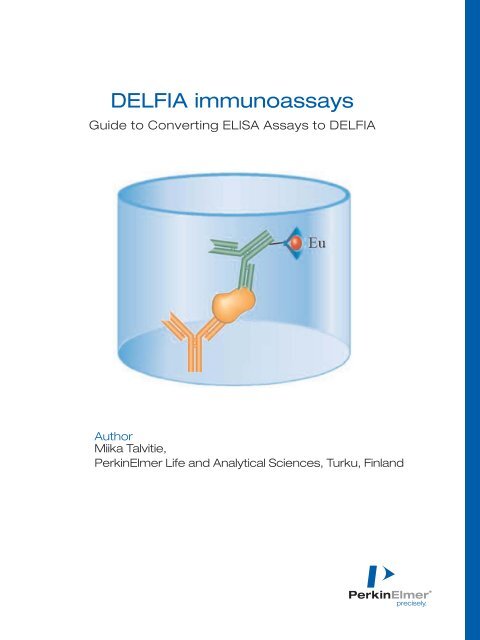
<strong>DELFIA</strong> immunoassays<br />
<strong>Guide</strong> <strong>to</strong> <strong>Converting</strong> <strong>ELISA</strong> Assays <strong>to</strong> <strong>DELFIA</strong><br />
Author<br />
Miika Talvitie,<br />
<strong>PerkinElmer</strong> Life and Analytical Sciences, Turku, Finland
Contents<br />
Contents . . . . . . . . . . . . . . . . . . . . . . . . . . . . . . . . . . . . . . . . . . . . . . . . . . . . . . . . . . . . . . . . . . . . . . . . . . . . . . . . . . . . . . .2<br />
1. Enhanced performance for your applications with <strong>DELFIA</strong> ® . . . . . . . . . . . . . . . . . . . . . . . . . . . . . . . . . . . . . . . . . . . . . . . . .3<br />
2. <strong>Converting</strong> <strong>ELISA</strong> assays <strong>to</strong> <strong>DELFIA</strong> . . . . . . . . . . . . . . . . . . . . . . . . . . . . . . . . . . . . . . . . . . . . . . . . . . . . . . . . . . . . . . . . . . .4<br />
2.1 Materials and methods . . . . . . . . . . . . . . . . . . . . . . . . . . . . . . . . . . . . . . . . . . . . . . . . . . . . . . . . . . . . . . . . . . . . . . . . . .5<br />
2.2 Results and discussion . . . . . . . . . . . . . . . . . . . . . . . . . . . . . . . . . . . . . . . . . . . . . . . . . . . . . . . . . . . . . . . . . . . . . . . . .6<br />
3. Optimizing <strong>DELFIA</strong> assays . . . . . . . . . . . . . . . . . . . . . . . . . . . . . . . . . . . . . . . . . . . . . . . . . . . . . . . . . . . . . . . . . . . . . . . . . . .7<br />
3.1 Microplates . . . . . . . . . . . . . . . . . . . . . . . . . . . . . . . . . . . . . . . . . . . . . . . . . . . . . . . . . . . . . . . . . . . . . . . . . . . . . . . . . . .7<br />
3.2 Capture antibody . . . . . . . . . . . . . . . . . . . . . . . . . . . . . . . . . . . . . . . . . . . . . . . . . . . . . . . . . . . . . . . . . . . . . . . . . . . . . .8<br />
3.2.1 Immobilizing the capture antibody . . . . . . . . . . . . . . . . . . . . . . . . . . . . . . . . . . . . . . . . . . . . . . . . . . . . . . . . . . . .8<br />
3.2.2 Selecting the capture antibody . . . . . . . . . . . . . . . . . . . . . . . . . . . . . . . . . . . . . . . . . . . . . . . . . . . . . . . . . . . . . .9<br />
3.2.3 Optimizing the amount of capture antibody . . . . . . . . . . . . . . . . . . . . . . . . . . . . . . . . . . . . . . . . . . . . . . . . . . .10<br />
3.3 Tracer . . . . . . . . . . . . . . . . . . . . . . . . . . . . . . . . . . . . . . . . . . . . . . . . . . . . . . . . . . . . . . . . . . . . . . . . . . . . . . . . . . . . . .10<br />
3.4 Optimizing assay conditions . . . . . . . . . . . . . . . . . . . . . . . . . . . . . . . . . . . . . . . . . . . . . . . . . . . . . . . . . . . . . . . . . . . . .11<br />
3.4.1 Suitable assay buffer . . . . . . . . . . . . . . . . . . . . . . . . . . . . . . . . . . . . . . . . . . . . . . . . . . . . . . . . . . . . . . . . . . . . .11<br />
3.4.2 Washing . . . . . . . . . . . . . . . . . . . . . . . . . . . . . . . . . . . . . . . . . . . . . . . . . . . . . . . . . . . . . . . . . . . . . . . . . . . . . . .11<br />
3.4.3 Shaking . . . . . . . . . . . . . . . . . . . . . . . . . . . . . . . . . . . . . . . . . . . . . . . . . . . . . . . . . . . . . . . . . . . . . . . . . . . . . . .11<br />
3.5 Sample matrix . . . . . . . . . . . . . . . . . . . . . . . . . . . . . . . . . . . . . . . . . . . . . . . . . . . . . . . . . . . . . . . . . . . . . . . . . . . . . . .12<br />
3.6 Detection . . . . . . . . . . . . . . . . . . . . . . . . . . . . . . . . . . . . . . . . . . . . . . . . . . . . . . . . . . . . . . . . . . . . . . . . . . . . . . . . . . .13<br />
3.6.1 Enhancement . . . . . . . . . . . . . . . . . . . . . . . . . . . . . . . . . . . . . . . . . . . . . . . . . . . . . . . . . . . . . . . . . . . . . . . . . . .13<br />
3.6.2 Detection Instruments . . . . . . . . . . . . . . . . . . . . . . . . . . . . . . . . . . . . . . . . . . . . . . . . . . . . . . . . . . . . . . . . . . . .14<br />
4. Dos and Don’ts . . . . . . . . . . . . . . . . . . . . . . . . . . . . . . . . . . . . . . . . . . . . . . . . . . . . . . . . . . . . . . . . . . . . . . . . . . . . . . . . . .14<br />
5. Troubleshooting . . . . . . . . . . . . . . . . . . . . . . . . . . . . . . . . . . . . . . . . . . . . . . . . . . . . . . . . . . . . . . . . . . . . . . . . . . . . . . . . . .15<br />
6. Supplemental material . . . . . . . . . . . . . . . . . . . . . . . . . . . . . . . . . . . . . . . . . . . . . . . . . . . . . . . . . . . . . . . . . . . . . . . . . . . . .16<br />
6.1 Assay reagents . . . . . . . . . . . . . . . . . . . . . . . . . . . . . . . . . . . . . . . . . . . . . . . . . . . . . . . . . . . . . . . . . . . . . . . . . . . . . . .16<br />
6.2 Additional information . . . . . . . . . . . . . . . . . . . . . . . . . . . . . . . . . . . . . . . . . . . . . . . . . . . . . . . . . . . . . . . . . . . . . . . . .16<br />
2
1. Enhanced performance for your<br />
applications with <strong>DELFIA</strong><br />
The first part of this guide presents a simple<br />
conversion of an existing TNF- (Tumor Necrosis<br />
Fac<strong>to</strong>r-) <strong>ELISA</strong> (Enzyme-Linked Immunosorbent<br />
Assay) <strong>to</strong> <strong>DELFIA</strong> (Dissociation Enhanced<br />
Lanthanide Fluoroimmunoassay). The following<br />
sections describe how <strong>to</strong> set up and further<br />
optimize <strong>DELFIA</strong> assays.<br />
<strong>DELFIA</strong><br />
Time-resolved fluorometry (TRF) is a well-established<br />
technique in drug discovery as well as in<br />
other research areas. Providing high sensitivity<br />
and wide dynamic range, the method is characterized<br />
by lack of sample interference during<br />
measurement and is easy <strong>to</strong> au<strong>to</strong>mate. The TRFbased<br />
<strong>DELFIA</strong> system from <strong>PerkinElmer</strong> is an<br />
optimal <strong>to</strong>ol for bio-affinity assays.<br />
<strong>DELFIA</strong> features and benefits:<br />
• Well-proven technology – published data in<br />
many application areas.<br />
• Very high sensitivity – minimizes reagent use<br />
and very sensitive <strong>to</strong> smaller analyte quantities.<br />
• Wide dynamic range – no need for additional<br />
dilutions.<br />
• Multi-label assay capability – enables costeffect<br />
assays with high information value.<br />
• High signal stability over 24 hours – improves<br />
reliability and ease of use.<br />
The experience of <strong>PerkinElmer</strong> in clinical diagnostics<br />
provides a solid background for <strong>DELFIA</strong><br />
research reagents and proves the versatility of this<br />
technology. There are over 1,000 published<br />
reference articles describing immunoassays<br />
alone. Some example articles in Table 1 from<br />
many different assay fields highlight the advantages<br />
of <strong>DELFIA</strong> over competing methods.<br />
Table 1. Examples of scientific references highlighting the advantages of <strong>DELFIA</strong> over traditional techniques.<br />
Analyte Article title Extract from Article abstract Reference<br />
A new highly sensitive immunoassay Compared <strong>to</strong> <strong>ELISA</strong>, the sensitivity and range Ogata A , J<br />
for cy<strong>to</strong>kines by dissociation-enhanced of measurement were significantly increased Immunol Methods, , 1992<br />
lanthanide fluoroimmunoassay (<strong>DELFIA</strong>). in <strong>DELFIA</strong> TNF alpha and IL-6. Apr 8;148(1-2):15-22.<br />
Cy<strong>to</strong>kines A time-resolved fluorescence The <strong>DELFIA</strong> enhanced the sensitivity of a mouse Allicotti G, J<br />
immunoassay (<strong>DELFIA</strong>) increases the IL-2 assay 8- <strong>to</strong> 27-fold, and a human GM-CSF Immunoassay Immunochem.<br />
sensitivity of antigen-driven assay 10-fold, as compared <strong>to</strong> colorimetric <strong>ELISA</strong>. 2003;24(4):345-58.<br />
cy<strong>to</strong>kine detection.<br />
Validation of an ultrasensitive and The mFSH IFMA lowered the detection limit 34-fold Jimenez M,<br />
specific immunofluorometric assay for (5 vs. 170 pg/sample) compared with standard Biol Reprod.<br />
mouse follicle-stimulating hormone. mFSH RIA. The IFMA has a wide analytical range, 2005 Jan;72(1):78-85.<br />
with a good precision profile. The greatly enhanced Epub 2004 Sep 1.<br />
sensitivity, specificity, and technical convenience<br />
will allow wider application of FSH measurements<br />
<strong>to</strong> very small blood samples in immature and mature<br />
mice as well as transgenic models<br />
Development of a sensitive and specific The main advantages of TR-FIA assay were its Fiet J, Steroids.<br />
new plasma 4-androstene-3.17- dione greater sensitivity compared <strong>to</strong> radioimmunoassay 2001 Aug;66(8):609-14.<br />
time-resolved fluoroimmunoassay (TR-FIA). and its higher precision.<br />
Hormone and A new specific and sensitive time More sensitive than RIA, thus well-suited <strong>to</strong> Fiet J, J Steroid Biochem<br />
protein detection resolved-fluoroimmunoassay of accurate measurement in endocrinological Mol Biol. 2001 May;77<br />
11-deoxycortisol in serum. studies. Moreover, this non-iso<strong>to</strong>pic assay is (2-3):143-50.<br />
cheaper than RIA.<br />
A time-resolved fluorescence immunoassay The TR-FIA method had higher sensitivity and Daijo JE, J Pharm<br />
for insulin in rodent plasma. required only one-tenth as much sample Biomed Anal. 1999<br />
as in <strong>ELISA</strong>.<br />
Mar;19(3-4):335-42.<br />
A sensitive time-resolved fluorescent The method allows measurement of low MT Butcher H, J<br />
immunoassay for metallothionein protein. levels that are undetectable using current RIA Immunol Methods. 2003<br />
and <strong>ELISA</strong> pro<strong>to</strong>cols. Jan 15;272(1-2):247-56.<br />
Rapid and sensitive detection of biological <strong>DELFIA</strong> TRF assays are significantly better in Peruski AH, J<br />
Biological warfare warfare agents using time-resolved terms of sensitivity, linear range, and run time Immunol Methods.<br />
agents fluorescence assays. than standard capture <strong>ELISA</strong>s and should facilitate 2002 May 1;263(1-2):35-41.<br />
early detection of potential biological warfare<br />
agents in clinical and environmental samples.<br />
www.perkinelmer.com 3
2. <strong>Converting</strong> <strong>ELISA</strong> assays <strong>to</strong> <strong>DELFIA</strong><br />
As shown in Table 2, the steps in a <strong>DELFIA</strong> assay<br />
are very similar <strong>to</strong> those in an <strong>ELISA</strong> assay. But<br />
the <strong>DELFIA</strong> format offers excellent sensitivity<br />
over a wider dynamic range (1 – 10,000 pg/ml),<br />
eliminating the need <strong>to</strong> use multiple <strong>ELISA</strong> kits<br />
<strong>to</strong> span a wide concentration range. This section<br />
demonstrates a straightforward conversion of<br />
an <strong>ELISA</strong> TNF- assay <strong>to</strong> the <strong>DELFIA</strong> format.<br />
Conversion of other assays can be adapted from<br />
this example by referring <strong>to</strong> Table 3, which<br />
provides a specific recipe for the TNF- assay.<br />
Table 2. Principles of <strong>ELISA</strong> and <strong>DELFIA</strong> TNF-α immunoassays<br />
Assay schematic<br />
<strong>ELISA</strong><br />
<strong>DELFIA</strong><br />
Assays An hTNF- <strong>ELISA</strong> assay was The hTNF- <strong>DELFIA</strong> assay used most of the<br />
performed using a commercial kit<br />
same components, though substituting Eu-SA<br />
(Biosource, part number KHC3012) as<br />
and some optimized <strong>DELFIA</strong> buffers<br />
directed in the kit insert.<br />
(see section 2.1) for SA-HRP.<br />
Principles of assays<br />
Both assays used a monoclonal antibody specific for human tumor necrosis fac<strong>to</strong>r alpha<br />
(hTNF-) coated in microplate wells. Samples, including calibration standards of known<br />
concentration of hTNF-, are added <strong>to</strong> these wells, followed by a biotinylated<br />
monoclonal antibody <strong>to</strong> bind <strong>to</strong> the hTNF- antigen. A four-member sandwich forms<br />
upon the addition of the detection molecule.<br />
Detection molecule Streptavidin-Peroxidase (SA-HRP) Europium – Streptavidin (Eu-SA)<br />
Signal development During the substrate solution incubation, In the Enhancement Solution, non-fluorescent<br />
a yellow color forms in the presence of HRP.<br />
<strong>DELFIA</strong> chelates are converted <strong>to</strong> stable<br />
The enzymatic reaction must<br />
fluorescent chelates, developing a signal in<br />
be s<strong>to</strong>pped after 30 min.<br />
5 minutes that is stable up <strong>to</strong> 8 hours.<br />
Measurement Absorbance was measured with a Time-resolved fluorescence was measured<br />
VICTOR at 450 nm using a fac<strong>to</strong>ry-set<br />
with a VICTOR using the fac<strong>to</strong>ry-set<br />
absorbance pro<strong>to</strong>col.<br />
<strong>DELFIA</strong> Europium pro<strong>to</strong>col<br />
(excitation at 340 and emission at 615 nm).<br />
4
Table 3. Comparison of <strong>ELISA</strong> and <strong>DELFIA</strong> assay steps.<br />
Step <strong>ELISA</strong> <strong>DELFIA</strong><br />
1 Add standards and samples 50 µl incubation buffer and 100 µl calibration standards<br />
on<strong>to</strong> coated microplate<br />
and samples<br />
2 Incubate 2 hours (slow shaking) at RT<br />
3 Wash 4 wash cycles<br />
4 Add a biotinylated component 100 µl of antigen specific biotinylated antibody<br />
5 Incubate 1 hour (slow shaking) at RT<br />
6 Wash 4 wash cycles<br />
7 Add a tracer Add 100 µl Streptavidin-Horse Add 100 µl Europium-Streptavidin<br />
Radish Peroxidase (SA-HRP)<br />
(Eu-SA) (100 ng/ml) in assay buffer<br />
8 Incubate 30 minutes (slow shaking) at RT<br />
9 Wash 6 wash cycles<br />
10 Add a detection solution 100 µl of substrate 200 µl Enhancement Solution<br />
11 Incubate 30 min at RT in the dark 5 min slow shake at RT<br />
12 S<strong>to</strong>p the enzyme reaction 100 µl S<strong>to</strong>p solution –<br />
13 Measure Absorbance at 450 nm (within two hours) TR-fluorescence at 615 nm<br />
2.1 Materials and methods<br />
Materials<br />
• <strong>ELISA</strong> TNF- kit (Biosource, cat. no. KHC3012)<br />
• <strong>DELFIA</strong> Eu (europium) - labeled streptavidin<br />
(Eu-streptavidin ) (<strong>PerkinElmer</strong> cat. no.<br />
1244-360)<br />
• <strong>DELFIA</strong> Assay buffer (<strong>PerkinElmer</strong> cat. no.<br />
1244-111)<br />
• <strong>DELFIA</strong> Enhancement Solution (<strong>PerkinElmer</strong><br />
cat. no. 1244-104)<br />
• <strong>DELFIA</strong> Wash solution (<strong>PerkinElmer</strong> cat. no.<br />
1244-114)<br />
• TNF- antigen (PeproTech EC, Cat. #300-01A)<br />
• <strong>DELFIA</strong>-compatible microplate (See <strong>PerkinElmer</strong>’s<br />
Microplate overview guide 007097_01)<br />
Instruments<br />
• Time-resolved fluorometer e.g. <strong>PerkinElmer</strong><br />
1420 VICTOR or 2102 EnVision<br />
• Au<strong>to</strong>matic shaker e.g. <strong>PerkinElmer</strong> <strong>DELFIA</strong><br />
Plateshake (1296-001/002 or 1296-003/004).<br />
• Au<strong>to</strong>matic washer e.g. <strong>PerkinElmer</strong> <strong>DELFIA</strong><br />
Platewash (1296-026).<br />
Methods<br />
• The two assays were done in parallel as<br />
presented in Table 3.<br />
• <strong>DELFIA</strong> Wash Solution and <strong>DELFIA</strong> Assay<br />
Buffer were used instead of equivalent ones<br />
in <strong>ELISA</strong>.<br />
• All the <strong>ELISA</strong> component dilutions (except the<br />
TNF- calibration standard) were done according<br />
<strong>to</strong> the kit instructions.<br />
• The <strong>ELISA</strong> kit recommends a range of calibration<br />
standards from 15.6 pg/ml <strong>to</strong> 1000 pg/ml.<br />
However, <strong>to</strong> obtain a wider assay range, the<br />
first dilution (2000 pg/ml) of TNF- was spiked<br />
with 191 µl of TNF- antigen diluted <strong>to</strong><br />
0.1 µl/ml with 1% TSA-BSA, raising the<br />
concentration <strong>to</strong> 20,000 pg/ml. This was<br />
diluted 1:2 providing a final calibration point<br />
of 10,000 pg/ml. Other calibration standards<br />
diluted with the buffer provided with the<br />
<strong>ELISA</strong> kit were 4641, 1000, 500, 125, 62.5,<br />
31.2, 15.6, and 0 pg/ml.<br />
• Eu-SA was diluted <strong>to</strong> 100 ng/ml with <strong>DELFIA</strong><br />
Assay buffer.<br />
• <strong>DELFIA</strong> Wash Concentrate (25x) was diluted<br />
25-fold by adding distilled water <strong>to</strong> give a<br />
buffered wash solution (pH 7.8).<br />
www.perkinelmer.com 5
2.2 Results and discussion<br />
Using two replicates, background values from the<br />
blank calibration standard were subtracted from<br />
the average counts (<strong>DELFIA</strong>) and absorbance values<br />
(<strong>ELISA</strong>). These corrected values are plotted in<br />
Figure 1 against standard concentration on a<br />
log-log scale with a best line fit provided by<br />
GraphPad Prism (GraphPad Software, Inc.)<br />
As shown, the TNF- <strong>ELISA</strong> assay was successfully<br />
converted <strong>to</strong> an equivalent <strong>DELFIA</strong> assay;<br />
in fact, even without parameter optimization,<br />
the <strong>DELFIA</strong> assay displays substantially wider<br />
dynamic range. The <strong>DELFIA</strong> assay accommodated<br />
very high TNF- concentrations without additional<br />
dilutions while the <strong>ELISA</strong> signal saturated<br />
after 1000 pg/ml. At the other end, the sensitivity<br />
of the TNF- <strong>DELFIA</strong> assay (15 pg/ml) was at<br />
least equivalent <strong>to</strong> the sensitivity obtained with<br />
the commercial <strong>ELISA</strong> kit. With the optimization<br />
steps taught in the following sections, the sensitivity<br />
of the <strong>DELFIA</strong> assay was improved <strong>to</strong> 0.4<br />
pg/mL and dynamic range extended <strong>to</strong> span 1 –<br />
100,000 pg/mL. This enhanced sensitivity could<br />
be used <strong>to</strong> detect smaller concentrations of<br />
analyte or <strong>to</strong> minimize reaction volumes <strong>to</strong><br />
reduce reagent costs.<br />
The <strong>DELFIA</strong> signal develops quickly and is stable<br />
over long periods of time, providing flexibility in<br />
work flow planning. In contrast, in <strong>ELISA</strong> assays,<br />
the color-producing reaction requires the addition<br />
Response (Counts)<br />
of s<strong>to</strong>p solution followed by measurement within<br />
30 min. Enzyme-based assays are also sensitive <strong>to</strong><br />
sample contamination since both horse radish<br />
peroxidase (HRP) and alkaline phosphatase (AP)<br />
enzymes are commonly found in biological<br />
samples. Some improvements in sensitivity and<br />
dynamic range over chromogenic <strong>ELISA</strong> assays<br />
are provided by the chemiluminescent immunoassay<br />
(CLIA). However CLIA assays also suffer from<br />
poor signal stability arising from their enzymatic<br />
approach. In contrast <strong>to</strong> <strong>ELISA</strong>, <strong>DELFIA</strong> technology<br />
also enables multiplexing assays using four<br />
different chelates (see 3.6.1). Table 4 succinctly<br />
summarizes the three approaches.<br />
100000<br />
10000<br />
1000<br />
100<br />
10 100 1000 10000<br />
TNF-α concentration (pg/ml)<br />
10<br />
1<br />
0.1<br />
0.01<br />
<strong>ELISA</strong><br />
<strong>DELFIA</strong><br />
Figure 1. Comparison of TNF-α <strong>DELFIA</strong> (left y-axis) and <strong>ELISA</strong> (right<br />
y-axis) standard curves.<br />
Response (Abs)<br />
Table 4. A comparison of <strong>DELFIA</strong> <strong>to</strong> colorimetric and chemiluminescent <strong>ELISA</strong> assays.<br />
Aspect <strong>DELFIA</strong> <strong>ELISA</strong> Colorimetric <strong>ELISA</strong> Chemiluminescence<br />
Dynamic range Over 4 logs < 3 logs < 4 logs<br />
Sensitivity Very good good Very good<br />
Signal stability 8 h 0.5 h 0.5 h<br />
Temperature Dependent Very dependent Dependent<br />
Assay time < 2.5 h > 3.5 h > 3 h<br />
Sample consumption Very small High Small<br />
Multiplexing Yes No No<br />
6
3. Optimizing <strong>DELFIA</strong> assays<br />
This chapter provides both additional theory and<br />
practical examples of <strong>DELFIA</strong> assays that demonstrate<br />
the better sensitivity, reproducibility and<br />
stability of the assay. The TNF- immunoassay is<br />
used as a specific and typical example <strong>to</strong> show<br />
how different assay designs (see Figure 2) influence<br />
the outcome of the assay. As in all immunoassays,<br />
several parameters for each design need <strong>to</strong><br />
be optimized.<br />
3.1 Microplates<br />
Minimizing the background fluorescence of the<br />
microplate containing the assay through careful<br />
selection of the plastic material is critical <strong>to</strong><br />
achieving high sensitivity and good precision.<br />
<strong>PerkinElmer</strong> provides microplates designed and<br />
tested for <strong>DELFIA</strong> assays (See <strong>PerkinElmer</strong><br />
Microplates: A Complete Overview, 007097_01)<br />
in both solid and strip formats. The <strong>DELFIA</strong><br />
technology is extremely flexible and suited <strong>to</strong><br />
both 96-well and 384-well coated and uncoated<br />
plate formats as well as filter assays. The <strong>DELFIA</strong><br />
TNF- assays described here were carried out in<br />
96-well format, but the 384-well plate would<br />
work as well. When using antibodies with limited<br />
availability, <strong>DELFIA</strong> Streptavidin-coated 384-well<br />
plates (CC11-H10) may be the best format as they<br />
require less antibody than do directly coated<br />
plates. They are based on our proven low fluorescence<br />
background plates and all coating procedures<br />
are tested and optimized <strong>to</strong> give high<br />
sensitivity and good precision in <strong>DELFIA</strong> assays.<br />
The converted <strong>DELFIA</strong> assay was performed<br />
with microplate strips provided with <strong>ELISA</strong> kit,<br />
causing a high assay background signal (1300-<br />
1700 counts) and compromising the sensitivity<br />
of the assay. The background of the plate can<br />
easily be measured with a plain plate containing<br />
no coating, buffer, or Enhancement Solution.<br />
This should give less than 500 and preferably<br />
less than 200 counts with VICTOR and EnVision<br />
(100 flashes).<br />
Figure 2. Different <strong>DELFIA</strong> TNF-α immunoassay designs. The first schematic on the left (A) shows the assay<br />
performed in chapter 2.<br />
www.perkinelmer.com 7
3.2 Capture antibody<br />
3.2.1 Immobilizing the capture antibody<br />
Capture antibodies can be immobilized <strong>to</strong> a solid<br />
surface directly or indirectly. In direct immobilization,<br />
the monoclonal antibody specific for<br />
an antigen is bound on<strong>to</strong> a solid surface through<br />
non-covalent bonds (see Figure 2, A and B).<br />
Indirect immobilization uses a secondary antibody<br />
coating or Streptavidin-coated plates with<br />
biotinylated biomolecules; e.g. peptides, proteins,<br />
antibodies and haptens (see Figure 2, C). Indirect<br />
coating reduces the cost of the immobilized antibody<br />
and thus many <strong>DELFIA</strong> assays are based on<br />
Streptavidin-coated plates. This approach was<br />
optimized below <strong>to</strong> enhance the sensitivity of<br />
the TNF- assay.<br />
Table 5. Pro<strong>to</strong>cols and components used <strong>to</strong> evaluate different capture and detection antibody pairs for optimization of the <strong>DELFIA</strong> TNF-α assay.<br />
All part numbers are <strong>PerkinElmer</strong> unless otherwise noted.<br />
Plates<br />
Indirect coatings: <strong>DELFIA</strong> streptavidin coated yellow strip plates (AAAND-0005) Direct antibody coatings:<br />
uncoated <strong>DELFIA</strong> yellow plate (AAAND-0001)<br />
Biotinylation of Required TNF-α antibodies (used with streptavidin coated plates) were labeled with Biotin-NHS.<br />
capture<br />
The labeling pro<strong>to</strong>col:<br />
antibodies Label: Biotin-NHS<br />
Label excess:<br />
15x<br />
pH: 9.3<br />
Buffer:<br />
0.1 mol/L sodium carbonate<br />
Incubation time:<br />
2 h<br />
Incubation temperature:<br />
on ice<br />
MAb concentration during labeling: 1.5 mg/ml<br />
Purification:<br />
NAP-5 and NAP-10 gel filtration<br />
Tracer<br />
Antibodies specific for TNF-α were directly labeled with Eu-DTPA-ITC (AD0021). Labeling or label<br />
concentration in the assay was not optimized. The labeling was performed using the following labeling<br />
pro<strong>to</strong>col aiming <strong>to</strong> a labeling degree of 7-8 Eu/IgG.<br />
Label:<br />
Eu-DTPA-ITC (AD0021)<br />
Chelate excess:<br />
250x<br />
pH: 9.3<br />
Buffer:<br />
0.1 mol/L sodium carbonate<br />
Incubation time:<br />
overnight<br />
Incubation temperature:<br />
+4°C<br />
MAb concentration:<br />
1.2 mg/mL<br />
Purification:<br />
Superdex 200 HR10/30 LPLC gel filtration<br />
Calibration The TNF-α calibration standard was diluted in human male serum and the reaction buffer<br />
standards (hTSH Ultra Assay Buffer).<br />
Direct coating<br />
1. Treat the wells with 1 µg/100µl ml of antibody preparation in 0.2 M NaH2PO4 buffer<br />
pro<strong>to</strong>col<br />
2. Incubate 20 h at room temperature<br />
3. Incubate 5 min RT with saline solution (16 µl HCl/10 ml H2O), Wash three times.<br />
4. Saturate wells* with saturation buffer and incubate overnight (50 mM NaH2PO4, 6% trehalose,<br />
0.1% BSA-TSA, 0.1% Germal II). Aspirate the saturation solution and s<strong>to</strong>re strips humid at 4 ºC until used.<br />
* It is critical <strong>to</strong> avoid heavy metal contamination in the reagents, particularly, bovine serum albumin<br />
(BSA) or gelatine used for saturation of the plates. Use only purified BSA (e.g. CR84-100).<br />
The assay For streptavidin-coated plates:<br />
pro<strong>to</strong>cols<br />
1. add 50 µl of serum, plasma, cells or supernatant including TNF-α<br />
2. add 50 µl biotinylated TNF-alpha antibody (200 ng/well) + Eu-labeled TNF-α antibody (100 ng/well)<br />
3. incubate 2 h at RT with slow shaking<br />
4. wash 4 x with <strong>DELFIA</strong> wash solution with <strong>DELFIA</strong> plate wash<br />
5. add 100 µl <strong>DELFIA</strong> Inducer<br />
6. incubate 15 min at RT slow shaking<br />
7. measure with VICTOR<br />
For direct-coated plates:<br />
1. add 50 µl of serum, plasma, cells or supernatant including TNF-α<br />
2. add 50 µl Eu-labeled TNF-α antibody (100 ng/well)<br />
3. incubate 2 h at RT slow shaking<br />
4. wash 4 x with <strong>DELFIA</strong> wash solution<br />
5. add 100 µl <strong>DELFIA</strong> Inducer<br />
6. incubate 15 min at RT slow shaking<br />
7. measure with VICTOR<br />
8
3.2.2 Selecting the capture antibody<br />
Because the choice of the capture and detection<br />
antibody pair greatly influences ultimate assay<br />
performance, the suitability of different monoclonal<br />
antibody pairs for <strong>DELFIA</strong> TNF- were<br />
tested. All antibody pairs evaluated proved<br />
functional. Capture antibodies were immobilized<br />
in microplates both directly and indirectly.<br />
Though the composition of the coating solution<br />
affects antibody immobilization and should be<br />
optimized (pH, saturation protein, etc.), a general<br />
coating pro<strong>to</strong>col for <strong>DELFIA</strong>, shown in Table 5<br />
along with the full assay pro<strong>to</strong>col, was used<br />
without optimization.<br />
As shown in Table 6, the sensitivity of the TNF-<br />
<strong>DELFIA</strong> is typically better than 1 pg/ml and can<br />
be further improved by careful optimization of<br />
conditions (label concentration, for example).<br />
Typically, the <strong>to</strong>tal signal and sensitivity is better<br />
using plates directly coated with antibody rather<br />
than with streptavidin. However, antibody<br />
consumption is significantly higher in direct<br />
coating (e.g. 1 µg of an antibody / well) than in<br />
indirect coating (e.g. 200 ng of biotinylated<br />
antibody / well) and coating plates with secondary<br />
antibody or streptavidin may often be the<br />
only viable option due <strong>to</strong> cost or limited availability<br />
of a specific antibody.<br />
Table 6. Signal and sensitivity* of TNF-α assays with different antibody pairs and coating pro<strong>to</strong>cols.<br />
Antibody pair Counts (calibration standard 1000) Sensitivity (pg/ml)<br />
Coated Labeled Direct coating SA-coating Direct coating* SA-coating*<br />
AHC3912 (Biosource) 610 (R & D) 61 844 40 173 0.5 0.4<br />
610 (R & D) AHC3912 (Biosource) 75 487 52 337 0.4 1.0<br />
M303E (Pierce) F6C5 (HyTest) 85 427 37 844 0.5 1.4<br />
F6C5 (HyTest) CH8810 (Anogen) 58 045 37 629 1.6 2.4<br />
* The sensitivity was calculated using the following equation:<br />
Detection limit cut-off (counts) = Assay background + 2*standard deviation (background)<br />
Table 7. Measurement data obtained with the antibody pair AHC3912 + 610 (direct coating).<br />
TNF-α pg/ml Mean Counts CV% Specific signal* S/B**<br />
0 551 2.6<br />
3 732 1.5 181 1.3<br />
10 1158 5.1 607 2.1<br />
30 2375 0.0 1824 4.3<br />
50 3604 1.7 3053 6.5<br />
100 6788 0.7 6236 12.3<br />
500 31079 6.1 30527 56.4<br />
1000 61844 4.5 61293 112.2<br />
10000 592997 1.8 592445 1075.9<br />
* Specific signal is calculated by subtracting background (0) counts from results<br />
** Signal <strong>to</strong> background (S/B) is obtained by dividing the counts by the 0-calibration standard counts<br />
www.perkinelmer.com 9
3.2.3 Optimizing the amount of<br />
capture antibody<br />
After selecting the plate, coating method, and<br />
antibody pair, the concentrations for the reaction<br />
components need <strong>to</strong> be adjusted. In the example<br />
shown in Figure 3 and Table 8, the effect of<br />
different Bio-Mab (Ab5) concentrations was<br />
tested with the pro<strong>to</strong>col described in Table 5<br />
using 100 ng per well of Eu-labeled Mab (F6C5).<br />
The calibration standards were diluted in human<br />
male serum.<br />
COUNTS<br />
4000000<br />
3000000<br />
2000000<br />
1000000<br />
0<br />
0 25000 50000 75000 100000 125000<br />
TNF-α (pg/ml)<br />
50 ng/well<br />
100 ng/well<br />
200 ng/well<br />
400 ng/well<br />
Figure 3. Calibration curves of the <strong>DELFIA</strong> TNF-α assay for different<br />
concentrations of biotinylated TNF-α antibody.<br />
Table 8. Calibration curves of the <strong>DELFIA</strong> TNF-α assay for different concentrations of biotinylated TNF-α antibody.<br />
TNF-α pg/ml<br />
Concentration of Biotinylated antibody<br />
50 ng/well 100 ng/well 200 ng/well 400 ng/well<br />
COUNTS S/B COUNTS S/B COUNTS S/B COUNTS S/B<br />
0 262 318 268 298<br />
1 296 1.1 301 0.9 384 1.4 350 1.2<br />
10 399 1.5 495 1.6 649 2.4 674 2.3<br />
100 1551 6 2594 8 3723 14 3975 13<br />
1 000 12149 46 23332 73 33809 126 35615 120<br />
10 000 145969 557 238464 750 344871 1289 335370 1127<br />
50 000 760047 2901 1194040 3758 1495854 5592 1654447 5561<br />
100 000 1431434 5463 2390947 7525 3007722 11244 3252618 10933<br />
Sensitivity(pg/ml) 2.5 9.2 0.5 0.6<br />
All of the Bio-Mab concentrations tested worked<br />
well and the calibration standard curves were<br />
linear up <strong>to</strong> 100,000 pg/ml. The dynamic range of<br />
the <strong>DELFIA</strong> TNF-α assay proved <strong>to</strong> be 100 times<br />
better than that of the commercial <strong>ELISA</strong> assay<br />
(up <strong>to</strong> 1000 pg/ml). The background of the assays<br />
(with no TNF-α) was approximately 300 counts,<br />
independent of the Bio-Mab concentration. The<br />
Bio-Mab concentration had a significant contribution<br />
<strong>to</strong> S/B, especially when measuring higher<br />
calibration standards in the range of 100 –<br />
100,000 pg/ml. The S/B-values were much lower<br />
with 50 ng/well of Bio-Mab than with 200 or 400<br />
ng/well and yet 50 ng/well was sufficient <strong>to</strong><br />
provide assay linearity up <strong>to</strong> 100,000 pg/ml.<br />
3.3 Tracer<br />
In the simple TNF-α conversion from <strong>ELISA</strong> <strong>to</strong><br />
<strong>DELFIA</strong> presented at the beginning of this Note,<br />
Eu-streptavidin (1244-360) was used as a detection<br />
molecule because it provides a fast and<br />
straightforward way <strong>to</strong> benefit from the advantages<br />
of <strong>DELFIA</strong>. Eu-streptavidin is intended for<br />
use as a generic reagent <strong>to</strong> detect biotinylated<br />
compounds, e.g. antibodies or antigens.<br />
More often immunoassays are performed with<br />
directly labeled biomolecules. The direct labeling<br />
of the detection antibody with a lanthanide<br />
chelate can be performed exploiting <strong>PerkinElmer</strong>’s<br />
labeling service or by labeling the molecules in-<br />
10
house using <strong>PerkinElmer</strong>’s <strong>DELFIA</strong> labeling kits<br />
(e.g. <strong>DELFIA</strong> Eu-labeling kit 1244-302). For more<br />
information on this <strong>to</strong>pic, see brochure 1244-1126,<br />
Applications of time-resolved fluorometry with<br />
<strong>DELFIA</strong> methods. The labeling procedure is<br />
extremely simple providing high labeling yields<br />
and stable compounds. Labeling with lanthanides<br />
typically has minimal effect on biological activity.<br />
In this work, ITC-activated Eu-DTPA chelate<br />
(AD0021) was used for labeling of TNF-α specific<br />
antibodies using the pro<strong>to</strong>col presented in Table 5.<br />
The DTPA-chelate was chosen in order <strong>to</strong> provide<br />
an assay capable of analyzing EDTA-plasma<br />
samples. A labeling kit with N1-chelate is available<br />
and is recommended when there is no need<br />
<strong>to</strong> analyze EDTA-plasma samples. Labeling with<br />
Eu-N1-ITC chelate is performed exactly as<br />
described in Table 5 for Eu-DTPA-ITC chelate.<br />
Ideally, the amount of labeled antibody used<br />
should be optimized when developing a <strong>DELFIA</strong><br />
assay. As a general rule, 25-100 ng/well of labeled<br />
antibody is enough for a non-competitive sandwich<br />
assay, but the optimal level varies with the<br />
purity and affinity of the antibody and the signal<br />
level desired. In the TNF-α <strong>DELFIA</strong> assay presented<br />
here, the concentration of the labeled antibody was<br />
fixed at 100 ng/well, providing excellent dynamic<br />
range and sensitivity without further optimization.<br />
3.4 Optimizing assay conditions<br />
The effects on reaction rate of temperature,<br />
component concentration, dynamics, and quality<br />
of immunoreagents have been investigated in<br />
Application Note 1234-976 How <strong>to</strong> optimize<br />
rapid and simple Immunoassays. The optimization<br />
of other assay conditions are presented here.<br />
3.4.1 Suitable assay buffer<br />
Ready-made <strong>DELFIA</strong> buffers (1244-106, 1244-111<br />
and 4002-0010) designed <strong>to</strong> minimize non-specific<br />
background are a good starting point for the<br />
development of a new <strong>DELFIA</strong> assay. For more<br />
information see <strong>DELFIA</strong> Buffers <strong>Guide</strong> (P10978).<br />
When commercial <strong>DELFIA</strong> assay buffers are not<br />
suitable, Tris-HCl-based buffers at neutral or<br />
slightly alkaline pH are recommended. Buffers<br />
containing chelating agents, e.g. HEPES and<br />
phosphate can be used with N1-chelates if the<br />
incubation time is short. In cell based assays, the<br />
background fluorescence can be decreased by<br />
increasing the DTPA concentration <strong>to</strong> 50 µmol/l<br />
or by using 100 µmol/l EDTA in the assay buffer.<br />
3.4.2 Washing<br />
Because lanthanide detection is very sensitive, high<br />
background and significant sample-<strong>to</strong>-sample<br />
variation can occur if the plate is not washed<br />
adequately. Using a well-maintained au<strong>to</strong>matic<br />
plate washer (e.g. <strong>DELFIA</strong> Platewash, 1296-026)<br />
and an optimized wash pro<strong>to</strong>col is thus recommended<br />
for optimal results. Since the <strong>DELFIA</strong><br />
chelates are pH sensitive, buffered solutions like<br />
Tris-HCl with detergents are suitable for washing,<br />
though <strong>DELFIA</strong> Wash concentrate (1244-114) is<br />
optimized specifically for <strong>DELFIA</strong> assays. Before<br />
the addition of Enhancement Solution, six washing<br />
cycles are recommended.<br />
3.4.3 Shaking<br />
Shaking improves assay kinetics significantly<br />
and is an important contribu<strong>to</strong>r <strong>to</strong> a successful<br />
enhancement step in <strong>DELFIA</strong>. The time needed<br />
for efficient enhancement of lanthanide fluorescence<br />
should be separately checked for each<br />
shaker model. As an example, the <strong>DELFIA</strong><br />
Plateshake (1296-003/004) is a general-purpose<br />
microplate shaker specially optimized for use<br />
with <strong>DELFIA</strong> kits. Shaking speeds ranging from<br />
100 <strong>to</strong> 1350 rpm can be manually selected and<br />
there are two preset shaking speeds optimized<br />
<strong>to</strong> give best results for <strong>DELFIA</strong> assays. Overly<br />
vigorous shaking should be avoided as it may<br />
cause air bubbles which interfere with the<br />
measurement. Enhancement times with the<br />
Plateshake for different chelates are presented<br />
in Table 9.<br />
www.perkinelmer.com 11
Table 9. Shaking times for the <strong>DELFIA</strong> Plateshake <strong>to</strong> reach 98% of maximum signal.<br />
<strong>DELFIA</strong> Enhancement Solution<br />
<strong>DELFIA</strong> Inducer<br />
Chelate Shaking (slow) (min) Without shaking (min) Shaking (slow) (min) Without shaking (min)<br />
N1 5 45 5 30<br />
DTPA 30 180 5 30<br />
W2014 45 120 5 30<br />
3.5 Sample matrix<br />
The suitability of the assay for measuring different<br />
sample matrices was evaluated testing the<br />
assay linearity (a cell culture sample) and recovery<br />
(serum, heparin and EDTA samples).<br />
Linearity of the TNF-α assay was evaluated with<br />
a stimulated cell supernatant sample which was<br />
diluted with a cell culture medium RPMI (Figure<br />
4). The assay was performed using biotinylated<br />
610 on a Streptavidin-coated plate with Eulabeled<br />
AHC3912. The calibration standards (1 –<br />
100,000) were diluted in RPMI medium.<br />
The <strong>DELFIA</strong> TNF-α assay linearity was very good<br />
and the sensitivity was excellent (0.2 pg/ml).<br />
Together with the extremely broad dynamic range<br />
TNF-α (pg/ml)<br />
100000<br />
10000<br />
1000<br />
100<br />
0.01 0.1 1 10<br />
1 / Dilution fac<strong>to</strong>r<br />
Figure 4. The <strong>DELFIA</strong> TNF-α assay linearity tested with a cell<br />
culture sample.<br />
TNF-α pg/ml Mean Counts CV% Specific signal S/B<br />
0 363 8.8<br />
1 634 1.2 270 1.7<br />
10 1053 9.1 690 2.9<br />
100 5811 1.7 5447 16.0<br />
1,000 51253 4.9 50889 141.1<br />
10,000 494540 2.9 494177 1361.1<br />
25,000 1130044 1.0 1129680 3110.2<br />
50,000 2062236 0.1 2061872 5675.9<br />
75,000 2791579 1.8 2791216 7683.2<br />
100,000 3311597 0.4 3311234 9114.5<br />
Table 10. Values for the calibration curve of the linearity test.<br />
(1 – 100,000) the <strong>DELFIA</strong> TNF-α assay is an<br />
excellent method for measuring cell culture<br />
samples without any additional dilutions.<br />
The assay recovery was evaluated by adding<br />
known concentrations of analyte <strong>to</strong> the samples.<br />
Untreated serum, heparin and EDTA plasma<br />
samples were spiked with different concentrations<br />
of TNF-α (500, 100, 20 and 10 pg/ml). The<br />
assays were run using an 610 -coated plate and<br />
AHC3912-Eu. As shown in Table 11, recoveries<br />
were excellent in all of the assays.<br />
12
Table 11. The <strong>DELFIA</strong> TNF-α assay recovery was tested with untreated serum, heparin and EDTA plasma samples spiked with<br />
TNF-α concentration 500, 100, 20 and 10 pg/ml.<br />
Spiked samples TNF-α (pg/ml) Recovery %<br />
serum 2 + 500 pg/ml 491.8 97.1<br />
serum 2 + 100 pg/ml 106.9 100.5<br />
serum 2 + 20 pg/ml 26.8 102.0<br />
serum 2 + 10 pg/ml 16.4 100.0<br />
serum 2 6.4<br />
Heparin plasma 3 + 500 pg/ml 509.3 100.4<br />
Heparin plasma 3 + 100 pg/ml 107.7 100.3<br />
heparin plasma 3 + 20 pg/ml 25.6 91.0<br />
heparin plasma 3 + 10 pg/ml 18.6 112.0<br />
heparin plasma 3 7.4<br />
EDTA-plasma 3 + 500 pg/ml 553.6 109.0<br />
EDTA-plasma 3 + 100 pg/ml 107.1 98.5<br />
EDTA-plasma 3 + 20 pg/ml 28.8 101.0<br />
EDTA-plasma 3 + 10 pg/ml 18.0 94.0<br />
EDTA-plasma 3 8.6<br />
3.6 Detection<br />
3.6.1 Enhancement<br />
While no optimization of the enhancement step is<br />
needed, close adherence <strong>to</strong> the recommended<br />
pro<strong>to</strong>col (see the Dos and Don’ts section below) is<br />
critical <strong>to</strong> the assay’s sensitivity.<br />
In <strong>DELFIA</strong> assays, the native lanthanide chelate<br />
is essentially non-fluorescent. However, after the<br />
binding reaction is complete the lanthanide<br />
fluorescence is developed by the addition of either<br />
<strong>DELFIA</strong> Enhancement Solution or <strong>DELFIA</strong> Inducer.<br />
The lanthanide ions are released in<strong>to</strong> solution at<br />
low pH where they rapidly form new, highly<br />
fluorescent chelates inside a protective micelle<br />
with components of the Enhancement Solution or<br />
<strong>DELFIA</strong> Inducer. The fluorescence of the lanthanide<br />
chelate is amplified 1-10 million times by<br />
this enhancement step. <strong>DELFIA</strong> Inducer is used<br />
for all <strong>DELFIA</strong> chelates, while Enhancement<br />
Solution is recommended only for N1 chelates<br />
(see Table 9).<br />
Multianalyte assays may involve the use of samarium,<br />
dysprosium or terbium chelates as secondary,<br />
tertiary or quaternary labels. Like europium,<br />
samarium requires only <strong>DELFIA</strong> Enhancement<br />
Solution or Inducer <strong>to</strong> develop its lanthanide<br />
fluorescence. Dysprosium and terbium however<br />
require an additional secondary addition of<br />
<strong>DELFIA</strong> Enhancer (C 500-100) (See Figure 5).<br />
Figure 5. Europium and samarium can be measured by adding<br />
Enhancement Solution while dysprosium and terbium require the<br />
secondary addition of <strong>DELFIA</strong> Enhancer.<br />
The speed of the dissociation enhancement<br />
process will depend on the original chelate used,<br />
the choice of <strong>DELFIA</strong> Enhancement Solution or<br />
<strong>DELFIA</strong> Inducer, and whether shaking is used<br />
(See Table 9). If the plate cannot be measured<br />
after finishing the assay it should be s<strong>to</strong>red empty<br />
and Enhancement Solution added immediately<br />
prior <strong>to</strong> measurement.<br />
3.6.2 Detection Instruments<br />
Various models of EnVision and VICTOR plate<br />
readers are available from <strong>PerkinElmer</strong> <strong>to</strong> meet<br />
most speed and capacity requirements. When<br />
www.perkinelmer.com 13
measuring <strong>DELFIA</strong> Eu-fluorescence on a 96-well<br />
plate, both instruments allow a detection limit<br />
better than 10 amol Eu/well in a measurement<br />
time of approximately 2 minutes/plate. With a<br />
384-well plate a detection limit better than 5<br />
amol Eu/well is achieved with reading times of 3<br />
and 5 minutes/plate for EnVision and VICTOR<br />
respectively. The EnVision enables measurement<br />
of more than 90,000 wells per day. For ultra-fast<br />
processing of 384 well plates the <strong>PerkinElmer</strong><br />
ViewLux ultraHTS microplate imager measures<br />
all samples on a microplate simultaneously.<br />
<strong>PerkinElmer</strong> instruments can be calibrated <strong>to</strong> give<br />
approximately the same signal count, which is<br />
useful when different instruments are used in<br />
assay development and HTS.<br />
Aside from the obvious requirement of TRF<br />
capability, other plate readers may be used if they<br />
are of sufficient sensitivity. This can be easily<br />
tested by measuring 200 µl of a 1 nmol/L<br />
Europium standard solution (B119-100) in a clear<br />
96-well plate in TRF mode. This should provide a<br />
signal <strong>to</strong> background ratio (S/B) > 10,000:1.<br />
4. Dos and Don’ts<br />
Assay step Do Don’t<br />
General • Allow reagents <strong>to</strong> reach room temperature (+20 - +25 ºC) • Use microplates with high<br />
before performing an assay. fluorescent background (see 3.1).<br />
• Avoid europium contamination and resulting high<br />
fluorescent background through careful pipetting<br />
and washing techniques.<br />
• Use optimized <strong>DELFIA</strong> assay buffers <strong>to</strong> assure<br />
good results.<br />
Tracer • S<strong>to</strong>re labeled proteins and peptides at a high • Expose the tracer <strong>to</strong> chelating agents<br />
concentration and in the absence of chela<strong>to</strong>rs or<br />
like EDTA during incubations.<br />
competing metals in the buffer. In most cases,<br />
(Reagents labeled with N1 chelates<br />
50 mmol/L Tris-HCl buffered saline solution (pH 7.5-8.0) <strong>to</strong>lerate chelating agents up <strong>to</strong> 50 µM<br />
containing 0.1-0.5% purified BSA will ensure the stability<br />
and reagents labeled with DTPA<br />
of the labeled compound during s<strong>to</strong>rage.<br />
chelates <strong>to</strong>lerate chelating agents up<br />
• Avoid carry-over when pipeting a tracer solution by<br />
<strong>to</strong> 5 mM).<br />
holding the pipette tip slightly above the <strong>to</strong>p of the well and • S<strong>to</strong>re labeled proteins or<br />
avoid <strong>to</strong>uching the plastic strip or the surface of the liquid.<br />
peptides in <strong>DELFIA</strong> Assay Buffer<br />
(1244-106, 1244-111, 4002-0010)<br />
or phosphate buffers.<br />
• S<strong>to</strong>re diluted reagents.<br />
Enhancement • Use 100-200 µl of Enhancement Solution for 96 and 50 µl • Dispense Enhancement Solution from<br />
for 384-well plates.<br />
labware which might have been<br />
• Use a dedicated Eppendorf Multipette or other pipeting<br />
contaminated with Eu<br />
instrument for the Enhancement solution and discard<br />
• Seal the plate with tape after the<br />
the first aliquot.<br />
addition of the Enhancement Solution.<br />
• Dispense the Enhancement Solution slowly <strong>to</strong> avoid air bubbles. The adhesive might quench the signal.<br />
• Flush pipette or dispenser tips and tubing thoroughly<br />
• Use the same reservoir for<br />
with <strong>DELFIA</strong> Enhancement Solution before use.<br />
Enhancement Solution and tracer.<br />
• Protect the plate from dust with a plate lid.<br />
• Use glass reservoir for Enhancement<br />
• Add Enhancement Solution just prior <strong>to</strong> shaking<br />
Solution.<br />
and measuring the plate.<br />
Washing and • Optimize the number of wash cycles. • Shake <strong>to</strong>o vigorously, which may<br />
shaking • Use an au<strong>to</strong>matic plate washer for optimal results. cause air bubbles that interfere in the<br />
• Ensure that each well is filled up completely <strong>to</strong> the <strong>to</strong>p edge fluorescence measurement<br />
• Check that the wells are dry after washing. If not,<br />
invert the plate and tap it firmly against absorbent paper.<br />
• Check the time needed for efficient enhancement of lanthanide<br />
fluorescence for each shaker model.<br />
Measurement • Measure with a sensitive time-resolved fluorometer like the • Measure plates with their plate covers.<br />
VICTOR or EnVision.<br />
14
5. Troubleshooting<br />
Problem Possible cause/effect Solution<br />
High background signal Background of the plain plate • Change plates<br />
(no coating, no buffer, no Enhancement solution)<br />
> 500<br />
Inadequate washing prior <strong>to</strong> measurement • Use 4-6 washing cycles in a <strong>DELFIA</strong> Plate<br />
Wash after incubation with Eu-labeled compound<br />
Inadequate blocking of the plates • Saturate overnight at RT, or > 2 hours + 37ºC<br />
• Use a saturation volume greater than that of<br />
the coating solution<br />
Binding of Eu-labeled polyclonal Ab <strong>to</strong> the plate • Add 0.1 % Tween 20 or 40 <strong>to</strong> assay buffer<br />
Contamination of pipettes and tables with label • Clean pipettes and tables carefully<br />
Inadequate sensitivity High background • See above<br />
Low maximal signal<br />
• Increase sample volume<br />
• Use two different tracer antibodies<br />
• Increase antibody concentration<br />
in a sandwich assay<br />
• Change antibody used<br />
Poor reproducibility Inadequate incubation with • Use at least 5 minutes incubation on a shaker<br />
Enhancement Solution<br />
before measurement. Check time needed<br />
for maximum signal.<br />
Antibody aggregation<br />
• Filter the antibody or the antibody diluted in<br />
Assay buffer through 0.22 µM filter<br />
Low affinity of antibody<br />
• For immunometric assays, increase the amount<br />
of tracer per well, increase incubation time,<br />
check coating procedure, etc.<br />
Uneven coating<br />
• Test with <strong>PerkinElmer</strong> microplates<br />
Plate sealed with tape during<br />
• Do not cover a plate containing Enhancement<br />
Enhancement and measurements<br />
Solution with tape<br />
Trace amounts of Eu in sample<br />
• Add 50 µmol/l DTPA or EDTA <strong>to</strong> assay buffer<br />
Labeled antibody s<strong>to</strong>red at wrong temperature • Optimize s<strong>to</strong>rage conditions<br />
Decrease in assay Heavy metal contamination of BSA • Add purified stabilizing agents like glycerol,<br />
counts after s<strong>to</strong>rage of<br />
glucose or BSA (CR84-100)<br />
labeled compound.<br />
Unsuitable s<strong>to</strong>rage conditions<br />
• Do not expose Eu-labeled antibody <strong>to</strong> chelating<br />
agents like EDTA or phosphate-based buffers<br />
during s<strong>to</strong>rage. S<strong>to</strong>re Eu-Antibody undiluted in<br />
Tris-HCl buffer (50 mM Tris-HCl, 0.9% NaCl,<br />
0.05% NaN3, pH 7.8)<br />
Instability of antibody or other reagents<br />
• Optimize reagents<br />
www.perkinelmer.com 15
6. Supplemental material<br />
6.1 Assay reagents<br />
Cat.No. Product Package<br />
Plates:<br />
AAAND-0001 <strong>DELFIA</strong> Yellow Plate, 96-well 60 plates<br />
AAAND-0005 <strong>DELFIA</strong> Streptavidin-coated yellow plate, 96-well 10 plates<br />
Labels:<br />
1244-360 <strong>DELFIA</strong> Eu-labeled streptavidin 250 µg<br />
AD0021 <strong>DELFIA</strong> Eu-DTPA ITC chelate and Eu standard 1 mg<br />
Buffer:<br />
1244-111 <strong>DELFIA</strong> Assay Buffer 250 mL<br />
1244-114 <strong>DELFIA</strong> Wash Concentrate 250 mL<br />
Enhancement:<br />
1244-104 <strong>DELFIA</strong> Enhancement Solution 50 mL<br />
4013-0010 <strong>DELFIA</strong> Inducer 250 mL<br />
Other:<br />
CR84-100 Stabilizer (DTPA-purified BSA), 7.5% 50 mL<br />
#300-01A PeproTech EC, TNF-α<br />
<strong>ELISA</strong> Kit:<br />
KHC3012 Biosource, Human tumor necrosis fac<strong>to</strong>r alpha (hTNF-α)<br />
6.2 Additional information<br />
Material Cat.No. Description<br />
Brochure 006837 <strong>DELFIA</strong> Research Reagents<br />
Brochure 1244-1126 Applications of time-resolved fluorometry with <strong>DELFIA</strong> methods<br />
(Labeling booklet)<br />
Brochure 007097 <strong>PerkinElmer</strong> Microplates: A Complete Overview<br />
Brochure P10978 <strong>DELFIA</strong> Buffers <strong>Guide</strong><br />
Application Note 1234-965 Eu-labeled oligonucleotides are stable and sensitive as probes and primers<br />
Application Note 1234-966 <strong>DELFIA</strong> assays bring convenience in monoclonal antibody development:<br />
Immunoassay designs and a model assay pro<strong>to</strong>col.<br />
Application Note 1234-976 How <strong>to</strong> optimize rapid and simple immunoassays (<strong>DELFIA</strong>)<br />
Application Note 1234-9847 Multiplexing <strong>DELFIA</strong> assays using lanthanide-labeled probes<br />
Application Note 1420-1000 The Acrowell Plate: Low fluorescence background using the <strong>DELFIA</strong> system<br />
<strong>PerkinElmer</strong> Life and<br />
Analytical Sciences<br />
710 Bridgeport Avenue<br />
Shel<strong>to</strong>n, CT 06484-4794 USA<br />
Phone: (800) 762-4000 or<br />
(+1) 203-925-4602<br />
www.perkinelmer.com<br />
For a complete listing of our global offices, visit www.perkinelmer.com/lasoffices<br />
©2006 <strong>PerkinElmer</strong>, Inc. All rights reserved. The <strong>PerkinElmer</strong> logo and design are registered trademarks of <strong>PerkinElmer</strong>, Inc. VICTOR is a trademark and <strong>DELFIA</strong>, EnVision<br />
and ViewLux are registered trademarks of <strong>PerkinElmer</strong>, Inc. or its subsidiaries, in the United States and other countries. GraphPad Prism is a registered trademark of GraphPad<br />
Software, Inc. All other trademarks not owned by <strong>PerkinElmer</strong>, Inc. or its subsidiaries that are depicted herein are the property of their respective owners. <strong>PerkinElmer</strong> reserves<br />
the right <strong>to</strong> change this document at any time without notice and disclaims liability for edi<strong>to</strong>rial, pic<strong>to</strong>rial or typographical errors.<br />
007650_01 Printed in USA