Congenital Ichthyosis in Severe Type II Gaucher Disease with a ...
Congenital Ichthyosis in Severe Type II Gaucher Disease with a ...
Congenital Ichthyosis in Severe Type II Gaucher Disease with a ...

You also want an ePaper? Increase the reach of your titles
YUMPU automatically turns print PDFs into web optimized ePapers that Google loves.
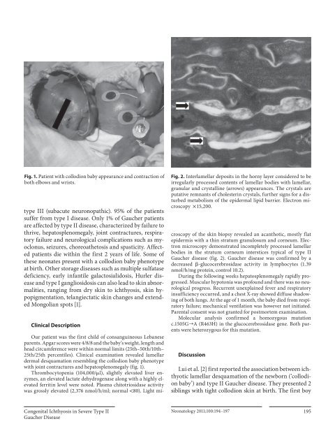
Fig. 1. Patient <strong>with</strong> collodion baby appearance and contraction of<br />
both elbows and wrists.<br />
type <strong>II</strong>I (subacute neuronopathic). 95% of the patients<br />
suffer from type I disease. Only 1% of <strong>Gaucher</strong> patients<br />
are affected by type <strong>II</strong> disease, characterized by failure to<br />
thrive, hepatosplenomegaly, jo<strong>in</strong>t contractures, respiratory<br />
failure and neurological complications such as myoclonus,<br />
seizures, choreoathetosis and spasticity. Affected<br />
patients die <strong>with</strong><strong>in</strong> the first 2 years of life. Some of<br />
these neonates present <strong>with</strong> a collodion baby phenotype<br />
at birth. Other storage diseases such as multiple sulfatase<br />
deficiency, early <strong>in</strong>fantile galactosialidosis, Hurler disease<br />
and type I gangliosidosis can also lead to sk<strong>in</strong> abnormalities,<br />
rang<strong>in</strong>g from dry sk<strong>in</strong> to ichthyosis, sk<strong>in</strong> hypopigmentation,<br />
telangiectatic sk<strong>in</strong> changes and extended<br />
Mongolian spots [1] .<br />
Cl<strong>in</strong>ical Description<br />
Fig. 2. Interlamellar deposits <strong>in</strong> the horny layer considered to be<br />
irregularly processed contents of lamellar bodies <strong>with</strong> lamellar,<br />
granular and crystall<strong>in</strong>e (arrows) appearances. The crystals are<br />
putative remnants of cholester<strong>in</strong> crystals, further signs for a disturbed<br />
metabolism of the epidermal lipid barrier. Electron microscopy<br />
!15,200.<br />
Our patient was the first child of consangu<strong>in</strong>eous Lebanese<br />
parents. Apgar scores were 4/8/8 and the baby’s weight, length and<br />
head circumference were <strong>with</strong><strong>in</strong> normal limits (25th–50th/10th–<br />
25th/25th percentiles). Cl<strong>in</strong>ical exam<strong>in</strong>ation revealed lamellar<br />
dermal desquamation resembl<strong>in</strong>g the collodion baby phenotype<br />
<strong>with</strong> jo<strong>in</strong>t contractures and hepatosplenomegaly ( fig. 1 ).<br />
Thrombocytopenia (104,000/ l), slightly elevated liver enzymes,<br />
an elevated lactate dehydrogenase along <strong>with</strong> a highly elevated<br />
ferrit<strong>in</strong> level were noted. Plasma chitotriosidase activity<br />
was grossly elevated (2,376 nmol/h/ml; normal ! 80). Light microscopy<br />
of the sk<strong>in</strong> biopsy revealed an acanthotic, mostly flat<br />
epidermis <strong>with</strong> a th<strong>in</strong> stratum granulosum and corneum. Electron<br />
microscopy demonstrated <strong>in</strong>completely processed lamellar<br />
bodies <strong>in</strong> the stratum corneum <strong>in</strong>terstices typical of type <strong>II</strong><br />
<strong>Gaucher</strong> disease ( fig. 2 ). <strong>Gaucher</strong> disease was confirmed by a<br />
decreased -glucocerebrosidase activity <strong>in</strong> lymphocytes (1.39<br />
nmol/h/mg prote<strong>in</strong>, control 10.2).<br />
Dur<strong>in</strong>g the follow<strong>in</strong>g weeks hepatosplenomegaly rapidly progressed.<br />
Muscular hypotonia was profound and there was no neurological<br />
progress. Recurrent unexpla<strong>in</strong>ed fever and respiratory<br />
<strong>in</strong>sufficiency occurred, and a chest X-ray showed diffuse shadow<strong>in</strong>g<br />
of both lungs. At the age of 1 month, the baby died from respiratory<br />
failure; mechanical ventilation was however not <strong>in</strong>itiated.<br />
Parental consent was not granted for postmortem exam<strong>in</strong>ation.<br />
Molecular analysis confirmed a homozygous mutation<br />
c.1505G ] A (R463H) <strong>in</strong> the glucocerebrosidase gene. Both parents<br />
were heterozygous for this mutation.<br />
Discussion<br />
Lui et al. [2] first reported the association between ichthyotic<br />
lamellar desquamation of the newborn (‘collodion<br />
baby’) and type <strong>II</strong> <strong>Gaucher</strong> disease. They presented 2<br />
sibl<strong>in</strong>gs <strong>with</strong> tight collodion sk<strong>in</strong> at birth. The first boy<br />
<strong>Congenital</strong> <strong>Ichthyosis</strong> <strong>in</strong> <strong>Severe</strong> <strong>Type</strong> <strong>II</strong><br />
<strong>Gaucher</strong> <strong>Disease</strong><br />
Neonatology 2011;100:194–197 195