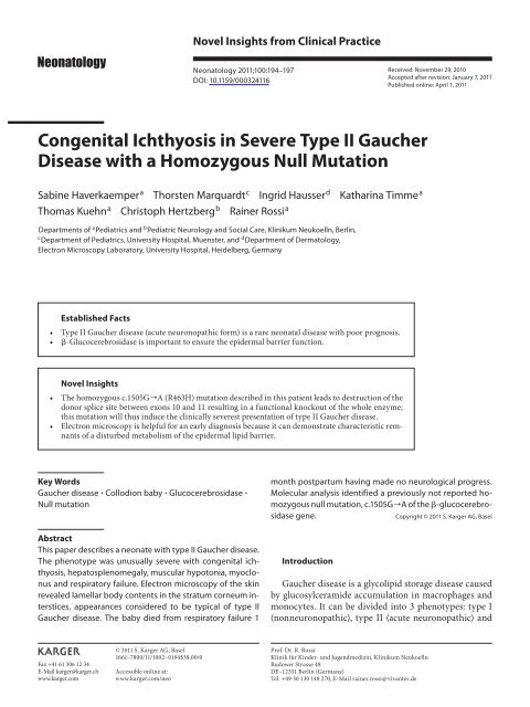
Congenital Ichthyosis in Severe Type II Gaucher Disease with a ...
Congenital Ichthyosis in Severe Type II Gaucher Disease with a ...
Congenital Ichthyosis in Severe Type II Gaucher Disease with a ...

You also want an ePaper? Increase the reach of your titles
YUMPU automatically turns print PDFs into web optimized ePapers that Google loves.
Novel Insights from Cl<strong>in</strong>ical Practice<br />
Neonatology 2011;100:194–197<br />
DOI: 10.1159/000324116<br />
Received: November 29, 2010<br />
Accepted after revision: January 7, 2011<br />
Published onl<strong>in</strong>e: April 1, 2011<br />
<strong>Congenital</strong> <strong>Ichthyosis</strong> <strong>in</strong> <strong>Severe</strong> <strong>Type</strong> <strong>II</strong> <strong>Gaucher</strong><br />
<strong>Disease</strong> <strong>with</strong> a Homozygous Null Mutation<br />
Sab<strong>in</strong>e Haverkaemper<br />
a<br />
Thorsten Marquardt c Ingrid Hausser d Kathar<strong>in</strong>a Timme a<br />
Thomas Kuehn a Christoph Hertzberg b Ra<strong>in</strong>er Rossi a<br />
Departments of a Pediatrics and b Pediatric Neurology and Social Care, Kl<strong>in</strong>ikum Neukoelln, Berl<strong>in</strong> ,<br />
c<br />
Department of Pediatrics, University Hospital, Muenster , and d Department of Dermatology,<br />
Electron Microscopy Laboratory, University Hospital, Heidelberg , Germany<br />
E s t a b l i s h e d Fa c t s<br />
• <strong>Type</strong> <strong>II</strong> <strong>Gaucher</strong> disease (acute neuronopathic form) is a rare neonatal disease <strong>with</strong> poor prognosis.<br />
• -Glucocerebrosidase is important to ensure the epidermal barrier function.<br />
N ove l I n s i g h t s<br />
• The homozygous c.1505G ] A (R463H) mutation described <strong>in</strong> this patient leads to destruction of the<br />
donor splice site between exons 10 and 11 result<strong>in</strong>g <strong>in</strong> a functional knockout of the whole enzyme;<br />
this mutation will thus <strong>in</strong>duce the cl<strong>in</strong>ically severest presentation of type <strong>II</strong> <strong>Gaucher</strong> disease.<br />
• Electron microscopy is helpful for an early diagnosis because it can demonstrate characteristic remnants<br />
of a disturbed metabolism of the epidermal lipid barrier.<br />
Key Words<br />
<strong>Gaucher</strong> disease Collodion baby Glucocerebrosidase <br />
Null mutation<br />
Abstract<br />
This paper describes a neonate <strong>with</strong> type <strong>II</strong> <strong>Gaucher</strong> disease.<br />
The phenotype was unusually severe <strong>with</strong> congenital ichthyosis,<br />
hepatosplenomegaly, muscular hypotonia, myoclonus<br />
and respiratory failure. Electron microscopy of the sk<strong>in</strong><br />
revealed lamellar body contents <strong>in</strong> the stratum corneum <strong>in</strong>terstices,<br />
appearances considered to be typical of type <strong>II</strong><br />
<strong>Gaucher</strong> disease. The baby died from respiratory failure 1<br />
month postpartum hav<strong>in</strong>g made no neurological progress.<br />
Molecular analysis identified a previously not reported homozygous<br />
null mutation, c.1505G ] A of the -glucocerebrosidase<br />
gene.<br />
Copyright © 2011 S. Karger AG, Basel<br />
Introduction<br />
<strong>Gaucher</strong> disease is a glycolipid storage disease caused<br />
by glucosylceramide accumulation <strong>in</strong> macrophages and<br />
monocytes. It can be divided <strong>in</strong>to 3 phenotypes: type I<br />
(nonneuronopathic), type <strong>II</strong> (acute neuronopathic) and<br />
Fax +41 61 306 12 34<br />
E-Mail karger@karger.ch<br />
www.karger.com<br />
© 2011 S. Karger AG, Basel<br />
1661–7800/11/1002–0194$38.00/0<br />
Accessible onl<strong>in</strong>e at:<br />
www.karger.com/neo<br />
Prof. Dr. R. Rossi<br />
Kl<strong>in</strong>ik für K<strong>in</strong>der- und Jugendmediz<strong>in</strong>, Kl<strong>in</strong>ikum Neukoelln<br />
Rudower Strasse 48<br />
DE–12351 Berl<strong>in</strong> (Germany)<br />
Tel. +49 30 130 148 270, E-Mail ra<strong>in</strong>er.rossi @ vivantes.de
Fig. 1. Patient <strong>with</strong> collodion baby appearance and contraction of<br />
both elbows and wrists.<br />
type <strong>II</strong>I (subacute neuronopathic). 95% of the patients<br />
suffer from type I disease. Only 1% of <strong>Gaucher</strong> patients<br />
are affected by type <strong>II</strong> disease, characterized by failure to<br />
thrive, hepatosplenomegaly, jo<strong>in</strong>t contractures, respiratory<br />
failure and neurological complications such as myoclonus,<br />
seizures, choreoathetosis and spasticity. Affected<br />
patients die <strong>with</strong><strong>in</strong> the first 2 years of life. Some of<br />
these neonates present <strong>with</strong> a collodion baby phenotype<br />
at birth. Other storage diseases such as multiple sulfatase<br />
deficiency, early <strong>in</strong>fantile galactosialidosis, Hurler disease<br />
and type I gangliosidosis can also lead to sk<strong>in</strong> abnormalities,<br />
rang<strong>in</strong>g from dry sk<strong>in</strong> to ichthyosis, sk<strong>in</strong> hypopigmentation,<br />
telangiectatic sk<strong>in</strong> changes and extended<br />
Mongolian spots [1] .<br />
Cl<strong>in</strong>ical Description<br />
Fig. 2. Interlamellar deposits <strong>in</strong> the horny layer considered to be<br />
irregularly processed contents of lamellar bodies <strong>with</strong> lamellar,<br />
granular and crystall<strong>in</strong>e (arrows) appearances. The crystals are<br />
putative remnants of cholester<strong>in</strong> crystals, further signs for a disturbed<br />
metabolism of the epidermal lipid barrier. Electron microscopy<br />
!15,200.<br />
Our patient was the first child of consangu<strong>in</strong>eous Lebanese<br />
parents. Apgar scores were 4/8/8 and the baby’s weight, length and<br />
head circumference were <strong>with</strong><strong>in</strong> normal limits (25th–50th/10th–<br />
25th/25th percentiles). Cl<strong>in</strong>ical exam<strong>in</strong>ation revealed lamellar<br />
dermal desquamation resembl<strong>in</strong>g the collodion baby phenotype<br />
<strong>with</strong> jo<strong>in</strong>t contractures and hepatosplenomegaly ( fig. 1 ).<br />
Thrombocytopenia (104,000/ l), slightly elevated liver enzymes,<br />
an elevated lactate dehydrogenase along <strong>with</strong> a highly elevated<br />
ferrit<strong>in</strong> level were noted. Plasma chitotriosidase activity<br />
was grossly elevated (2,376 nmol/h/ml; normal ! 80). Light microscopy<br />
of the sk<strong>in</strong> biopsy revealed an acanthotic, mostly flat<br />
epidermis <strong>with</strong> a th<strong>in</strong> stratum granulosum and corneum. Electron<br />
microscopy demonstrated <strong>in</strong>completely processed lamellar<br />
bodies <strong>in</strong> the stratum corneum <strong>in</strong>terstices typical of type <strong>II</strong><br />
<strong>Gaucher</strong> disease ( fig. 2 ). <strong>Gaucher</strong> disease was confirmed by a<br />
decreased -glucocerebrosidase activity <strong>in</strong> lymphocytes (1.39<br />
nmol/h/mg prote<strong>in</strong>, control 10.2).<br />
Dur<strong>in</strong>g the follow<strong>in</strong>g weeks hepatosplenomegaly rapidly progressed.<br />
Muscular hypotonia was profound and there was no neurological<br />
progress. Recurrent unexpla<strong>in</strong>ed fever and respiratory<br />
<strong>in</strong>sufficiency occurred, and a chest X-ray showed diffuse shadow<strong>in</strong>g<br />
of both lungs. At the age of 1 month, the baby died from respiratory<br />
failure; mechanical ventilation was however not <strong>in</strong>itiated.<br />
Parental consent was not granted for postmortem exam<strong>in</strong>ation.<br />
Molecular analysis confirmed a homozygous mutation<br />
c.1505G ] A (R463H) <strong>in</strong> the glucocerebrosidase gene. Both parents<br />
were heterozygous for this mutation.<br />
Discussion<br />
Lui et al. [2] first reported the association between ichthyotic<br />
lamellar desquamation of the newborn (‘collodion<br />
baby’) and type <strong>II</strong> <strong>Gaucher</strong> disease. They presented 2<br />
sibl<strong>in</strong>gs <strong>with</strong> tight collodion sk<strong>in</strong> at birth. The first boy<br />
<strong>Congenital</strong> <strong>Ichthyosis</strong> <strong>in</strong> <strong>Severe</strong> <strong>Type</strong> <strong>II</strong><br />
<strong>Gaucher</strong> <strong>Disease</strong><br />
Neonatology 2011;100:194–197 195
developed recurrent laryngospasms, convulsions and<br />
thrombocytopenia. A brother was dependent on assisted<br />
ventilation from birth and had hepatosplenomegaly as<br />
well as generalized jo<strong>in</strong>t contractures. Both patients were<br />
characterized by a lack of any normal neurological reactivity<br />
and early death (3 months/11 days). Diagnosis was<br />
confirmed by a decreased glucocerebrosidase activity <strong>in</strong><br />
leukocytes. In addition, <strong>Gaucher</strong> cells were discovered<br />
<strong>in</strong> different organs by postmortem exam<strong>in</strong>ation <strong>in</strong> patient<br />
2.<br />
<strong>Ichthyosis</strong> <strong>in</strong> <strong>Gaucher</strong> type <strong>II</strong> patients demonstrates<br />
the importance of -glucocerebrosidase for ceramide<br />
production and supports the assumption that ichthyosis<br />
based on other molecular defects may be caused by a decreased<br />
ceramide content due to secondary -glucocerebrosidase<br />
deficiency. Lamellar bodies conta<strong>in</strong> glucosylceramides<br />
and phospholipids that are cleaved by glucocerebrosidase<br />
and sph<strong>in</strong>gomyel<strong>in</strong>ase to ceramides and free<br />
fatty acids. They form <strong>in</strong>tercellular lamellar membranes<br />
between the corneocytes <strong>in</strong> the stratum corneum. Ceramides<br />
are a major component of the stratum corneum<br />
and make up to 40% of the total lipids [3] . -Glucoce<br />
rebrosidase metabolizes ceramide from glucosylceramide.<br />
The pH gradient <strong>in</strong> the stratum corneum (pH<br />
from 7.4 to 5.0) is essential for the enzymatic activity of<br />
-glucocerebrosidase [4] . A disturbance of the epidermal<br />
barrier <strong>in</strong> disorders like ichthyosis vulgaris leads to <strong>in</strong>creased<br />
pH and <strong>in</strong>duces impaired -glucocerebrosidase<br />
function.<br />
In ultrastructural and functional studies <strong>in</strong> type <strong>II</strong><br />
<strong>Gaucher</strong> disease null allele mice [5], -glucocerebrosidase<br />
was shown to be necessary to ensure the epidermal<br />
barrier function. Sidransky et al. [6] exam<strong>in</strong>ed sk<strong>in</strong> biopsies<br />
of all 3 types of <strong>Gaucher</strong> patients and type <strong>II</strong> <strong>Gaucher</strong><br />
mice to f<strong>in</strong>d out whether epidermal abnormalities are<br />
unique to this disease entity. Only patients <strong>with</strong> type <strong>II</strong><br />
disease had an <strong>in</strong>creased ratio of epidermal glucosylceramide<br />
to ceramide and ultrastructural abnormalities,<br />
such as <strong>in</strong>completely processed lamellar body-derived<br />
contents throughout the stratum corneum <strong>in</strong>tersitices.<br />
These human f<strong>in</strong>d<strong>in</strong>gs were comparable to those found<br />
<strong>in</strong> type <strong>II</strong> mice and were therefore considered to be characteristic<br />
of type <strong>II</strong> <strong>Gaucher</strong> disease.<br />
Like other <strong>Gaucher</strong> patients, type <strong>II</strong> patients <strong>with</strong> congenital<br />
ichthyosis demonstrate a genotypic diversity [7] .<br />
On the other hand, there is a large phenotypic variability<br />
among patients <strong>with</strong> the same mutations [8] . The<br />
c.1505G ] A (R463H) mutation described <strong>in</strong> this patient<br />
leads to destruction of the donor splice site between exons<br />
10 and 11. In a patient described by Ohshima et al.<br />
[9] , an alternative donor splice site is used result<strong>in</strong>g <strong>in</strong> a<br />
12-bp <strong>in</strong>sertion and the generation of a premature stop<br />
codon follow<strong>in</strong>g codon 463 and delet<strong>in</strong>g the term<strong>in</strong>al 34<br />
am<strong>in</strong>o acids <strong>with</strong> a complete loss of activity. This patient<br />
was compound heterogeneous for the c.1505G ] A mutation<br />
but carried a second mutation lead<strong>in</strong>g to <strong>Gaucher</strong><br />
disease type <strong>II</strong>I. In a series of 6 patients <strong>with</strong> 5 new mutations,<br />
Beutler et al. [10] described a <strong>Gaucher</strong> type I patient<br />
carry<strong>in</strong>g another mutation <strong>in</strong> the same splice donor<br />
site [c.1505, IVS10(+2)], but this patient’s second mutation<br />
was only a mild one (c.1604G ] A). In contrast, our<br />
patient was homozygous for the splice donor site mutation,<br />
result<strong>in</strong>g <strong>in</strong> a functional knockout of the whole enzyme.<br />
The residual activity of -glucocerebrosidase<br />
measured <strong>in</strong> our patient was determ<strong>in</strong>ed <strong>with</strong> an artificial<br />
4 MU substrate and is no <strong>in</strong>dication of genu<strong>in</strong>e residual<br />
activity.<br />
An animal model was established by Tybulewicz et al.<br />
[11] from targeted disruption of the mouse glucocerebrosidase<br />
gene. Comparable to our patient, these mice typically<br />
show ichthyosiform sk<strong>in</strong> and they rapidly deteriorate<br />
<strong>with</strong> decreased movements, reduced respiratory<br />
drive along <strong>with</strong> feed<strong>in</strong>g difficulties lead<strong>in</strong>g to death<br />
<strong>with</strong><strong>in</strong> 24 h of birth. The <strong>in</strong>duced genetic defect <strong>in</strong> these<br />
mice resulted <strong>in</strong> a complete functional knockout of the<br />
-glucocerebrosidase gene like the homozygous splice<br />
site mutation found <strong>in</strong> our patient.<br />
In summary, our patient not only revealed the previously<br />
described collodion baby phenotype and characteristic<br />
horny layer deposits, seen at the ultrastructural level<br />
but also carried a homozygous functional knockout mutation<br />
lead<strong>in</strong>g to an extremely severe and early lethal<br />
form of <strong>Gaucher</strong> type <strong>II</strong> disease. Comparable to knockout<br />
mice, lead<strong>in</strong>g cl<strong>in</strong>ical signs were a collodion-baby-like<br />
appearance <strong>with</strong> hepatosplenomegaly and lack of neurological<br />
progress. Although <strong>Gaucher</strong> disease is and <strong>in</strong> this<br />
case was ultimately confirmed by reduced -glucocerebrosidase<br />
activity and a mutation <strong>in</strong> its gene, sk<strong>in</strong> biopsy<br />
contributed to the early diagnosis. Management <strong>in</strong>cluded<br />
limitation to supportive care.<br />
Acknowledgements<br />
This work was supported by a grant (to I.H.) from the German<br />
BMBF (NIRK, 01GM0610). T.M. was supported by a research<br />
grant from Genzyme. Ingrid Du Chesne is acknowledged for the<br />
molecular analysis.<br />
196<br />
Neonatology 2011;100:194–197<br />
Haverkaemper /Marquardt /Hausser /<br />
Timme /Kuehn /Hertzberg /Rossi
Laboratories<br />
Electron microscopy: Dr. I. Hausser, Department of Dermatology,<br />
Electron Microscopy Laboratory, University Hos -<br />
pital, Heidelberg, Germany; E-Mail Ingrid.hausser@med.uniheidelberg.de.<br />
Chitotriosidase and -glucocerebrosidase assay as<br />
well as molecular biology (<strong>in</strong>clud<strong>in</strong>g prenatal diagnosis): Prof. Dr.<br />
T. Marquardt, Department of Pediatrics, University Hospital,<br />
Muenster, Germany; E-Mail marquat@uni-muenster.de.<br />
Disclosure Statement<br />
No conflict of <strong>in</strong>terest.<br />
References<br />
1 Staretz-Chacham O, Lang TC, La Marca ME,<br />
Krasnewich D, Sidransky E: Lysosomal storage<br />
disorders <strong>in</strong> the newborn. Pediatrics<br />
2009; 123: 1191–1207.<br />
2 Lui K, Commens C, Choong R, Jaworski R:<br />
Collodion babies <strong>with</strong> <strong>Gaucher</strong>’s disease.<br />
Arch Dis Child 1988; 63: 854–856.<br />
3 Hamanaka S, Hara M, Nishio H, Otsuka F,<br />
Suzuki A, Uchida Y: Human epidermal glycosylceramides<br />
are major precursors of stratum<br />
corneum ceramides. J Invest Dermatol<br />
2002; 119: 416–423.<br />
4 Takagi Y, Kriehuber E, Imokawa G, Elias<br />
PM, Holleran WM: Beta-glucocerebrosidase<br />
activity <strong>in</strong> mammalian stratum corneum. J<br />
Lipid Res 1999; 40: 861–869.<br />
5 Holleran WM, Takagi Y, Menon GK, Legler<br />
G, Fe<strong>in</strong>gold KR, Elias PM: Consequences of<br />
-glucocerebrosidase deficiency <strong>in</strong> epidermis:<br />
ultrastructure and permeability barrier<br />
alterations <strong>in</strong> <strong>Gaucher</strong> disease. J Cl<strong>in</strong> Invest<br />
1994; 93: 1756–1764.<br />
6 Sidransky E, Fartasch M, Lee RE, Metlay LA,<br />
Abella S, Zimran A, et al: Epidermal abnormalities<br />
may dist<strong>in</strong>guish type 2 from type 1<br />
and 3 of <strong>Gaucher</strong> disease. Pediatr Res 1996;<br />
39: 134–141.<br />
7 Stone DL, Carey WF, Christodoulou J, Sillence<br />
D, Nelson P, Callahan M, et al: <strong>Type</strong> 2<br />
<strong>Gaucher</strong> disease: the collodion baby revisited.<br />
Arch Dis Child Fetal Neonatal Ed 2000;<br />
82:F163–F166.<br />
8 Sidransky E: <strong>Gaucher</strong> disease: complexity <strong>in</strong><br />
a ‘simple’ disorder. Mol Genet Metabol 2004;<br />
83: 6–15.<br />
9 Ohshima T, Sasaki M, Matsuzaka T,<br />
Sakuragawa N: A novel splic<strong>in</strong>g abnormality<br />
<strong>in</strong> a Japanese patient <strong>with</strong> <strong>Gaucher</strong>’s disease.<br />
Hum Mol Genet 1993; 2: 1497–1498.<br />
10 Beutler E, Gelbart T, Dem<strong>in</strong>a A, Zimran A,<br />
LeCoutre P: Five new <strong>Gaucher</strong> disease mutations.<br />
Blood Cells Mol Dis 1995; 21: 20–24.<br />
11 Tybulewicz V, Tremblay ML, La Marca ME,<br />
Willemsen R, Stubblefield BK, W<strong>in</strong>field S, et<br />
al: Animal model of <strong>Gaucher</strong>’s disease from<br />
targeted disruption of the mouse glucocerebrosidase<br />
gene. Nature 1992; 357: 40 7– 410 .<br />
<strong>Congenital</strong> <strong>Ichthyosis</strong> <strong>in</strong> <strong>Severe</strong> <strong>Type</strong> <strong>II</strong><br />
<strong>Gaucher</strong> <strong>Disease</strong><br />
Neonatology 2011;100:194–197 197