Microwave-assisted Protein Preparation and Enzymatic Digestion in ...
Microwave-assisted Protein Preparation and Enzymatic Digestion in ...
Microwave-assisted Protein Preparation and Enzymatic Digestion in ...

Create successful ePaper yourself
Turn your PDF publications into a flip-book with our unique Google optimized e-Paper software.
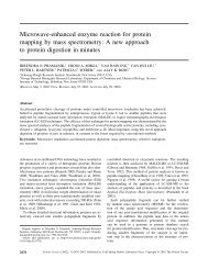
<strong>Microwave</strong>-<strong>assisted</strong> <strong>Prote<strong>in</strong></strong> <strong>Preparation</strong> <strong>and</strong><br />
<strong>Enzymatic</strong> <strong>Digestion</strong> <strong>in</strong> Proteomics* □S<br />
Wei Sun‡, Shijuan Gao, L<strong>in</strong>jie Wang, Yong Chen, Shuzhen Wu, Xiaorong Wang,<br />
Dexian Zheng, <strong>and</strong> Youhe Gao§<br />
The comb<strong>in</strong>ations of gel electrophoresis or LC <strong>and</strong> mass<br />
spectrometry are two popular approaches for large scale<br />
prote<strong>in</strong> identification. However, the throughput of both<br />
approaches is limited by the speed of the prote<strong>in</strong> digestion<br />
process. Present research <strong>in</strong>to fast prote<strong>in</strong> enzymatic<br />
digestion has been focused ma<strong>in</strong>ly on known prote<strong>in</strong>s,<br />
<strong>and</strong> it is unclear whether these results can be extrapolated<br />
to complex prote<strong>in</strong> mixtures. In this study microwave<br />
technology was used to develop a fast prote<strong>in</strong> preparation<br />
<strong>and</strong> enzymatic digestion method for prote<strong>in</strong><br />
mixtures. The prote<strong>in</strong> mixtures <strong>in</strong> solution or <strong>in</strong> gel were<br />
prepared <strong>and</strong> digested by microwave-<strong>assisted</strong> prote<strong>in</strong> enzymatic<br />
digestion, which rapidly produces peptide fragments.<br />
The peptide fragments were further analyzed by<br />
capillary LC <strong>and</strong> ESI-ion trap-MS or MALDI-TOF-MS. The<br />
technique was optimized us<strong>in</strong>g bov<strong>in</strong>e serum album<strong>in</strong> <strong>and</strong><br />
then applied to human ur<strong>in</strong>ary prote<strong>in</strong>s <strong>and</strong> yeast lysate.<br />
The method enabled preparation <strong>and</strong> digestion of prote<strong>in</strong><br />
mixtures <strong>in</strong> solution (human ur<strong>in</strong>ary prote<strong>in</strong>s) or <strong>in</strong> gel<br />
(yeast lysate) <strong>in</strong> 6 or 25 m<strong>in</strong>, respectively. Equivalent (<strong>in</strong>solution)<br />
or better (<strong>in</strong>-gel) digestion efficiency was obta<strong>in</strong>ed<br />
us<strong>in</strong>g microwave-<strong>assisted</strong> prote<strong>in</strong> enzymatic digestion<br />
compared with the st<strong>and</strong>ard overnight digestion<br />
method. This new application of microwave technology to<br />
prote<strong>in</strong> mixture preparation <strong>and</strong> enzymatic digestion will<br />
hasten the application of proteomic techniques to biological<br />
<strong>and</strong> cl<strong>in</strong>ical research. Molecular & Cellular Proteomics<br />
5:769–776, 2006.<br />
Proteomics aims to characterize a large number of prote<strong>in</strong>s<br />
extracted from a cell, tissue, or organism so that a global<br />
perspective of changes <strong>in</strong> prote<strong>in</strong> expression can be obta<strong>in</strong>ed<br />
<strong>in</strong> a rapid fashion (1, 2). Proteomic analysis of complex mixtures<br />
of prote<strong>in</strong>s usually proceeds along either a bottom-up or<br />
top-down approach. In the bottom-up approach, the entire<br />
proteome is digested <strong>in</strong>to a pool of possibly thous<strong>and</strong>s of<br />
peptides (3–5). Two-dimensional (2D) 1 LC <strong>and</strong> MS are used to<br />
From the Proteomics Research Center <strong>and</strong> National Key Laboratory<br />
of Medical Molecular Biology, Institute of Basic Medical Sciences,<br />
Pek<strong>in</strong>g Union Medical College/Ch<strong>in</strong>ese Academy of Medical Sciences,<br />
Beij<strong>in</strong>g 100005, Ch<strong>in</strong>a<br />
Received, September 1, 2005, <strong>and</strong> <strong>in</strong> revised form, November 1,<br />
2005<br />
Published, MCP Papers <strong>in</strong> Press, December 12, 2005, DOI<br />
10.1074/mcp.T500022-MCP200<br />
1 The abbreviations used are: 2D, two-dimensional; 1D, one-dimen-<br />
Technology<br />
resolve <strong>and</strong> identify peptide components <strong>in</strong> the mixture. This<br />
approach, referred to as shotgun proteomics, affords the<br />
advantages of automation <strong>and</strong> sensitivity but at the loss of<br />
<strong>in</strong>formation regard<strong>in</strong>g the <strong>in</strong>tact prote<strong>in</strong>. In the top-down approach,<br />
the characteristics of a prote<strong>in</strong>, such as molecular<br />
weight, isoelectric po<strong>in</strong>t, <strong>and</strong> hydrophobicity, are used to<br />
isolate the <strong>in</strong>tact prote<strong>in</strong> (6, 7). Gel electrophoresis is the most<br />
commonly used top-down approach (8). Each slice or spot <strong>in</strong><br />
the gels is excised, digested, <strong>and</strong> identified by MALDI-MS or<br />
ESI-ion trap-MS. Another top-down approach uses chromatofocus<strong>in</strong>g<br />
<strong>and</strong> reverse phase chromatography <strong>in</strong> an HPLC<br />
format to separate prote<strong>in</strong>s (9–12). Each fraction from LC is<br />
also digested <strong>and</strong> identified by MS. LC offers an advantage<br />
over gel electrophoresis <strong>in</strong> terms of ease of automation <strong>and</strong><br />
prote<strong>in</strong> recovery.<br />
Although prote<strong>in</strong> <strong>and</strong> peptide separation <strong>and</strong> identification<br />
can be made highly automated <strong>and</strong> rapid, sample preparation<br />
<strong>and</strong> digestion <strong>in</strong> contrast are considerably slower (more than<br />
16 h) <strong>and</strong> limit the speed of large scale prote<strong>in</strong> identification.<br />
Recently several approaches have been developed for fast<br />
prote<strong>in</strong> digestion. One approach is the use of modified tryps<strong>in</strong><br />
for <strong>in</strong>-gel digestion of prote<strong>in</strong>s <strong>in</strong>stead of native tryps<strong>in</strong> (13).<br />
Another approach uses on-l<strong>in</strong>e prote<strong>in</strong> digestion dur<strong>in</strong>g LC<br />
us<strong>in</strong>g a proteolytic reactor (14, 15) or an immobilized enzyme<br />
column (16, 17). Other promis<strong>in</strong>g approaches <strong>in</strong>clude microwave-<strong>assisted</strong><br />
prote<strong>in</strong> enzymatic digestion (MAPED) or acid<br />
hydrolysis (18–22).<br />
Several recent reports have highlighted the speed <strong>and</strong> convenience<br />
of MAPED. Juan et al. (18) used microwave technology<br />
to digest several known prote<strong>in</strong>s <strong>in</strong> gel with tryps<strong>in</strong> <strong>in</strong> 5<br />
m<strong>in</strong>. Pramanik et al. (19) also applied microwave technique to<br />
digest known prote<strong>in</strong>s <strong>in</strong> solution or <strong>in</strong> gel with tryps<strong>in</strong> <strong>in</strong> 10<br />
m<strong>in</strong> <strong>in</strong>clud<strong>in</strong>g a prote<strong>in</strong> that was tightly folded <strong>and</strong> extremely<br />
resistant to denaturation (bov<strong>in</strong>e ubiquit<strong>in</strong>). L<strong>in</strong> et al. (20) <strong>in</strong>vestigated<br />
microwave-<strong>assisted</strong> enzyme-catalyzed reactions<br />
<strong>in</strong> various solvent systems. They found that digestion efficiency<br />
<strong>and</strong> sequence coverage were <strong>in</strong>creased when the<br />
tryps<strong>in</strong> digestion occurred <strong>in</strong> ACN-, methanol-, <strong>and</strong> chloroform-conta<strong>in</strong><strong>in</strong>g<br />
solutions under microwave irradiation. These<br />
authors found that ACN did not deactivate the proteolytic<br />
enzyme dur<strong>in</strong>g the irradiation period, whereas methanol did.<br />
sional; MAPED, microwave-<strong>assisted</strong> prote<strong>in</strong> enzymatic digestion; RP,<br />
reverse phase; SCX, strong cation exchange; Tric<strong>in</strong>e, N-[2-hydroxy-<br />
1,1-bis(hydroxymethyl)ethyl]glyc<strong>in</strong>e; W, watts.<br />
© 2006 by The American Society for Biochemistry <strong>and</strong> Molecular Biology, Inc. Molecular & Cellular Proteomics 5.4 769<br />
This paper is available on l<strong>in</strong>e at http://www.mcponl<strong>in</strong>e.org
<strong>Microwave</strong>-<strong>assisted</strong> <strong>Prote<strong>in</strong></strong> <strong>Enzymatic</strong> <strong>Digestion</strong><br />
There are also some reports on microwave-<strong>assisted</strong> prote<strong>in</strong><br />
acid hydrolysis. Chen et al. (21) <strong>and</strong> Zhong et al. (22) used<br />
microwave irradiation to accelerate the hydrolysis of peptides<br />
<strong>and</strong> prote<strong>in</strong>s with 6 M HCl. Zhong et al. (23) also described a<br />
microwave-<strong>assisted</strong> acid hydrolysis method for rapid prote<strong>in</strong><br />
degradation with trifluoroacetic acid. They applied the method<br />
to analyze a membrane prote<strong>in</strong>-enriched fraction of cell lysates<br />
<strong>and</strong> identified 41 membrane prote<strong>in</strong>s.<br />
Published studies us<strong>in</strong>g MAPED have ma<strong>in</strong>ly focused on<br />
one or several known prote<strong>in</strong>s, <strong>and</strong> the microwave technique<br />
is only used for prote<strong>in</strong> enzymatic digestion (18–20). Whether<br />
the microwave technique can be applied to prote<strong>in</strong> mixture<br />
preparation <strong>and</strong> digestion, especially for prote<strong>in</strong> recovery<br />
from the gel, has still not been well <strong>in</strong>vestigated. For microwave-<strong>assisted</strong><br />
prote<strong>in</strong> acid hydrolysis, Zhong et al. (22, 23)<br />
have developed the method for known prote<strong>in</strong>s as well as<br />
prote<strong>in</strong> mixtures. But prote<strong>in</strong> acid hydrolysis is known to lack<br />
cleavage specificity (21, 22) <strong>and</strong> is generally not used <strong>in</strong><br />
proteomic studies. Therefore it is necessary to develop the<br />
proper MAPED method for prote<strong>in</strong> mixtures.<br />
In this study, we utilized a known prote<strong>in</strong> (bov<strong>in</strong>e serum<br />
album<strong>in</strong>) as well as complex mixtures of prote<strong>in</strong>s derived from<br />
human ur<strong>in</strong>e <strong>and</strong> yeast lysate to address the problems noted<br />
above. In our experiments MAPED could prepare <strong>and</strong> digest<br />
prote<strong>in</strong> mixtures <strong>in</strong> solution or <strong>in</strong> gel <strong>in</strong> 6 or 25 m<strong>in</strong>, respectively,<br />
<strong>and</strong> its peptide yield efficiency was the same (<strong>in</strong> solution)<br />
or better (<strong>in</strong> gel) than the present st<strong>and</strong>ard method (16 h<br />
or overnight). This new application of microwave technology<br />
speeds up prote<strong>in</strong> sample preparation <strong>and</strong> enzymatic digestion<br />
<strong>in</strong> proteomic studies of biological <strong>and</strong> cl<strong>in</strong>ical samples.<br />
MATERIALS AND METHODS<br />
Apparatus<br />
An LCQ Deca XP plus ion trap mass spectrometer was purchased<br />
from Thermo F<strong>in</strong>nigan (San Jose, CA). A Voyager-DE Pro MALDI-TOF<br />
mass spectrometer was purchased from Applied Biosystems (Foster<br />
City, CA). Accessories for capillary liquid chromatography were from<br />
Upchurch Scientific Inc. (Oak Harbor, WA). C 18 reverse phase (RP)<br />
<strong>and</strong> strong cation exchange (SCX) res<strong>in</strong>s (5 �m, 300 Å) were from<br />
Merck <strong>and</strong> PolyLC Inc. (Columbia, MD), respectively. A vertical m<strong>in</strong>igel<br />
system (M<strong>in</strong>i-Protean II) was purchased from Bio-Rad. The microwave<br />
oven used <strong>in</strong> this study was solid-state Whirlpool model VIP271<br />
(Shanghai, Ch<strong>in</strong>a), <strong>and</strong> the maximum output power was 850 W.<br />
TABLE I<br />
St<strong>and</strong>ard <strong>and</strong> MAPED <strong>in</strong>-solution preparation <strong>and</strong> digestion protocols<br />
Protocol Solution<br />
St<strong>and</strong>ard,<br />
sample 1 Sample 2<br />
MAPED<br />
Sample 3 Sample 4 Sample 5<br />
h m<strong>in</strong><br />
Reduce 10 mM DTT 1 60 60 5 0<br />
Alkylate 55 mM iodoacetamide 0.5 30 0 0 0<br />
Digest 0.5 �M tryps<strong>in</strong>,<br />
25 mM ammonium bicarbonate<br />
16 1 1 1 1<br />
Total time 17.5 91 61 6 1<br />
770 Molecular & Cellular Proteomics 5.4<br />
Reagents<br />
Deionized water from a MilliQ RG ultrapure water system (Millipore,<br />
Bedford, MA) was used at all times. HPLC grade ACN <strong>and</strong> formic<br />
acid, trifluoroacetic acid, ammonium bicarbonate, iodoacetamide,<br />
<strong>and</strong> DTT were purchased from Merck. Sequenc<strong>in</strong>g grade modified<br />
tryps<strong>in</strong>, protease <strong>in</strong>hibitor PMSF, <strong>and</strong> �-cyano-4-hydroxyc<strong>in</strong>namic<br />
acid were purchased from Sigma.<br />
<strong>Prote<strong>in</strong></strong> Sample<br />
BSA used <strong>in</strong> this study was purchased from LianX<strong>in</strong>g (Beij<strong>in</strong>g,<br />
Ch<strong>in</strong>a). Human ur<strong>in</strong>ary prote<strong>in</strong>s were obta<strong>in</strong>ed from five healthy male<br />
mixed morn<strong>in</strong>g ur<strong>in</strong>e samples by acetone precipitation as described<br />
previously (24). The five ur<strong>in</strong>ary samples were centrifuged at 5000 �<br />
g for 30 m<strong>in</strong>, <strong>and</strong> the precipitates were removed. The supernatants<br />
from five donors were mixed with the same volume, <strong>and</strong> the mixed<br />
ur<strong>in</strong>ary samples were precipitated by 50% acetone for 10 m<strong>in</strong> follow<strong>in</strong>g<br />
centrifugation at 12,000 � g for 30 m<strong>in</strong>. The pellets were resuspended<br />
<strong>in</strong> 25 mM ammonium bicarbonate. Yeast lysate was obta<strong>in</strong>ed<br />
with the method described previously (25). Stra<strong>in</strong> CG1945 was grown<br />
to log phase <strong>in</strong> yeast, peptone, dextrose medium. The cells were<br />
collected by centrifugation <strong>and</strong> lysed with glass beads <strong>in</strong> lysis buffer<br />
(25 mM ammonium bicarbonate, pH 8.0, 0.5 mM EDTA, 1 mM PMSF).<br />
The ur<strong>in</strong>e <strong>and</strong> yeast prote<strong>in</strong> mixtures described above were quantitated<br />
by Bradford method.<br />
One-dimensional Electrophoresis<br />
BSA (10 �g) <strong>and</strong> yeast lysate (20 �g) were mixed with the same<br />
volume of glyc<strong>in</strong>e load<strong>in</strong>g buffer (50 mM Tris-HCl, pH 6.8, 100 mM<br />
DTT, 2% SDS, 0.1% bromphenol blue, 10% glycerol), heated at<br />
100 °C for 3 m<strong>in</strong>, cooled to room temperature, <strong>and</strong> loaded onto the<br />
gel. Electrophoresis was performed on 12% polyacrylamide, 100 �<br />
80-mm slab gels with 0.75-mm spacers us<strong>in</strong>g M<strong>in</strong>i-Protean II. Human<br />
ur<strong>in</strong>ary prote<strong>in</strong>s (10 �g) <strong>and</strong> two tryptic digested ur<strong>in</strong>ary peptide<br />
mixtures from microwave irradiation (Table I, Column 6) <strong>and</strong> st<strong>and</strong>ard<br />
(Table I, Column 3) digestion methods were mixed with Tric<strong>in</strong>e load<strong>in</strong>g<br />
buffer (125 mM Tris-HCl, pH 6.8, 100 mM DTT, 5% SDS, 0.1%<br />
bromphenol blue, 10% glycerol) <strong>and</strong> run on a 15% Tric<strong>in</strong>e gel as<br />
described above.<br />
A microwave-<strong>assisted</strong> Coomassie Blue sta<strong>in</strong><strong>in</strong>g protocol was followed<br />
(26). The gel was <strong>in</strong>cubated <strong>in</strong> the microwave oven at 850 W for<br />
1 m<strong>in</strong> <strong>in</strong> the methanol/acetic acid fixative followed by 1-m<strong>in</strong> sta<strong>in</strong><strong>in</strong>g<br />
<strong>in</strong> the sta<strong>in</strong><strong>in</strong>g solution (0.1% Coomassie Blue R-250 (w/v), 40%<br />
methanol, 5% acetic acid) <strong>and</strong> 3-m<strong>in</strong> desta<strong>in</strong><strong>in</strong>g <strong>in</strong> the desta<strong>in</strong><strong>in</strong>g<br />
solution (40% methanol, 5% acetic acid).<br />
<strong>Prote<strong>in</strong></strong> <strong>Digestion</strong><br />
In-solution <strong>Digestion</strong>—BSA was used as a model prote<strong>in</strong> to study<br />
<strong>in</strong>-solution digestion. 1 mg of BSA was dissolved <strong>in</strong> 1 ml of ammo-
TABLE II<br />
St<strong>and</strong>ard <strong>and</strong> MAPED <strong>in</strong>-gel preparation, digestion, <strong>and</strong> peptide<br />
extraction protocols<br />
Protocol Solution<br />
St<strong>and</strong>ard,<br />
sample 6<br />
MAPED,<br />
sample 7<br />
h m<strong>in</strong><br />
Reduce 10 mM DTT 1 0<br />
Alkylate 55 mM iodoacetamide 0.5 0<br />
Dehydrate 100% ACN 0.5 5<br />
Rehydrate 0.5 �M tryps<strong>in</strong>, 25 mM<br />
ammonium bicarbonate<br />
0.5 5<br />
Digest 0.5 �M tryps<strong>in</strong>, 25 mM<br />
ammonium bicarbonate<br />
16 5<br />
Extract 5% formic acid, 50% ACN 0.5 � 2 5 � 2<br />
Total time 19.5 25<br />
nium bicarbonate buffer (25 mM, pH 8.5) to a concentration of 1<br />
mg/ml. It was divided <strong>in</strong>to five parts, <strong>and</strong> each was 200 �l. An<br />
<strong>in</strong>-solution digestion procedure was performed on the five BSA samples<br />
(samples 1–5) follow<strong>in</strong>g the five protocols outl<strong>in</strong>ed <strong>in</strong> Table I.<br />
Sample 1 was digested accord<strong>in</strong>g to the st<strong>and</strong>ard protocol (Table I,<br />
Column 3). It was reduced by DTT at 57 °C for 1 h, alkylated by<br />
iodoacetamide at room temperature <strong>in</strong> the dark for 0.5 h, <strong>and</strong> digested<br />
by tryps<strong>in</strong> (1:50) at 37 °C for 16 h. Samples 2–4 were prepared<br />
<strong>and</strong> digested accord<strong>in</strong>g to MAPED protocols (Table I, Columns 4–7).<br />
Sample 2 was reduced <strong>and</strong> alkylated the same as sample 1, sample<br />
3 was reduced <strong>in</strong> DTT for 60 m<strong>in</strong> but not alkylated, sample 4 was<br />
reduced with DTT by heat<strong>in</strong>g at 100 °C for 5 m<strong>in</strong> <strong>and</strong> not alkylated,<br />
<strong>and</strong> sample 5 was neither reduced nor alkylated. Samples 2–5 were<br />
all digested with tryps<strong>in</strong> (1:50, wt/wt) <strong>in</strong> the microwave oven at 850 W<br />
us<strong>in</strong>g the follow<strong>in</strong>g method. Samples 2–5 were each put <strong>in</strong>to 0.6-ml<br />
polypropylene vials. As described previously (27, 28), a conta<strong>in</strong>er with<br />
1,000 ml of water was placed beside the sample vials to absorb the<br />
extra microwave energy. The microwave oven was turned on for 1<br />
m<strong>in</strong>. After microwave irradiation, the vials were removed from the<br />
microwave oven <strong>and</strong> prepared for further MS analysis.<br />
Of the five protocols described above, the fastest <strong>and</strong> most effective<br />
sample preparation <strong>and</strong> digestion method was further retested<br />
us<strong>in</strong>g human ur<strong>in</strong>ary prote<strong>in</strong>s. The digested ur<strong>in</strong>ary peptide mixtures<br />
were lyophilized for one-dimensional Tric<strong>in</strong>e gel <strong>and</strong> 2D LC-ESI-MS<br />
analysis. As control, the same ur<strong>in</strong>ary prote<strong>in</strong>s were also digested by<br />
st<strong>and</strong>ard protocol <strong>and</strong> analyzed by Tric<strong>in</strong>e gel <strong>and</strong> 2D LC-ESI-MS.<br />
In-gel <strong>Digestion</strong>—BSA was also used to develop the <strong>in</strong>-gel digestion<br />
method. The BSA b<strong>and</strong> <strong>in</strong> the one-dimensional glyc<strong>in</strong>e gel was<br />
excised manually from the gel slab, cut <strong>in</strong>to pieces, divided <strong>in</strong>to two<br />
parts, <strong>and</strong> put <strong>in</strong>to 1.5-ml polypropylene vials. An <strong>in</strong>-gel digestion<br />
procedure was performed to the two BSA samples (6, 7) follow<strong>in</strong>g the<br />
protocols outl<strong>in</strong>ed <strong>in</strong> Table II. Sample 6 was prepared <strong>and</strong> digested<br />
us<strong>in</strong>g the st<strong>and</strong>ard protocol (29) (Table II, Column 3). Briefly BSA was<br />
<strong>in</strong>-gel reduced <strong>and</strong> alkylated <strong>and</strong> then dehydrated by ACN. Gel pieces<br />
were further rehydrated <strong>and</strong> digested for 16 h. Sample 7 was prepared<br />
<strong>and</strong> digested us<strong>in</strong>g the MAPED protocol (Table II, Column 4)<br />
without prior reduction <strong>and</strong> alkylation (30). The gel pieces were dehydrated,<br />
rehydrated, <strong>and</strong> digested by tryps<strong>in</strong> for 5 m<strong>in</strong>, <strong>and</strong> the<br />
peptides were twice extracted from the gel by microwave irradiation.<br />
The microwave irradiation method was the same as that used for the<br />
<strong>in</strong>-solution MAPED protocol, <strong>and</strong> the irradiation time was 5 m<strong>in</strong> for<br />
each step.<br />
To <strong>in</strong>vestigate the <strong>in</strong>-gel digestion efficacy of MAPED for prote<strong>in</strong><br />
mixtures, five b<strong>and</strong>s of yeast lysate <strong>in</strong> one-dimensional electrophoresis<br />
were selectively excised from the gel, <strong>and</strong> each b<strong>and</strong> was divided<br />
<strong>in</strong>to two parts, prepared, <strong>and</strong> digested as described above us<strong>in</strong>g the<br />
protocols outl<strong>in</strong>ed <strong>in</strong> Table II. The BSA <strong>and</strong> yeast lysate extracted<br />
<strong>Microwave</strong>-<strong>assisted</strong> <strong>Prote<strong>in</strong></strong> <strong>Enzymatic</strong> <strong>Digestion</strong><br />
peptides from one-dimensional gel were further resolved by MALDI-<br />
TOF-MS <strong>and</strong> 1D LC-ESI-MS.<br />
Peptide Mass F<strong>in</strong>gerpr<strong>in</strong>t<strong>in</strong>g<br />
After digestion BSA samples 1–7 were directly spotted on the<br />
sample plate for MALDI-TOF-MS. �-Cyano-4-hydroxyc<strong>in</strong>namic acid<br />
(0.5 �l of 10 mg/ml) was applied to each spot, <strong>and</strong> the spots were<br />
air-dried at room temperature prior to acquir<strong>in</strong>g mass spectra. All<br />
mass spectra were acquired <strong>in</strong> positive ion, reflectron mode, <strong>and</strong> the<br />
peptide masses were measured as monoisotopic masses.<br />
LC-ESI-MS/MS<br />
All lyophilized samples were redissolved <strong>in</strong> 0.1% formic acid (buffer<br />
A) before ESI-MS analysis. For BSA samples, 100 fmol each of<br />
samples 1–5 <strong>and</strong> 300 fmol of samples 6 <strong>and</strong> 7 were loaded onto<br />
self-packed C 18 RP capillary columns (100 mm � 0.17-mm <strong>in</strong>ner<br />
diameter) with buffer A (0.1% formic acid, 99.9% H 2O). The BSA<br />
peptides were eluted with 5–30% buffer B (0.1% formic acid, 99.9%<br />
ACN; flow rate, 2 �l/m<strong>in</strong>) for 60 m<strong>in</strong>. Each BSA sample was run three<br />
times. For ur<strong>in</strong>ary prote<strong>in</strong>s, 100 �g of digested prote<strong>in</strong> mixture was<br />
resolved us<strong>in</strong>g an on-l<strong>in</strong>e SCX capillary column (150 mm � 0.32-mm<br />
<strong>in</strong>ner diameter) <strong>and</strong> RP capillary column (150 mm � 0.17-mm <strong>in</strong>ner<br />
diameter). The ammonia acetate concentration of elution steps for the<br />
SCX capillary column were 0 mM,25mM,50mM,75mM, 100 mM, 125<br />
mM, 150 mM, <strong>and</strong> 1 M. The elution gradient for the RP column was<br />
from 5 to 30% buffer B for 3 h. Yeast lysate peptides extracted from<br />
gels were separated by a self-packed C 18 capillary column (150<br />
mm � 0.17-mm <strong>in</strong>ner diameter) with 5–30% elution gradient (buffer B)<br />
for3h.<br />
Eluted peptides were detected <strong>in</strong> a survey scan from 400 to 1500<br />
amu (three microscans) followed by five data-dependent MS/MS<br />
scans (five microscans each; isolation width, 3 amu; 35% normalized<br />
collision energy; dynamic exclusion for 3 m<strong>in</strong>) <strong>in</strong> a completely automated<br />
fashion on an LCQ-DECA XP plus ESI mass spectrometer. All<br />
MS/MS spectra were respectively searched us<strong>in</strong>g SEQUEST algorithm-based<br />
Bioworks 3.1 SR1 (Thermo F<strong>in</strong>nigan) aga<strong>in</strong>st a bov<strong>in</strong>e<br />
database created from a non-redundant database, a yeast database<br />
from the National Center for Biotechnology Information website<br />
(ftp.ncbi.nih.gov/), <strong>and</strong> the International <strong>Prote<strong>in</strong></strong> Index human database<br />
(version 3.07) from the European Bio<strong>in</strong>formatics Institute website<br />
(www.ebi.ac.uk/IPI/). The search criteria used were those reported<br />
<strong>in</strong> Ref. 3.<br />
Accord<strong>in</strong>g to previous reports (31–33), the number of identified<br />
MS/MS spectra for a prote<strong>in</strong> can be used as the measure of that<br />
prote<strong>in</strong>’s abundance. In this study the MS/MS spectra number from<br />
prote<strong>in</strong> samples was used to evaluate the efficiency of each digestion<br />
protocol. For the same sample amount, the more MS/MS spectra<br />
identified, the higher the digestion efficiency of the protocol was.<br />
Paired t test was utilized to def<strong>in</strong>e the statistical significance between<br />
the identified MS/MS spectral numbers from the two protocols for the<br />
same sample amount. A level of p � 0.05 was considered to be<br />
significant.<br />
RESULTS AND DISCUSSION<br />
In-solution <strong>Digestion</strong><br />
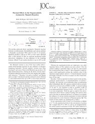
BSA In-solution <strong>Digestion</strong>—Fig. 1 (A–E) shows the<br />
MALDI-MS results of five BSA samples (samples 1–5). The<br />
mass spectrum of sample 1 was almost the same as that of<br />
sample 2, <strong>in</strong>dicat<strong>in</strong>g that similar peptides were formed despite<br />
the approximately 100� difference <strong>in</strong> process<strong>in</strong>g time. The<br />
results from sample 3 <strong>and</strong> sample 4 showed the same trend.<br />
Molecular & Cellular Proteomics 5.4 771
<strong>Microwave</strong>-<strong>assisted</strong> <strong>Prote<strong>in</strong></strong> <strong>Enzymatic</strong> <strong>Digestion</strong><br />
772 Molecular & Cellular Proteomics 5.4
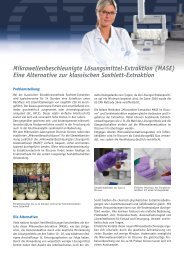
FIG. 2.Comparison of the number of identified MS/MS spectra<br />
for BSA samples 1–7 by 1D LC-ESI-MS analysis. The amount<br />
loaded on-column for samples 1–5 was 100 fmol, <strong>and</strong> 300 fmol was<br />
loaded for samples 6 <strong>and</strong> 7. Samples 1 <strong>and</strong> 6 were prepared <strong>and</strong><br />
digested us<strong>in</strong>g a st<strong>and</strong>ard protocol, whereas the other five samples<br />
were prepared us<strong>in</strong>g the MAPED protocol (Tables I <strong>and</strong> II). *, p � 0.05.<br />
To compare the digestion efficiency of the five protocols<br />
described above, the five samples were further analyzed by<br />
1D LC-ESI-MS, <strong>and</strong> the results are shown <strong>in</strong> Fig. 2. The<br />
numbers of identified MS/MS spectra for samples 1–4 (100<br />
fmol) were similar, suggest<strong>in</strong>g that the digestion efficiency of<br />
these four protocols was the same (31, 32). However, the<br />
MS/MS spectral number for sample 5 was much less than that<br />
of the other four samples, <strong>in</strong>dicat<strong>in</strong>g reduced digestion<br />
efficiency.<br />
Sample 1 <strong>and</strong> sample 2 were prepared by the same method<br />
but digested us<strong>in</strong>g the st<strong>and</strong>ard <strong>and</strong> MAPED protocols, respectively.<br />
The MALDI <strong>and</strong> ESI results described above<br />
showed that their MS peptide peak pattern <strong>and</strong> MS/MS spectrum<br />
numbers were similar, <strong>in</strong>dicat<strong>in</strong>g that MAPED could obta<strong>in</strong><br />
the same digestion efficiency as the st<strong>and</strong>ard protocol.<br />
Before enzymatic digestion, prote<strong>in</strong> samples are usually<br />
reduced <strong>and</strong> alkylated. DTT is used to reduce prote<strong>in</strong> disulfide<br />
bond, <strong>and</strong> iodoacetamide is utilized to alkylate prote<strong>in</strong>s. If free<br />
sulfhydryl groups are not alkylated dur<strong>in</strong>g the course of 16-h<br />
enzymatic digestion there is a high probability that free sulfhydryl<br />
groups will rejo<strong>in</strong> <strong>in</strong>to disulfide bonds, so alkylation is<br />
the essential step for the st<strong>and</strong>ard protocol. For the MAPED<br />
protocol, prote<strong>in</strong> digestion time was only 1 m<strong>in</strong>, <strong>and</strong> the<br />
chance for free sulfhydryl groups to rejo<strong>in</strong> <strong>in</strong>to disulfide bonds<br />
is much reduced so that alkylation might not be necessary.<br />
Based on this hypothesis, sample 3 was only reduced without<br />
alkylation. MALDI <strong>and</strong> LC-MS/MS results showed that although<br />
the MALDI peptide peak pattern of sample 3 was<br />
<strong>Microwave</strong>-<strong>assisted</strong> <strong>Prote<strong>in</strong></strong> <strong>Enzymatic</strong> <strong>Digestion</strong><br />
different from those of samples 1 <strong>and</strong> 2, there was no statistical<br />
significance <strong>in</strong> the MS/MS spectrum number for the three<br />
protocols. So the MAPED protocol could get the same digestion<br />
efficiency as the st<strong>and</strong>ard protocol without alkylation.<br />
Recently it was reported that prote<strong>in</strong>s could be reduced <strong>in</strong><br />
boiled water with DTT <strong>in</strong> 5 m<strong>in</strong> (34), so we tried this method<br />
<strong>and</strong> digested BSA us<strong>in</strong>g the MAPED protocol for sample 4.<br />
The MALDI <strong>and</strong> ESI results of sample 4 were similar to those<br />
of sample 3, which provided an even more convenient sample<br />
preparation protocol.<br />
Sample 5 was digested us<strong>in</strong>g the MAPED protocol without<br />
reduction <strong>and</strong> alkylation. The MALDI <strong>and</strong> ESI results showed<br />
that although BSA could be digested <strong>in</strong>to peptide fragments<br />
the number of MS/MS spectra obta<strong>in</strong>ed by LC-ESI-MS was<br />
significantly lower than for the other four samples. One possible<br />
reason was prote<strong>in</strong> partial enzymatic digestion. Without<br />
disulfide bond reduction, specific Lys or Arg residues might<br />
not be exposed <strong>and</strong> cleaved by tryps<strong>in</strong>, so the number of<br />
tryptic peptides decreased, <strong>and</strong> correspondently the number<br />
of identified MS/MS spectra was decreased. Another possible<br />
reason for the decrease might be the database-search<strong>in</strong>g<br />
algorithm. SEQUEST identifies l<strong>in</strong>ear peptides from MS/MS<br />
spectra but not peptides l<strong>in</strong>ked together with a disulfide bond.<br />
So with the SEQUEST algorithm only a part of the digested<br />
peptide pool could be identified. Regardless even without<br />
reduction <strong>and</strong> alkylation a prote<strong>in</strong> could at least be partially<br />
digested by tryps<strong>in</strong> with the MAPED protocol.<br />
Human Ur<strong>in</strong>ary <strong>Prote<strong>in</strong></strong>s In-solution <strong>Digestion</strong>—Based on<br />
the BSA results the most convenient <strong>and</strong> fastest protocol was<br />
the fourth one (Table I, Column 6), which was to reduce<br />
prote<strong>in</strong>s <strong>in</strong> boiled water <strong>and</strong> to digest us<strong>in</strong>g the MAPED protocol.<br />
As a prote<strong>in</strong> mixture, human ur<strong>in</strong>ary prote<strong>in</strong>s were used<br />
to retest the enzymatic digestion efficiency of the MAPED<br />
protocol described above.<br />
Fig. 3 shows the one-dimensional Tric<strong>in</strong>e gel results of<br />
human ur<strong>in</strong>ary prote<strong>in</strong>s before <strong>and</strong> after tryps<strong>in</strong> digestion.<br />
With the MAPED protocol most of the ur<strong>in</strong>ary prote<strong>in</strong>s higher<br />
than 14 kDa disappeared, <strong>and</strong> some b<strong>and</strong>s appeared below<br />
14 kDa. But around 31 kDa there were still a few b<strong>and</strong>s,<br />
<strong>in</strong>dicat<strong>in</strong>g that part of the prote<strong>in</strong> pool had not been fully<br />
digested. With the st<strong>and</strong>ard protocol some b<strong>and</strong>s appeared<br />
below 14 kDa, but there were much more <strong>in</strong>tense b<strong>and</strong>s<br />
higher than 14 kDa. So for prote<strong>in</strong> mixtures the MAPED protocol<br />
seemed to obta<strong>in</strong> higher digestion efficiency than the<br />
st<strong>and</strong>ard protocol.<br />
To further def<strong>in</strong>e the digestion efficiency, the two ur<strong>in</strong>e<br />
FIG. 1.The MALDI mass spectra of BSA samples 1–7 prepared us<strong>in</strong>g st<strong>and</strong>ard <strong>and</strong> MAPED protocols (Table I <strong>and</strong> text). A–E were<br />
obta<strong>in</strong>ed after <strong>in</strong>-solution digestion, <strong>and</strong> F <strong>and</strong> G were obta<strong>in</strong>ed after <strong>in</strong>-gel digestion. A, st<strong>and</strong>ard protocol, reduced at 57 °C, alkylated, <strong>and</strong><br />
digested for 16 h. B, MAPED protocol, reduced at 57 °C, alkylated, <strong>and</strong> digested with microwave irradiation for 1 m<strong>in</strong>. C, MAPED protocol,<br />
reduced at 57 °C, <strong>and</strong> digested with microwave irradiation for 1 m<strong>in</strong>. D, MAPED protocol, reduced at 100 °C, <strong>and</strong> digested with microwave<br />
irradiation for 1 m<strong>in</strong>. E, MAPED protocol, digested with microwave irradiation for 1 m<strong>in</strong>. F, st<strong>and</strong>ard protocol, prepared <strong>and</strong> digested for 19.5 h.<br />
G, MAPED protocol, prepared <strong>and</strong> digested with microwave irradiation for 25 m<strong>in</strong>.<br />
Molecular & Cellular Proteomics 5.4 773
<strong>Microwave</strong>-<strong>assisted</strong> <strong>Prote<strong>in</strong></strong> <strong>Enzymatic</strong> <strong>Digestion</strong><br />
FIG. 3.One-dimensional Tric<strong>in</strong>e gel of human ur<strong>in</strong>ary prote<strong>in</strong>s<br />
before <strong>and</strong> after tryps<strong>in</strong> digestion us<strong>in</strong>g the st<strong>and</strong>ard <strong>and</strong> MAPED<br />
protocols <strong>in</strong> Table I (Columns 1 <strong>and</strong> 4), respectively. With the<br />
MAPED protocol, most of the ur<strong>in</strong>ary prote<strong>in</strong>s with molecular mass<br />
greater than 14 kDa disappeared, <strong>and</strong> some b<strong>and</strong>s appeared below<br />
14 kDa. With the st<strong>and</strong>ard protocol there were still many b<strong>and</strong>s higher<br />
than 14 kDa.<br />
samples digested by the st<strong>and</strong>ard <strong>and</strong> MAPED protocols<br />
were analyzed by 2D LC-ESI-MS. The number of prote<strong>in</strong>s<br />
identified us<strong>in</strong>g two or more t<strong>and</strong>em mass spectra were 153<br />
for the st<strong>and</strong>ard protocol <strong>and</strong> 139 for the MAPED protocol,<br />
<strong>and</strong> the total numbers of MS/MS spectra were 2,333 <strong>and</strong><br />
2,541, respectively (detailed data are shown <strong>in</strong> the supplemental<br />
table). Although there was about an 8.5% difference <strong>in</strong><br />
the identified MS/MS spectrum number for the two samples,<br />
there was not a statistical significance between them. So we<br />
conclude that the ESI-MS identification efficiency from the<br />
two protocols was similar. We repeated the above experiments,<br />
<strong>in</strong>clud<strong>in</strong>g sample digestion <strong>and</strong> one-dimensional<br />
Tric<strong>in</strong>e gel <strong>and</strong> 2D LC-ESI-MS analysis but obta<strong>in</strong>ed similar<br />
results (data not shown). The reason why the identified<br />
MS/MS number from the two samples was similar but the<br />
Tric<strong>in</strong>e gel results showed different patterns for this phenomenon<br />
is still unknown.<br />
Because some of the prote<strong>in</strong>s appeared to be only partially<br />
digested with the MAPED protocol (Fig. 3), we attempted to<br />
modify the MAPED protocol to improve digestion efficiency.<br />
774 Molecular & Cellular Proteomics 5.4<br />
FIG. 4.One-dimensional glyc<strong>in</strong>e gel for BSA (10 �g) <strong>and</strong> yeast<br />
lysate (20 �g). The BSA b<strong>and</strong> at 66 kDa <strong>and</strong> yeast lysate b<strong>and</strong>s 1–5<br />
were excised <strong>and</strong> divided <strong>in</strong>to two parts, prepared, <strong>and</strong> digested<br />
accord<strong>in</strong>g to the protocols <strong>in</strong> Table II.<br />
For example, we tested prolong<strong>in</strong>g the reduction time (from 5<br />
to 30 m<strong>in</strong>), <strong>in</strong>creas<strong>in</strong>g the digestion time (from 1 to 15 m<strong>in</strong>),<br />
<strong>and</strong> digest<strong>in</strong>g the prote<strong>in</strong> mixtures first with Lys-C then by<br />
tryps<strong>in</strong>, but none of these methods showed any improvement<br />
(data not shown). From the above results we concluded that<br />
the MAPED protocol could obta<strong>in</strong> digestion efficiency similar<br />
to that of the st<strong>and</strong>ard protocol.<br />
In-gel <strong>Digestion</strong><br />
BSA In-gel <strong>Digestion</strong>—Previously a microwave technique<br />
was applied to tryps<strong>in</strong> <strong>in</strong>-gel digestion <strong>and</strong> showed high efficiency<br />
(18, 19). Whether it could be used for prote<strong>in</strong> mixture<br />
<strong>in</strong>-gel preparation <strong>and</strong> digestion, especially for <strong>in</strong>-gel peptide<br />
extraction, was still unknown. In this study BSA was used to<br />
address this question. Fig. 1 (F <strong>and</strong> G) shows the MALDI<br />
results for BSA samples 6 <strong>and</strong> 7. The peptide peak patterns<br />
were almost the same, <strong>in</strong>dicat<strong>in</strong>g that the digested peptide<br />
fragments from the two protocols were similar.<br />
To evaluate the <strong>in</strong>-gel prote<strong>in</strong> digestion <strong>and</strong> extraction efficiency<br />
for the two protocols, the two samples (samples 6 <strong>and</strong><br />
7) were analyzed by 1D LC-ESI-MS, <strong>and</strong> the results are shown<br />
<strong>in</strong> Fig. 2. It has been reported that with the st<strong>and</strong>ard <strong>in</strong>-gel<br />
digestion protocol only about 50% of total peptide could be<br />
recovered from the gel (35). Accord<strong>in</strong>g to previous analysis<br />
(31), the identified MS/MS spectrum number from 300 fmol of<br />
BSA by 1D LC-ESI-MS should be about 20. In this study, with<br />
the st<strong>and</strong>ard protocol an average 11 spectra (55%) were<br />
extracted from the gel, similar to the previous report (35). With
the MAPED protocol about 14 spectra (70%) were recovered<br />
from the gel. There was statistical significance between the<br />
results from the two samples (p � 0.05), so we concluded that<br />
the MAPED protocol peptide extraction efficiency from the gel<br />
was higher than that of the st<strong>and</strong>ard protocol.<br />
Yeast Lysate In-gel <strong>Digestion</strong>—Further experiments with<br />
yeast lysate were used to prove the <strong>in</strong>-gel peptide extraction<br />
<strong>and</strong> digestion efficiency of MAPED protocol for prote<strong>in</strong> mixtures.<br />
Five cont<strong>in</strong>uous b<strong>and</strong>s of yeast lysate (Fig. 4) were cut,<br />
<strong>in</strong>-gel prepared, digested, <strong>and</strong> resolved by 1D LC-ESI-MS.<br />
The identified prote<strong>in</strong>s with more than three MS/MS spectra<br />
are shown <strong>in</strong> Table III. The number of prote<strong>in</strong>s identified with<br />
MAPED versus the st<strong>and</strong>ard protocol was approximately the<br />
same for b<strong>and</strong>s 1–3 <strong>and</strong> higher <strong>in</strong> the case of b<strong>and</strong>s 4 <strong>and</strong> 5.<br />
The number of identified MS/MS spectra obta<strong>in</strong>ed with the<br />
MAPED protocol was also more than that with the st<strong>and</strong>ard<br />
protocol (about 96, 200, 29, 188, <strong>and</strong> 29% for the five b<strong>and</strong>s,<br />
respectively). These results <strong>in</strong>dicate that the MAPED <strong>in</strong>-gel<br />
protocol could significantly improve peptide recovery efficiency<br />
from gel for prote<strong>in</strong> mixtures compared with st<strong>and</strong>ard<br />
<strong>in</strong>-gel digestion protocols.<br />
Conclusion<br />
In this study we developed a MAPED protocol for prote<strong>in</strong><br />
preparation <strong>and</strong> digestion useful for both <strong>in</strong>-solution <strong>and</strong> <strong>in</strong>gel<br />
digestion of prote<strong>in</strong>. The <strong>in</strong>-solution MAPED protocol enabled<br />
prote<strong>in</strong> mixtures to be prepared <strong>and</strong> digested <strong>in</strong> 6 m<strong>in</strong><br />
<strong>Microwave</strong>-<strong>assisted</strong> <strong>Prote<strong>in</strong></strong> <strong>Enzymatic</strong> <strong>Digestion</strong><br />
TABLE III<br />
The identified prote<strong>in</strong>s from five yeast lysate b<strong>and</strong>s by 1D LC-ESI-MS with st<strong>and</strong>ard <strong>and</strong><br />
MAPED <strong>in</strong>-gel preparation, digestion, <strong>and</strong> peptide extraction protocols<br />
GI no. <strong>Prote<strong>in</strong></strong> name Molecular mass pI<br />
Number of MS/MS<br />
spectra<br />
St<strong>and</strong>ard MAPED<br />
kDa<br />
B<strong>and</strong> 1<br />
6319279 Pyruvate k<strong>in</strong>ase 54.9 7.57 22 46<br />
6323073 Pyruvate decarboxylase 61.7 5.76 4 4<br />
6321631<br />
B<strong>and</strong> 2<br />
Glyceraldehyde-3-phosphate dehydrogenase 3 35.9 6.51 2 5<br />
6321693 Enolase I 46.9 6.16 3 8<br />
6321631<br />
B<strong>and</strong> 3<br />
Glyceraldehyde-3-phosphate dehydrogenase 3 35.9 6.51 1 4<br />
6321693 Enolase I 46.9 6.16 17 24<br />
6321968 Enolase 47.0 5.61 9 12<br />
6323333<br />
B<strong>and</strong> 4<br />
O-Acetylhomoser<strong>in</strong>e-O-acetylser<strong>in</strong>e sulfhydrylase 48.7 5.97 9 9<br />
6324486 Alcohol dehydrogenase 37.3 6.22 3 4<br />
6321693 Enolase I 46.9 6.16 2 4<br />
6323073 Pyruvate decarboxylase 61.7 5.76 2 3<br />
6321631 Glyceraldehyde-3-phosphate dehydrogenase 3 35.9 6.51 1 4<br />
6319279 Pyruvate k<strong>in</strong>ase 54.9 7.57 0 5<br />
6319314<br />
B<strong>and</strong> 5<br />
Heat shock prote<strong>in</strong> of HSP70 family, cytoplasmic 69.9 4.84 0 3<br />
6324486 Alcohol dehydrogenase 37.3 6.22 12 16<br />
6323961 Alcohol dehydrogenase II 37.2 6.28 6 6<br />
6321693 Enolase I 46.9 6.16 3 3<br />
6321631 Glyceraldehyde-3-phosphate dehydrogenase 3 35.9 6.51 3 2<br />
6321968 Enolase 47.0 5.61 0 4<br />
<strong>and</strong> showed prote<strong>in</strong> digestion efficacy similar to that us<strong>in</strong>g<br />
st<strong>and</strong>ard protocols. Us<strong>in</strong>g the <strong>in</strong>-gel MAPED protocol, the<br />
total time for prote<strong>in</strong> mixture preparation, digestion, <strong>and</strong> peptide<br />
extraction could be shortened to 25 m<strong>in</strong>, <strong>and</strong> higher<br />
peptide recovery efficiency could be achieved than with the<br />
st<strong>and</strong>ard protocol.<br />
For large scale cl<strong>in</strong>ical proteomic research one of the important<br />
tasks is to prepare <strong>and</strong> digest tens or hundreds of<br />
prote<strong>in</strong> samples. Present st<strong>and</strong>ard sample preparation <strong>and</strong><br />
digestion protocols are labor-<strong>in</strong>tensive <strong>and</strong> time-consum<strong>in</strong>g.<br />
The MAPED protocol we present here simplifies the sample<br />
preparation <strong>and</strong> digestion procedure <strong>and</strong> will be helpful for the<br />
application of proteomics to biological cl<strong>in</strong>ical research.<br />
* This work was supported <strong>in</strong> part by National Basic Research<br />
Program (973) Grant 2004CB520804 <strong>and</strong> National Natural Science<br />
Foundation Grants 30270657, 30230150, <strong>and</strong> 3037030. The costs of<br />
publication of this article were defrayed <strong>in</strong> part by the payment of<br />
page charges. This article must therefore be hereby marked “advertisement”<br />
<strong>in</strong> accordance with 18 U.S.C. Section 1734 solely to <strong>in</strong>dicate<br />
this fact.<br />
□S The on-l<strong>in</strong>e version of this article (available at http://www.<br />
mcponl<strong>in</strong>e.org) conta<strong>in</strong>s supplemental material.<br />
‡ To whom correspondence may be addressed: Inst. of Basic<br />
Medical Sciences, Pek<strong>in</strong>g Union Medical College/Ch<strong>in</strong>ese Academy<br />
of Medical Sciences, 5 Dong Dan San Tiao, Beij<strong>in</strong>g 100005, Ch<strong>in</strong>a.<br />
Tel.: 86-10-6529-6407; Fax: 86-10-6521-2284; E-mail: sunwei1018@<br />
hotmail.com.<br />
§ To whom correspondence may be addressed: Inst. of Basic<br />
Medical Sciences, Pek<strong>in</strong>g Union Medical College/Ch<strong>in</strong>ese Academy<br />
Molecular & Cellular Proteomics 5.4 775
<strong>Microwave</strong>-<strong>assisted</strong> <strong>Prote<strong>in</strong></strong> <strong>Enzymatic</strong> <strong>Digestion</strong><br />
of Medical Sciences, 5 Dong Dan San Tiao, Beij<strong>in</strong>g 100005, Ch<strong>in</strong>a.<br />
Tel.: 86-10-6529-6407; Fax: 86-10-6521-2284; E-mail: gaoyouhe@<br />
pumc.edu.cn.<br />
REFERENCES<br />
1. Was<strong>in</strong>ger, V. C., Cordwell, S. J., Cerpa-Poljak, A., Yan, J. X., Gooley, A. A.,<br />
Wilk<strong>in</strong>s, M. R., Duncan, M. W., Harris, R., Williams, K. L., <strong>and</strong> Humphery-<br />
Smith, I. (1995) Progress with gene-product mapp<strong>in</strong>g of the Mollicutes:<br />
Mycoplasma genitalium. Electrophoresis 16, 1090–1094<br />
2. Kahn, P. (1995) From genome to proteome: look<strong>in</strong>g at a cell’s prote<strong>in</strong>s.<br />
Science 270, 369–370<br />
3. Washburn, M. P., Wolters, D., <strong>and</strong> Yates, J. R. (2001) Large-scale analysis<br />
of the yeast proteome by multidimensional prote<strong>in</strong> identification technology.<br />
Nat. Biotechnol. 19, 242–247<br />
4. Wolters, D. A., Washburn, M. P., <strong>and</strong> Yates, J. R. (2001) An automated<br />
multidimensional prote<strong>in</strong> identification technology for shotgun proteomics.<br />
Anal. Chem. 73, 5683–5690<br />
5. Zhu, H., Bilg<strong>in</strong>, M., <strong>and</strong> Snyder, M. (2003) Proteomics. Annu. Rev. Biochem.<br />
72, 783–812<br />
6. Kelleher, N. L. (2004) Top-down proteomics. Anal. Chem. 76, 197–203<br />
7. Yates, J. R. (2004) Mass spectral analysis <strong>in</strong> proteomics. Annu. Rev. Biophys.<br />
Biomol. Struct. 33, 297–316<br />
8. Rabilloud, T. (2002) Two-dimensional gel electrophoresis <strong>in</strong> proteomics:<br />
old, old fashioned, but it still climbs up the mounta<strong>in</strong>s. Proteomics 2,<br />
3–10<br />
9. Opiteck, G. J., Lewis, K. C., Jorgenson, J. W., <strong>and</strong> Anderegg, R. J. (1997)<br />
Comprehensive on-l<strong>in</strong>e LC/LC/MS of prote<strong>in</strong>s. Anal. Chem. 69,<br />
1518–1524<br />
10. Kachman, M. T., Wang, H. X., Schwartz, D. R., Cho, K. R., <strong>and</strong> Lubman,<br />
D. M. (2002) A 2-D liquid separations/mass mapp<strong>in</strong>g method for <strong>in</strong>terlysate<br />
comparison of ovarian cancers. Anal. Chem. 74, 1779–1791<br />
11. Liu, H. J., Berger, S. J., Chakraborty, A. B., Plumb, R. S., <strong>and</strong> Cohen, S. A.<br />
(2002) Multidimensional chromatography coupled to electrospray ionization<br />
time-of-flight mass spectrometry as an alternative to two-dimensional<br />
gels for the identification <strong>and</strong> analysis of complex mixtures of<br />
<strong>in</strong>tact prote<strong>in</strong>s. J. Chromatogr. B 782, 267–289<br />
12. VerBerkmoes, N. C., Bundy, J. L., Hauser, L., Asano, K. G., Razumovskaya,<br />
J., Larimer, F., Hettich, R. L., <strong>and</strong> Stephenson, J. L. (2002) Integrat<strong>in</strong>g<br />
“top-down” <strong>and</strong> “bottom-up” mass spectrometric approaches for proteomic<br />
analysis of Shewanella oneidensis. J. Proteome Res. 1, 239–252<br />
13. Havlis, J., Thomas, H., Sebela, M., <strong>and</strong> Shevchenko, A. (2003) Fast-response<br />
proteomics by accelerated <strong>in</strong>-gel digestion of prote<strong>in</strong>s. Anal.<br />
Chem. 75, 1300–1306<br />
14. Cooper, J. W., Chen, J. Z., Li, Y., <strong>and</strong> Lee, C. S. (2003) Membrane-based<br />
nanoscale proteolytic reactor enabl<strong>in</strong>g prote<strong>in</strong> digestion, peptide separation,<br />
<strong>and</strong> prote<strong>in</strong> identification us<strong>in</strong>g mass spectrometry. Anal. Chem.<br />
75, 1067–1074<br />
15. Ye, M. L., Hu, S., Schoenherr, R. M., <strong>and</strong> Dovichi, N. J. (2004) On-l<strong>in</strong>e<br />
prote<strong>in</strong> digestion <strong>and</strong> peptide mapp<strong>in</strong>g by capillary electrophoresis with<br />
post-column label<strong>in</strong>g for laser-<strong>in</strong>duced fluorescence detection. Electrophoresis<br />
25, 1319–1326<br />
16. Kato, M., Sakai-Kato, K., J<strong>in</strong>, H. M., Kubota, K., Miyano, H., Toyooka, T.,<br />
Dulay, M. T., <strong>and</strong> Zare, R. N. (2004) Integration of on-l<strong>in</strong>e prote<strong>in</strong> digestion,<br />
peptide separation, <strong>and</strong> prote<strong>in</strong> identification us<strong>in</strong>g peps<strong>in</strong>-coated<br />
photopolymerized sol-gel columns <strong>and</strong> capillary electrophoresis/mass<br />
spectrometry. Anal. Chem. 76, 1896–1902<br />
17. Slysz, G. W., <strong>and</strong> Schriemer, D. C. (2005) Blend<strong>in</strong>g prote<strong>in</strong> separation <strong>and</strong><br />
peptide analysis through real-time proteolytic digestion. Anal. Chem. 77,<br />
1572–1579<br />
776 Molecular & Cellular Proteomics 5.4<br />
18. Juan, H. F., Chang, S. C., Huang, H. C., <strong>and</strong> Chen, S. T. (2005) A new<br />
application of microwave technology to proteomics. Proteomics 5,<br />
840–842<br />
19. Pramanik, N. B., Mirza, U. A., N<strong>in</strong>g, Y. H., Liu, Y. H., Bartner, P. L., Weber,<br />
P. C., <strong>and</strong> Bose, A. K. (2002) <strong>Microwave</strong>-enhanced enzyme reaction for<br />
prote<strong>in</strong> mapp<strong>in</strong>g by mass spectrometry: a new approach to prote<strong>in</strong><br />
digestion <strong>in</strong> m<strong>in</strong>utes. <strong>Prote<strong>in</strong></strong> Sci. 11, 2676–2687<br />
20. L<strong>in</strong>, S. S., Wu, C. H., Sun, M. C., Sun, C. M., <strong>and</strong> Ho, Y. P. (2005)<br />
<strong>Microwave</strong>-<strong>assisted</strong> enzyme-catalyzed reactions <strong>in</strong> various solvent systems.<br />
J. Am. Soc. Mass. Spectrom. 16, 581–588<br />
21. Chen, S. T., Chiou, S. H., <strong>and</strong> Wang, K. T. (1991) Enhancement of chemical<br />
reactions by microwave irradiation. J. Ch<strong>in</strong>. Chem. Soc. 38, 85–91<br />
22. Zhong, H. Y., Zhang, Y., Wen, Z. H., <strong>and</strong> Li, L. (2004) <strong>Prote<strong>in</strong></strong> sequenc<strong>in</strong>g by<br />
mass analysis of polypeptide ladders after controlled prote<strong>in</strong> hydrolysis.<br />
Nat. Biotech. 22, 1291–1296<br />
23. Zhong, H., Marcus, S. L., <strong>and</strong> Li, L. (2005) <strong>Microwave</strong>-<strong>assisted</strong> acid hydrolysis<br />
of prote<strong>in</strong>s comb<strong>in</strong>ed with liquid chromatography MALDI MS/MS for<br />
prote<strong>in</strong> identification. J. Am. Soc. Mass Spectrom. 16, 471–481<br />
24. Sun, W., Li, F. X., Wu, S. Z., Wang, X. R., Zheng, D. X., <strong>and</strong> Gao, Y. H. (2005)<br />
Human ur<strong>in</strong>ary proteome analysis by three separation approaches. Proteomics<br />
5, 4994–5001<br />
25. Peng, J., Elias, J. E., Thoreen, C. C., Licklider, L. J., <strong>and</strong> Gygi, S. P. (2003)<br />
Evaluation of multidimensional chromatography coupled with t<strong>and</strong>em<br />
mass spectrometry (LC/LC-MS/MS) for large-scale prote<strong>in</strong> analysis: the<br />
yeast proteome. J. Proteome Res. 2, 43–50<br />
26. Nesatyy, V. J., Dacanay, A., Kelly, J. F., <strong>and</strong> Ross, N. W. (2002) <strong>Microwave</strong><strong>assisted</strong><br />
prote<strong>in</strong> sta<strong>in</strong><strong>in</strong>g: mass spectrometry compatible methods for<br />
rapid prote<strong>in</strong> visualization. Rapid Commun. Mass Spectrom. 16,<br />
270–280<br />
27. Bose, A. K., Banik, B. K., Lavl<strong>in</strong>skaia, N., Jayaraman, M., <strong>and</strong> Manhas, M. S.<br />
(1997) MORE chemistry <strong>in</strong> a microwave. Chemtech 27, 18–24<br />
28. Giguere, R. J., Bray, T. L., Duncan, S. M., <strong>and</strong> Majeich, G. (1986) Application<br />
of commercial microwave ovens to organic synthesis. Tetrahedron<br />
Lett. 27, 4945–4948<br />
29. Shevchenko, A., Wilm, M., Vorm, O., <strong>and</strong> Mann, M. (1996) Mass spectrometric<br />
sequenc<strong>in</strong>g of prote<strong>in</strong>s silver-sta<strong>in</strong>ed polyacrylamide gels. Anal.<br />
Chem. 68, 850–858<br />
30. Borchers, C., Peter, J. F., Hall, M. C., Kunkel, T. A., <strong>and</strong> Tomer, K. B. (2000)<br />
Identification of <strong>in</strong>-gel digested prote<strong>in</strong>s by complementary peptide<br />
mass f<strong>in</strong>gerpr<strong>in</strong>t<strong>in</strong>g <strong>and</strong> t<strong>and</strong>em mass spectrometry data obta<strong>in</strong>ed on an<br />
electrospray ionization quadrupole time-of-flight mass spectrometer.<br />
Anal. Chem. 72, 1163–1168<br />
31. Sun, W., Wu, S. Z., Wang, X. R., Zheng, D. X., <strong>and</strong> Gao, Y. H. (2004) A<br />
systematical analysis of tryptic peptide identification with reverse phase<br />
liquid chromatography <strong>and</strong> electrospray ion trap mass spectrometry.<br />
Genomics Proteomics Bio<strong>in</strong>formatics 2, 174–183<br />
32. Sun, W., Wang, X. R., Wu, S. Z., Zheng, D. X., <strong>and</strong> Gao, Y. H. (2005) An<br />
analysis of prote<strong>in</strong> abundance suppression <strong>in</strong> proteomics research with<br />
five prote<strong>in</strong> digestion mixtures. Eur. J. Mass Spectrom. 11, 575–580<br />
33. Liu, H., Sadygov, R. G., <strong>and</strong> Yates, J. R., III (2004) A model for r<strong>and</strong>om<br />
sampl<strong>in</strong>g <strong>and</strong> estimation of relative prote<strong>in</strong> abundance <strong>in</strong> shotgun proteomics.<br />
Anal. Chem. 76, 4193–4201<br />
34. Wierenga, S. K., Zocher, M. J., Mirus, M. M., Conrads, T. P., Goshe, M. B.,<br />
<strong>and</strong> Veenstra, T. D. (2002) A method to evaluate tryptic digestion efficiency<br />
for high-throughput proteome analysis. Rapid Commun. Mass<br />
Spectrom. 16, 1404–1408<br />
35. Havlis, J., <strong>and</strong> Shevchenko, A. (2004) Absolute quantification of prote<strong>in</strong>s <strong>in</strong><br />
solutions <strong>and</strong> <strong>in</strong> polyacrylamide gels by mass spectrometry. Anal. Chem.<br />
76, 3029–3036