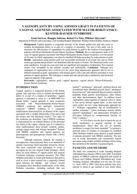
vaginoplasty by using amnion graft in patients of vaginal agenesis ...
vaginoplasty by using amnion graft in patients of vaginal agenesis ...
vaginoplasty by using amnion graft in patients of vaginal agenesis ...

Create successful ePaper yourself
Turn your PDF publications into a flip-book with our unique Google optimized e-Paper software.
J Ayub Med Coll Abbottabad 2010;22(1)<br />
VAGINOPLASTY BY USING AMNION GRAFT IN PATIENTS OF<br />
VAGINAL AGENESIS ASSOCIATED WITH MAYOR-ROKITANSKY-<br />
KUSTER-HAUSER SYNDROME<br />
Iram Sarwar, Ruqqia Sultana, Rahat Un Nisa, Iftikhar Qayyum*<br />
Department <strong>of</strong> Obstetric and Gynaecology, Ayub Teach<strong>in</strong>g Hospital Abbottabad. *Rehman Medical Institute, Peshawar, Pakistan<br />
Background: Vag<strong>in</strong>al <strong>agenesis</strong> is congenital anomaly <strong>of</strong> the female genital tract and may occur as<br />
isolated developmental defect or as part <strong>of</strong> a complex <strong>of</strong> anomalies. The aim <strong>of</strong> this study was to<br />
determ<strong>in</strong>e the effectiveness <strong>of</strong> <strong>vag<strong>in</strong>oplasty</strong> <strong>by</strong> <strong>us<strong>in</strong>g</strong> <strong>amnion</strong> as <strong>graft</strong> <strong>in</strong> the creation <strong>of</strong> neovag<strong>in</strong>a for<br />
<strong>patients</strong> with Mayor-Rokitansky-Kuster-Hauser Syndrome. Methods: this is a retrospective study <strong>of</strong> 28<br />
cases <strong>of</strong> vag<strong>in</strong>al <strong>agenesis</strong> associated with Mayor-Rokitansky-Kuster-Hauser Syndrome, over the period<br />
<strong>of</strong> 20 years, <strong>in</strong> which <strong>vag<strong>in</strong>oplasty</strong> was done <strong>by</strong> modified McIndoe procedure <strong>by</strong> <strong>us<strong>in</strong>g</strong> <strong>amnion</strong> as <strong>graft</strong>.<br />
Results: <strong>vag<strong>in</strong>oplasty</strong> <strong>us<strong>in</strong>g</strong> <strong>amnion</strong> <strong>graft</strong> was successfully performed <strong>in</strong> all except one case <strong>in</strong> which<br />
rectum got opened and procedure was abandoned after the repair <strong>of</strong> rectum. The functional results were<br />
quite satisfactory. Except one case none had any significant peri-operative complication. Post surgical<br />
results were acceptable to the <strong>patients</strong> sexually and aesthetically. Conclusion: Although new<br />
techniques <strong>of</strong> <strong>vag<strong>in</strong>oplasty</strong> have evolved over the years <strong>us<strong>in</strong>g</strong> laparoscopic approach and <strong>by</strong> use <strong>of</strong><br />
different materials as <strong>graft</strong>, <strong>vag<strong>in</strong>oplasty</strong> with <strong>amnion</strong> <strong>graft</strong> is still a safe and effective procedure to treat<br />
<strong>patients</strong> <strong>of</strong> vag<strong>in</strong>al <strong>agenesis</strong>. The technique is simple and safe and provides a satisfactory and functional<br />
vag<strong>in</strong>a <strong>in</strong> majority <strong>of</strong> the <strong>patients</strong>.<br />
Keywords: <strong>vag<strong>in</strong>oplasty</strong>, <strong>amnion</strong> <strong>graft</strong>, vag<strong>in</strong>al <strong>agenesis</strong>, vag<strong>in</strong>al atresia, Mayor-Rokitansky-<br />
Kuster-Hauser Syndrome<br />
INTRODUCTION<br />
Vag<strong>in</strong>al <strong>agenesis</strong> is congenital anomaly <strong>of</strong> the female<br />
genital tract and may occur as isolated developmental<br />
defect or as part <strong>of</strong> a complex <strong>of</strong> anomalies. 1 vag<strong>in</strong>al<br />
<strong>agenesis</strong> is estimated to occur <strong>in</strong> 1 <strong>in</strong> 4000-5000 live<br />
female births. 2 vag<strong>in</strong>al <strong>agenesis</strong> is most commonly<br />
associated with Mayor-Rokitansky-Kuster-Hauser<br />
(MRKH) syndrome and androgen <strong>in</strong>sensitivity<br />
syndrome. 2 MRKH syndrome is a congenital<br />
malformation characterised <strong>by</strong> an absence <strong>of</strong> the vag<strong>in</strong>a<br />
associated with a variable abnormality <strong>of</strong> the uterus and<br />
the ur<strong>in</strong>ary tract but functional ovaries. 3 Two types <strong>of</strong><br />
this syndrome are described. Type-I MRKH syndrome<br />
is characterised <strong>by</strong> an isolated absence <strong>of</strong> the proximal<br />
two thirds <strong>of</strong> the vag<strong>in</strong>a, whereas type II is marked <strong>by</strong><br />
other malformations; these <strong>in</strong>clude vertebral, cardiac,<br />
urologic (upper tract), and otologic anomalies. 4 Patients<br />
with MRKH syndrome and vag<strong>in</strong>al <strong>agenesis</strong> are<br />
phenotypically and genotypically female with a 46XX<br />
karyotype. 1<br />
Although numerous methods for creat<strong>in</strong>g a<br />
neovag<strong>in</strong>a have been proposed, there is no unanimity <strong>of</strong><br />
op<strong>in</strong>ion concern<strong>in</strong>g which procedure should be chosen.<br />
The most commonly used techniques to create a<br />
neovag<strong>in</strong>a are the non surgical Frank technique, which<br />
relies on serial dilation <strong>of</strong> vag<strong>in</strong>al pouch and surgical<br />
Vecchietti technique (cont<strong>in</strong>ous pressure). The Abbe<br />
McIndoe procedure <strong>in</strong> which split thickness sk<strong>in</strong> <strong>graft</strong> is<br />
used to cover a stent <strong>in</strong>serted <strong>in</strong>to a surgically created<br />
space between the bladder and rectum. 4 several<br />
<strong>in</strong>vestigators have described modifications <strong>of</strong> the Abbe<br />
McIndoe procedure, <strong>in</strong>clud<strong>in</strong>g methods that use<br />
<strong>amnion</strong> 5,6 , peritoneum 7 , <strong>in</strong>tercede 8 , artificial dermis and<br />
recomb<strong>in</strong>ant basic fibroblast growth factor 9 , autologous<br />
buccal mucosa 10 and rotational flap procedures <strong>us<strong>in</strong>g</strong> the<br />
pudendal, thigh, gracilis myocutaneous, labia m<strong>in</strong>ora<br />
and other fasciocutaneous flaps 4 . In addition bowel<br />
<strong>vag<strong>in</strong>oplasty</strong> <strong>us<strong>in</strong>g</strong> segment <strong>of</strong> sigmoid colon or ileum<br />
to l<strong>in</strong>e newly formed vag<strong>in</strong>al canal is also used and<br />
some centres are now <strong>us<strong>in</strong>g</strong> laparoscopic approach for<br />
it. 11 Williams <strong>vag<strong>in</strong>oplasty</strong> and its modifications is<br />
another technique. 12 Latest techniques <strong>in</strong>clude robotic<br />
sigmoid <strong>vag<strong>in</strong>oplasty</strong> 13 and laparoscopic formation <strong>of</strong><br />
neovag<strong>in</strong>a followed <strong>by</strong> extraperitoneal traction on<br />
Foley’s catheter. 14<br />
In 1910 Davis was the first to report the use <strong>of</strong><br />
foetal membranes as surgical material <strong>in</strong> sk<strong>in</strong><br />
transplantation. S<strong>in</strong>ce then the use <strong>of</strong> amniotic<br />
membrane <strong>in</strong> surgery has been expanded. 15 we present a<br />
personal series <strong>of</strong> creation <strong>of</strong> neovag<strong>in</strong>a <strong>by</strong> modified<br />
Abbe McIndoe method <strong>us<strong>in</strong>g</strong> <strong>amnion</strong> as <strong>graft</strong> material<br />
<strong>in</strong> <strong>patients</strong> presented with MRKH syndrome. The aim<br />
was to create functionally and cosmetically normal<br />
neovag<strong>in</strong>a <strong>us<strong>in</strong>g</strong> simple available technique and to br<strong>in</strong>g<br />
this operation to the attention <strong>of</strong> gynaecologists.<br />
PATIENTS AND METHODS<br />
The study was conducted at Ayub Teach<strong>in</strong>g Hospital<br />
Abbottabad from January 2009 to June 2009. It <strong>in</strong>cluded<br />
<strong>patients</strong> <strong>of</strong> MRKH syndrome diagnosed and treated at<br />
Women and Children Hospital Abbottabad and Ayub<br />
Teach<strong>in</strong>g Hospital Abbottabad over the last 20 years,<br />
i.e., from 1989 to 2009. Only those <strong>patients</strong> who were<br />
married or about to get married <strong>in</strong> near future (three<br />
http://www.ayubmed.edu.pk/JAMC/PAST/22-1/Iram.pdf 7
J Ayub Med Coll Abbottabad 2010;22(1)<br />
months before marriage) were operated and <strong>in</strong>cluded <strong>in</strong><br />
study due to our social setup. It is a descriptive study<br />
with data collected retrospectively. All <strong>patients</strong><br />
exhibited primary amenorrhoea, normal female<br />
secondary sex characteristics and a vag<strong>in</strong>al dimple<br />
without vag<strong>in</strong>al orifice. Patients underwent preoperative<br />
workup which <strong>in</strong>cluded apart from rout<strong>in</strong>e<br />
<strong>in</strong>vestigations karyotyp<strong>in</strong>g, abdom<strong>in</strong>opelvic ultrasound<br />
and diagnostic laparoscopy. Patients and their parents<br />
were thoroughly counselled before operation about the<br />
optimal operation time, method as well as the possible<br />
complications <strong>of</strong> the procedure. All <strong>patients</strong> were<br />
followed for at least 6 months.<br />
Amniotic membranes were obta<strong>in</strong>ed under<br />
sterile conditions from elective Caesarean deliveries.<br />
Amnion donors (mothers) were screened for hepatitis B<br />
and C as well as HIV viral <strong>in</strong>fections and syphilis. Inner<br />
amniotic membrane was separated from outer<br />
membrane and r<strong>in</strong>sed <strong>in</strong> sterile normal sal<strong>in</strong>e solution<br />
conta<strong>in</strong><strong>in</strong>g cephalospor<strong>in</strong> <strong>in</strong>jection.<br />
Under general anaesthesia, the patient was<br />
placed <strong>in</strong> lithotomy position after catheterisation and<br />
per<strong>in</strong>eal area cleaned and draped. A transverse <strong>in</strong>cision<br />
was made just below the dimple and a potential space<br />
was created <strong>in</strong> between the bladder and urethra and<br />
rectum <strong>by</strong> blunt dissection, carefully palpat<strong>in</strong>g the<br />
catheter <strong>in</strong> front and a f<strong>in</strong>ger <strong>in</strong> the rectum to guard<br />
aga<strong>in</strong>st the <strong>in</strong>jury. A cavity size <strong>of</strong> depth 8–10 cm <strong>in</strong><br />
length and about 4–5 cm <strong>in</strong> diameter were achieved.<br />
A vag<strong>in</strong>al mould made with 50 ml syr<strong>in</strong>ge<br />
wrapped with foam, covered with latex condom and<br />
sterilised <strong>in</strong> cidex solution was then wrapped with<br />
<strong>amnion</strong> tent and placed <strong>in</strong> the constructed cavity. The<br />
<strong>amnion</strong> <strong>graft</strong> was fixed to mould <strong>by</strong> sutur<strong>in</strong>g the edges<br />
<strong>of</strong> <strong>amnion</strong> to the mould. The labia majora was then<br />
sutured together loosely with silk sutures to hold the<br />
mould <strong>in</strong> position and T bandage applied. Prophylactic<br />
antibiotics were given for 7 days. Mould was removed<br />
on day 8 along with catheter. The <strong>graft</strong> was reta<strong>in</strong>ed and<br />
well taken <strong>in</strong> all the cases. Vag<strong>in</strong>al douch<strong>in</strong>g was done<br />
with pyod<strong>in</strong>e and second mould made with 20 ml<br />
syr<strong>in</strong>ge (with upper dra<strong>in</strong>age hole created) was kept <strong>in</strong><br />
place. Patients were counselled about the method <strong>of</strong><br />
placement, removal and wash<strong>in</strong>g <strong>of</strong> mould to facilitate<br />
the further change <strong>of</strong> mould herself. Patients were<br />
discharged with the advice to wear the mould for 3<br />
months cont<strong>in</strong>uously followed <strong>by</strong> nightly <strong>in</strong>sertions for<br />
another 3 months to prevent contractions. Fortnightly<br />
follow up visits were advised. Physical relation was<br />
allowed after 3 months <strong>in</strong> married women.<br />
RESULTS<br />
A total <strong>of</strong> 28 females underwent the surgical procedure<br />
dur<strong>in</strong>g these 20 years. Their ages ranged from 16 to 22<br />
years; 26 (93%) were unmarried and 2 were married.<br />
The unmarried females presented with primary<br />
amenorrhoea; the married ones with primary<br />
amenorrhoea and <strong>in</strong>ability to have sexual <strong>in</strong>tercourse.<br />
On cl<strong>in</strong>ical exam<strong>in</strong>ation, all subjects had<br />
normal female secondary sexual characteristics and the<br />
external genitalia were normal female. However all <strong>of</strong><br />
them had absence <strong>of</strong> vag<strong>in</strong>a. Ultrasound and<br />
Diagnostic laparoscopy revealed a small nodular/<br />
rudimentary/absent uterus <strong>in</strong> all cases with normal<br />
ovaries and distal part <strong>of</strong> fallopian tubes.<br />
Associated renal tract anomalies were found<br />
<strong>in</strong> 4 <strong>patients</strong> (14%). These <strong>in</strong>cluded horse shoe shaped<br />
kidney <strong>in</strong> one (25%), double unilateral kidney <strong>in</strong> one<br />
(25%) and a s<strong>in</strong>gle kidney <strong>in</strong> 2 <strong>patients</strong> (50%).<br />
Karyotypes were performed <strong>in</strong> 18 <strong>patients</strong><br />
(64.3%) who showed a normal XX female pattern.<br />
Serum testosterone levels were done <strong>in</strong> 15 cases<br />
(53.6%) with normal female levels.<br />
The operation times ranged from 20–45<br />
m<strong>in</strong>utes. There was immediate per-operative<br />
complication <strong>of</strong> rectal <strong>in</strong>jury <strong>in</strong> 1 patient requir<strong>in</strong>g<br />
abandonment <strong>of</strong> <strong>vag<strong>in</strong>oplasty</strong>; the rema<strong>in</strong><strong>in</strong>g <strong>patients</strong><br />
underwent successful <strong>vag<strong>in</strong>oplasty</strong>. Outcome <strong>of</strong><br />
<strong>vag<strong>in</strong>oplasty</strong> at 3 months showed that 24/27 (89%) had<br />
normal recovery with vag<strong>in</strong>al depths <strong>of</strong> up to 7 cm.<br />
Three <strong>patients</strong> (11%) had vag<strong>in</strong>al constriction due to<br />
poor compliance with second mould placement<br />
protocol; digital dilation was performed under General<br />
Anaesthesia.<br />
At 6 months follow up, all <strong>patients</strong> had<br />
adequate vag<strong>in</strong>al lengths and diameters. All <strong>of</strong> them<br />
had normal sexual <strong>in</strong>tercourse after 3 months <strong>of</strong><br />
surgery (unmarried <strong>patients</strong> also got married <strong>by</strong> then)<br />
obviat<strong>in</strong>g the need for second mould placement.<br />
Figure-1: Before surgery<br />
Figure-2: Transverse <strong>in</strong>cision below dimple to<br />
create potential space<br />
8<br />
http://www.ayubmed.edu.pk/JAMC/PAST/22-1/Iram.pdf
J Ayub Med Coll Abbottabad 2010;22(1)<br />
Figure-3: Mould<br />
Figure-4: Mould removal on day 8<br />
Figure-5: Second mould placement<br />
DISCUSSION<br />
Figure-6: 3 months after surgery<br />
Amniotic membranes have been used as surgical<br />
material <strong>in</strong> different procedures <strong>in</strong>clud<strong>in</strong>g as dress<strong>in</strong>g<br />
for burned sk<strong>in</strong>, sk<strong>in</strong> wounds and chronic leg ulcers,<br />
surgical reconstruction <strong>of</strong> vag<strong>in</strong>a and repair <strong>of</strong><br />
omphalocoeles, and to prevent tissue adhesions <strong>in</strong><br />
surgical procedures. It has also been used <strong>in</strong> treat<strong>in</strong>g<br />
variety <strong>of</strong> ocular surface disorders. 15<br />
We selected <strong>amnion</strong> as <strong>graft</strong> for<br />
<strong>vag<strong>in</strong>oplasty</strong> over sk<strong>in</strong>/other <strong>graft</strong>s because it is<br />
easily available and its supply is nearly unlimited.<br />
Amniotic membranes do not express HLA-A, B or<br />
DR antigens hence immunological rejection does not<br />
occur. It is also believed to have antimicrobial<br />
properties reduc<strong>in</strong>g the risks <strong>of</strong> postoperative<br />
<strong>in</strong>fection. Antifibroblastic activity and cell<br />
migration/growth promot<strong>in</strong>g activity have also been<br />
demonstrated which stimulates epithelialisation. 15<br />
and lastly its preparation method and time did not<br />
pose any challenge. Other methods <strong>us<strong>in</strong>g</strong> sk<strong>in</strong> and<br />
buccal mucosa and peritoneum may scar the patient. 10<br />
Use <strong>of</strong> <strong>in</strong>test<strong>in</strong>e cause cont<strong>in</strong>uous pr<strong>of</strong>use secretions<br />
and unpleasant odour. 1 Laparoscopic techniques are<br />
lengthier and require specialised skills and tra<strong>in</strong><strong>in</strong>g.<br />
Dilation techniques although simple, require<br />
motivation and long term follow up.<br />
Twenty-eight cases were recorded for<br />
<strong>vag<strong>in</strong>oplasty</strong>. All <strong>patients</strong> except one (96.43%) had<br />
uneventful surgical procedures and successful<br />
outcomes. In one patient rectum got opened dur<strong>in</strong>g<br />
the procedure and then the procedure was abandoned.<br />
Follow up at 3 months was satisfactory <strong>in</strong> 89% <strong>of</strong><br />
<strong>patients</strong>, while 11% required a m<strong>in</strong>or second<br />
procedure <strong>in</strong> the form <strong>of</strong> digital dilation due to<br />
vag<strong>in</strong>al constriction secondary to poor compliance.<br />
Follow-up at 6 months was satisfactory <strong>in</strong> 100% <strong>of</strong><br />
<strong>patients</strong> <strong>in</strong> terms <strong>of</strong> anatomical and functional results.<br />
A study conducted at Lahore <strong>in</strong> Pakistan <strong>in</strong><br />
2006 on 10 <strong>patients</strong> over 4 years <strong>us<strong>in</strong>g</strong> <strong>amnion</strong> <strong>graft</strong>,<br />
had similar results. In that study one patient had rectal<br />
<strong>in</strong>jury dur<strong>in</strong>g surgery (90% operative success rate);<br />
however operation was carries out after rectal repair.<br />
At 6 months they had 80% success rate, one patient<br />
had cicatrisation and one was lost to follow-up. 5<br />
Another study from Germany conducted <strong>in</strong><br />
2009 on 7 <strong>patients</strong> also reported similar outcomes.<br />
Operative success was 85.71% and one patient had<br />
major operative complication. After 18 months follow<br />
up anatomical and functional results were 100%. 6<br />
Although few studies have used <strong>amnion</strong> as<br />
<strong>graft</strong> <strong>in</strong> the creation <strong>of</strong> neovag<strong>in</strong>a but the results are<br />
very satisfy<strong>in</strong>g. Advantages <strong>of</strong> this procedure is that<br />
it is safe, <strong>in</strong>expensive and easy to perform. Epithelial<br />
l<strong>in</strong><strong>in</strong>g <strong>of</strong> the neovag<strong>in</strong>a resembl<strong>in</strong>g normal vag<strong>in</strong>a is<br />
found, which facilitates comfortable sexual<br />
<strong>in</strong>tercourse. There is less emotional stress and better<br />
cosmetic and economic benefits.<br />
CONCLUSION<br />
The ideal method for <strong>vag<strong>in</strong>oplasty</strong> is not currently<br />
known and depends on numerous factors <strong>in</strong>clud<strong>in</strong>g<br />
http://www.ayubmed.edu.pk/JAMC/PAST/22-1/Iram.pdf 9
J Ayub Med Coll Abbottabad 2010;22(1)<br />
patient preparedness, surgeon experience, and patient<br />
and surgeon preference. Although new techniques <strong>of</strong><br />
<strong>vag<strong>in</strong>oplasty</strong> have evolved over the years <strong>us<strong>in</strong>g</strong><br />
laparoscopic approach and <strong>by</strong> use <strong>of</strong> different<br />
materials as <strong>graft</strong>, but <strong>in</strong> develop<strong>in</strong>g country like<br />
Pakistan where facilities and expertise for newer<br />
techniques are not available freely, <strong>vag<strong>in</strong>oplasty</strong> <strong>by</strong><br />
modified Abbe-McIndoe procedure <strong>us<strong>in</strong>g</strong> <strong>amnion</strong><br />
<strong>graft</strong> is still a safe and effective procedure to treat<br />
<strong>patients</strong> <strong>of</strong> vag<strong>in</strong>al <strong>agenesis</strong>.<br />
RECOMMENDATIONS<br />
Vag<strong>in</strong>oplasty <strong>by</strong> modified Abbe-McIndoe procedure<br />
<strong>us<strong>in</strong>g</strong> <strong>amnion</strong> <strong>graft</strong> should be recommended <strong>in</strong><br />
develop<strong>in</strong>g countries lack<strong>in</strong>g modern facilities as<br />
well as <strong>in</strong> developed countries because this procedure<br />
is simple, safe and effective and requires less<br />
expertise as compared to more modern and<br />
sophisticated procedures.<br />
REFERENCES<br />
1. Saxena AK, Herman MI. Vag<strong>in</strong>al Atresia. Webpage. Cited<br />
June 02, 2009. Available at: http://emedic<strong>in</strong>e.medscape.com/<br />
article/954110-pr<strong>in</strong>t.<br />
2. Saraf S, Saraf P. McIndoe <strong>vag<strong>in</strong>oplasty</strong>: revisited. Internet J<br />
Gynecol Obstetr 2007;6(2). Onl<strong>in</strong>e Journal. Cited August 18,<br />
2008. Available from: http://www.ispub.com/journal/the<br />
_<strong>in</strong>ternet_journal_<strong>of</strong>_gynecology_and_obstetrics/volume_6_<br />
number_2_6/article/mc<strong>in</strong>doe_<strong>vag<strong>in</strong>oplasty</strong>_revisited.htmls.<br />
3. Gupta NP, Ansari MS. Mayer-Rokitansky-Kuster-Hauser<br />
(MRKH) syndrome–a review. Indian J Urol 2002;18:111–6.<br />
4. Kirsch AJ, Kaye JD, Carter SM, Gross SJ. Mayer-Rokitansky<br />
syndrome. Webpage. Cited June 02, 2009. Available from:<br />
http://emedic<strong>in</strong>e.medscape.com/article/953492-pr<strong>in</strong>t.<br />
5. Chohan A, Burr F, Mansoor H, Falak T. Amnion <strong>graft</strong> <strong>in</strong><br />
<strong>vag<strong>in</strong>oplasty</strong>–an experience at 3 teach<strong>in</strong>g hospitals <strong>of</strong> Lahore.<br />
Biomedica 2006;22(1):21–4.<br />
6. Fotopoulou C, Sehouli J, Gehrmann N, Schoenborn I,<br />
Lichtenegger W. Functional and anatomical results <strong>of</strong><br />
<strong>amnion</strong> <strong>vag<strong>in</strong>oplasty</strong> <strong>in</strong> young women with Mayer-<br />
Rokitansky-Kuster-Hauser syndrome. Fertil Steril 2009;<br />
(Epub ahead <strong>of</strong> pr<strong>in</strong>t).<br />
7. Rothman D. The use <strong>of</strong> peritoneum <strong>in</strong> the construction <strong>of</strong> a<br />
vag<strong>in</strong>a. Obstet Gynecol 1972;40:835–8.<br />
8. Jackson ND, Rosenblatt PL. Use <strong>of</strong> <strong>in</strong>terceed absorbable<br />
adhesion barrier for <strong>vag<strong>in</strong>oplasty</strong>. Obstet Gynecol<br />
1994;84:1048–50.<br />
9. Noguchi S, Nakatsuka M, Sugiyama Y, Chekir C, Kamada<br />
Y, Hiramatsu Y. Use <strong>of</strong> artificial dermis and recomb<strong>in</strong>ant<br />
basic fibroblast growth factor for creat<strong>in</strong>g a neovag<strong>in</strong>a <strong>in</strong> a<br />
patient with Mayer-Rokitansky-Kuster-Hauser syndrome.<br />
Hum Reprod 2004;19:1629–32.<br />
10. L<strong>in</strong> WC, Chang CYY, Shen YY, Tsai HD. Use <strong>of</strong> autologous<br />
buccal mucosa for <strong>vag<strong>in</strong>oplasty</strong>: a study <strong>of</strong> eight cases. Hum<br />
Reprod 2003;18:604–7.<br />
11. Cai B, Zhang JR, Xi XW, Yan Q, Wan XP. Laparoscopically<br />
assisted sigmoid colon <strong>vag<strong>in</strong>oplasty</strong> <strong>in</strong> women with Mayer-<br />
Rokitansky-Kuster-Hauser syndrome: feasibility and short<br />
term results. BJOG 2007;114:1486–92.<br />
12. Creatsas G, Deligeoroglou E, Makrakis E, Kontoravdis A.<br />
Papadimitriou L. Creation <strong>of</strong> a neovag<strong>in</strong>a follow<strong>in</strong>g Williams<br />
<strong>vag<strong>in</strong>oplasty</strong> and Creatsas modification <strong>in</strong> 111 <strong>patients</strong> with<br />
Mayer-Rokitansky-Kuster-Hauser syndrome. Fertil Steril<br />
2001;76:1036–40.<br />
13. Kim C, Campbell B, Ferrer F. Robotic sigmoid <strong>vag<strong>in</strong>oplasty</strong>:<br />
a novel technique. Urology 2008;72:847–9.<br />
14. El-Saman AM. Retropubic balloon <strong>vag<strong>in</strong>oplasty</strong> for<br />
management <strong>of</strong> Mayer-Rokitansky-Kuster-Hauser syndrome.<br />
Fertil Steril 2009; (Epub ahead <strong>of</strong> pr<strong>in</strong>t).<br />
15. Dua HS, Azuara-Blanca A. Amniotic membrane<br />
transplantation. Br J Opthalmol 1999;83:748–52.<br />
Address for Correspondence:<br />
Dr. Iram Sarwar, Department <strong>of</strong> Obstetric and Gynaecology, Ayub Medical College, Abbottabad-22040, Pakistan.<br />
Cell: +92-333-5058286<br />
Email: iramsarwar@hotmail.com<br />
10<br />
http://www.ayubmed.edu.pk/JAMC/PAST/22-1/Iram.pdf