ABR Radiation Oncology Study Guide: Medical Physics for ...
ABR Radiation Oncology Study Guide: Medical Physics for ...
ABR Radiation Oncology Study Guide: Medical Physics for ...
Create successful ePaper yourself
Turn your PDF publications into a flip-book with our unique Google optimized e-Paper software.
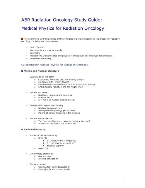
<strong>ABR</strong> <strong>Radiation</strong> <strong>Oncology</strong> <strong>Study</strong> <strong>Guide</strong>:<br />
<strong>Medical</strong> <strong>Physics</strong> <strong>for</strong> <strong>Radiation</strong> <strong>Oncology</strong><br />
This exam tests your knowledge of the principles of physics underlying the practice of radiation<br />
oncology. Included are questions on:<br />
• basic physics<br />
• instruments and measurements<br />
• dosimetry<br />
• radioactivity (radionuclides and physics of therapeutically employed radionuclides)<br />
• protection and safety<br />
Categories <strong>for</strong> <strong>Medical</strong> <strong>Physics</strong> <strong>for</strong> <strong>Radiation</strong> <strong>Oncology</strong><br />
Atomic and Nuclear Structure<br />
• Bohr model of the atom<br />
o Coulombic <strong>for</strong>ce and electron binding energy<br />
o Electron orbits (energy levels)<br />
o Electron transitions—absorption and emission of energy<br />
o Characteristic radiation and the Auger effect<br />
• Nuclear structure<br />
o Nucleons —protons and neutrons<br />
o Nuclear <strong>for</strong>ce<br />
o E = mc 2 and nuclear binding energy<br />
• Factors affecting nuclear stability<br />
o Neutron-to-proton ratio<br />
o Average binding energy per nucleon<br />
o Pairing of similar nucleons in the nucleus<br />
• Nuclear nomenclature<br />
o The four isos (isotopes, isotones, isobars, isomers)<br />
o Shorthand representation of isotopes<br />
Radioactive Decay<br />
• Modes of radioactive decay<br />
o Beta (ß)<br />
ƒ ß- (negative beta, negatron)<br />
ƒ ß+ (positive beta, positron)<br />
ƒ Electron capture<br />
o Alpha (α)<br />
• Other decay processes<br />
o Gamma rays<br />
o Internal conversion<br />
• Decay schemes<br />
o Construction and interpretation<br />
o Examples <strong>for</strong> each decay mode<br />
1
• Mathematics of radioactive decay<br />
o Units (SI Units)<br />
o Exponential decay equation<br />
ƒ Half-life<br />
ƒ Decay constant<br />
ƒ Mean life, average life, and effective half life<br />
ƒ Simple dose calculation <strong>for</strong> implants<br />
o Radioactive equilibrium<br />
ƒ Secular equilibrium<br />
ƒ Radium needles<br />
ƒ<br />
90 Sr applicators<br />
ƒ Transient equilibrium<br />
ƒ Nuclear medicine generators<br />
o Counting statistics<br />
• Naturally occurring radioisotopes<br />
• Manmade radioisotopes<br />
o Fission<br />
o Nuclear bombardment<br />
• Decay schemes and properties <strong>for</strong> therapeutic isotopes<br />
Properties and Production of Particulate and Electromagnetic <strong>Radiation</strong><br />
• Particulate radiation<br />
o Mass, charge<br />
o Relativistic energy equation<br />
• Electromagnetic radiation<br />
o Wave-particle duality<br />
o Wave equations<br />
o Electromagnetic spectrum<br />
• Production of radiation<br />
o Principles<br />
o Radioactive decay<br />
o X-ray tube<br />
• Linear accelerators<br />
o Operational theory of wave guides<br />
ƒ Standing wave guides<br />
ƒ Traveling wave guides<br />
o Bending magnet systems<br />
o Flattening filters<br />
o Electron scattering foils<br />
o Electron cones<br />
o Targets<br />
o Factors affecting<br />
ƒ Beam energy<br />
ƒ Entrance dose<br />
ƒ Depth of maximum dose<br />
ƒ Beam uni<strong>for</strong>mity<br />
ƒ Dose rate<br />
o Monitor chamber<br />
o Collimation systems<br />
2
ƒ Primary and secondary collimators<br />
ƒ Coupled and independent jaws (including virtual wedges)<br />
ƒ Multileaf collimators<br />
ƒ Other collimation systems (e.g., stereotactic systems)<br />
ƒ <strong>Radiation</strong> and light fields (including field size definition)<br />
o Mechanical and operational features<br />
• Cyclotron<br />
• Microtron<br />
• Cobalt units<br />
• Therapeutic x-ray (
o Elastic collisions with hydrogen nuclei<br />
o Depth dose characteristics vs charged particles and photons<br />
• Biological implications of particle therapy<br />
Quantification and Measurement of Dose (including SI units)<br />
• Exposure (air kerma)<br />
• Absorbed dose (kerma)<br />
• Dose equivalent<br />
• RBE dose<br />
• Calculation of absorbed dose from exposure (e.g., f factor)<br />
• Bragg-Gray cavity theory<br />
• Gas-filled detectors<br />
o Principles of operation<br />
o Uses<br />
• Ion chambers<br />
o Types<br />
o Exposure measurement<br />
o As a Bragg-Gray cavity<br />
o Correction factors (e.g., temperature and pressure)<br />
o Calibration of photon and electron beams (e.g., TG 21 and TG 25)<br />
• Thermoluminescent dosimetry<br />
• Calorimetry<br />
• Film<br />
• Chemical dosimetry<br />
• Solid state diodes<br />
• Scintillation detectors<br />
• Measurement techniques<br />
Characteristics of Photon Beams<br />
• Mathematics of exponential attenuation<br />
o Half-value thickness<br />
o Attenuation coefficients (linear, mass, partial, total)<br />
o Narrow beam vs broad beam geometry<br />
o Monoenergetic vs heteroenergetic<br />
o Parallel vs diverging beams<br />
• Beam quality <strong>for</strong> heteroenergetic beams<br />
o Energy distribution of accelerated electron beam<br />
4
o Filtration<br />
o Geometry<br />
o Effective energy<br />
o Energy spectra<br />
Dosimetry of Photon Beams in a Homogeneous Water Phantom<br />
• Dose distributions<br />
o Central axis percent depth dose<br />
o Isodose curves<br />
o Factors affecting dose distributions and penumbra<br />
ƒ Beam energy or quality (including patient dose from neutrons)<br />
ƒ Source size<br />
ƒ SSD and SAD<br />
ƒ Mayneord F factor<br />
ƒ Inverse square law<br />
ƒ Field size and shape<br />
ƒ Equivalent square<br />
ƒ Scatter effects<br />
ƒ Flattening filters<br />
ƒ Depth<br />
ƒ Surface dose<br />
ƒ Other<br />
o Dose distributions <strong>for</strong> multiple unshaped beams<br />
ƒ Open beams<br />
ƒ Wedged beams<br />
o Tissue-air ratio and backscatter factor<br />
o Tissue-maximum ratio<br />
o Tissue-phantom ratio<br />
o Relationships between PDD, TAR, TMR, TPR<br />
o Point dose and treatment time calculation methods <strong>for</strong> single unshaped fields<br />
ƒ Machine output factors (e.g., absolute and relative output, head scatter,<br />
patient scatter factors)<br />
ƒ Equivalent squares<br />
ƒ SSD vs SAD setups<br />
ƒ Beam modifier factors (e.g., wedge and tray factors)<br />
o Dose calculation at the isocenter of a rotating beam<br />
o Point dose and treatment time calculations <strong>for</strong> single shaped fields<br />
ƒ Separation and recombination of primary and scatter radiation (e.g.,<br />
Clarkson techniques)<br />
ƒ Off-axis factors<br />
ƒ Dose under blocks<br />
ƒ Equivalent squares <strong>for</strong> shaped fields<br />
o Isodose distributions <strong>for</strong> multiple fields, including arc therapy<br />
o Measurement of photon dose distributions<br />
Dosimetry of Photon Beams in a Patient<br />
• Dose specification (eg, ICRU 50)<br />
• Corrections <strong>for</strong> patient contour<br />
o Effective SSD method<br />
o TAR ratio method<br />
o Isodose shift method<br />
• Corrections <strong>for</strong> tissue inhomogeneities<br />
o TAR ratio method<br />
o Power law method<br />
o Isodose shift method<br />
5
Equivalent TAR<br />
Superposition/Convolution<br />
Monte Carlo<br />
• Dose within and around an inhomogeneity<br />
• Matching of adjacent fields<br />
• Using multiple wedged fields<br />
• Parallel opposed beams<br />
o Point of maximum dose<br />
o Uni<strong>for</strong>mity, dependence upon<br />
ƒ Energy<br />
ƒ Separation<br />
ƒ SSD<br />
• Entrance dose and exit dose, including beam modifying devices<br />
• Isodose distributions <strong>for</strong> multiple beams, including mixed modality and arc therapy<br />
• Compensation<br />
o Missing tissue<br />
o Dose compensation<br />
o Bolus<br />
• Off-axis factors<br />
• Practical/simple calculation of dose<br />
• Practical/simple 2D treatment planning<br />
• 3D con<strong>for</strong>mal treatment planning<br />
• Dose delivery accuracy and precision<br />
Dosimetry of Electron Beams<br />
• Dose distributions<br />
o Central axis percent depth dose<br />
o Isodose curves<br />
• Factors affecting dose distributions<br />
o Beam quality<br />
o Beam spreading systems<br />
o SSD and SDD<br />
ƒ Effective SSD techniques<br />
ƒ Inverse square<br />
o Field size and shape<br />
o X-ray contamination<br />
o Depth<br />
o Surface dose<br />
o Inhomogeneities (e.g., CET)<br />
o Other<br />
• Energy specification<br />
o Most probable energy<br />
o Mean energy<br />
6
o Energy at depth<br />
o Ranges (extrapolated, practical, R50)<br />
• Choice of energy and field size<br />
• Air gaps and oblique incidence<br />
• Tissue inhomogeneities<br />
• Bolus, absorbers, and spoilers<br />
• Matching adjacent fields<br />
• Point dose and treatment time calculations<br />
• Field shaping techniques<br />
• Electron arc<br />
• Total skin electron therapy<br />
Brachytherapy<br />
• Historical review—role of radium<br />
• Calculation of dose from a point source<br />
• Calculation of dose from a line source<br />
• Physical and dosimetric properties of commercial sealed sources and applicators<br />
• Implant instrumentation and techniques<br />
o Low dose rate<br />
o High dose rate (including PDR)<br />
o Biological considerations of dose, dose rate, and fractionation<br />
• Calibration and specification of sources<br />
• Disseminated (unsealed sources)<br />
• Acceptance testing and quality assurance<br />
• Dose specification, implantation dosimetry, and dosimetry systems<br />
o Patterson-Parker<br />
o Quimby<br />
o Paris<br />
o Other<br />
• Dose specification and dosimetry systems of intracavitary implants<br />
Advanced Treatment Planning <strong>for</strong> EBRT<br />
• Plane radiography and fluoroscopy <strong>for</strong> simulation<br />
• Portal imaging<br />
7
o Film-based<br />
o Electronic<br />
• Imaging <strong>for</strong> radiation therapy planning<br />
o CT<br />
o MRI<br />
o Ultrasound<br />
o Isotope imaging<br />
• Image processing<br />
o Image enhancement<br />
o 2D and 3D visualization of volumetric data (DRRs, volume rendering)<br />
o Image registration<br />
• Virtual simulation (including BEV techniques)<br />
• Treatment planning systems<br />
• 3D con<strong>for</strong>mal treatment planning<br />
o Plan evaluation (DVH, NTCP, TCP, etc)<br />
o Dose optimization techniques<br />
o Noncoplanar beams<br />
• Radiosurgery<br />
• Patient setup and alignment<br />
• IMRT<br />
Quality Assurance<br />
• Equipment-related<br />
o Regulations and recommendations<br />
o Measurement techniques<br />
• Patient related<br />
o Misadministration<br />
• External beam<br />
• Brachytherapy<br />
• Patient-specific QA<br />
• Brachytherapy source inventory<br />
<strong>Radiation</strong> Protection and Safety<br />
• Principles, biological effect models, personnel dose limits, rules, regulations<br />
• Structural shielding design <strong>for</strong> external beam therapy<br />
o Primary barriers<br />
o Secondary barriers<br />
o Machine shielding (beam stoppers and head shielding)<br />
o Neutrons<br />
• <strong>Radiation</strong> protection <strong>for</strong> brachytherapy procedures<br />
o Source storage and transport containers<br />
o Patient room<br />
o Special considerations <strong>for</strong> high dose rate brachytherapy<br />
o Special procedures and source prep rooms<br />
o Release of patients treated with temporary implants<br />
8
• Leak testing of sealed sources<br />
• Routine radiation surveys<br />
• Personnel monitoring<br />
• Protection against nonionizing radiation<br />
• Administrative requirements<br />
o <strong>Radiation</strong> Safety Officer<br />
o <strong>Radiation</strong> Safety Committee<br />
• Safety instructions and safety precautions<br />
o Sealed-source brachytherapy<br />
o Radiopharmaceutical therapy<br />
Quality Management Program<br />
• Written directive<br />
• Identification of patient<br />
• Plan and delivery in accordance with written directive<br />
• Unintended deviation<br />
o Recordable events<br />
o Misadministrations<br />
Special Topics<br />
• Hyperthermia<br />
• Computers<br />
9


