Oral Lesions at Birth
Oral Lesions at Birth
Oral Lesions at Birth

Create successful ePaper yourself
Turn your PDF publications into a flip-book with our unique Google optimized e-Paper software.
The Iranian Journal of<br />
Otorhinolaryngology<br />
Vol.17, No.41, Autumn-2005<br />
هجلِ گَش،گلَ،تیٌی ٍ حٌجرُ ایراى،<br />
دٍرُ ّفذّن،شوارُ چْل ٍیکن، پاییس1384<br />
<strong>Oral</strong> <strong>Lesions</strong> <strong>at</strong> <strong>Birth</strong><br />
1 Mohammadzadeh A.MD, 2 Mokhtari N.MD<br />
1 Neon<strong>at</strong>ologist , 2 Otolaryngologist -Mashhad University of Medical Sciences<br />
Abstract<br />
Objective: The aim of this study was to determine the frequency distribution of oral lesions <strong>at</strong><br />
birth.<br />
Method: All babies delivered in Mashhad sina hospital were examined for oral lesions<br />
immedi<strong>at</strong>ely after birth for one year since 1.1.82.<br />
In this cross sectional descriptive study , frequency distribution was detected From d<strong>at</strong>a.<br />
Results: During the study period 3298 babies were delivered. <strong>Oral</strong> lesions included 76%<br />
Epstein pearls , 35% Bone nodule , 0.33% ankyloglossia, 0.12% n<strong>at</strong>al teeth, 0.03%<br />
congenital epulis, 0.03% cleft lip , 0.06% cleft pal<strong>at</strong>e , 0.03% cleft lip and pal<strong>at</strong>e.<br />
Conclusion: In this study Epstein pearls were the most common lesion <strong>at</strong> birth and cleft lip,<br />
with or without cleft pal<strong>at</strong>e and congenital epulis, were the lowest ones.<br />
Keyword: Newborn, <strong>Oral</strong> lesion , Routine examin<strong>at</strong>ion of the newborn<br />
Introduction<br />
O<br />
ral structures should be examined<br />
routinely <strong>at</strong> birth and <strong>at</strong> Coneeguext<br />
child visits. Early examin<strong>at</strong>ion can reveal<br />
abnormalities th<strong>at</strong> require tre<strong>at</strong>ment or serve<br />
as baseline against which to compare l<strong>at</strong>er<br />
development.<br />
The oral lesions th<strong>at</strong> present in newborn<br />
periods include n<strong>at</strong>al and neon<strong>at</strong>al teeth,<br />
clefts, dental lamina cyst, Epstein pearls,<br />
Bohn nodules, alveolar lymphangioma,<br />
Tumors (congenital epulis), ankyloglossia,<br />
geographic tongue, ranula, mucocele and<br />
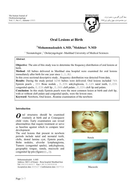
congenital lip pits (figure) (1,2).<br />
Ranula<br />
Mohammadzadeh A.MD<br />
Address: NICU of Emam – Reza hospital Mashhad Iran<br />
Tel: 09153146917 Fax:+ 98 5118593038<br />
Accept<strong>at</strong>ion d<strong>at</strong>e: 83/5/27 Confirm<strong>at</strong>ion d<strong>at</strong>e: 83/7/14<br />
Mucocele<br />
Page 3-7
The Iranian Journal of Otorhinolaryngology<br />
2005<br />
.،،،<br />
The aim of this study was to determine the<br />
frequency of the lesions in first exam in<br />
newborn.<br />
This d<strong>at</strong>a is important in order to detect<br />
differences in geographic areas, diagnosis<br />
line tendencies and for clinicians to perform<br />
judgment to evalu<strong>at</strong>e the pedi<strong>at</strong>ric p<strong>at</strong>ients<br />
before the biopsy and management of<br />
pedi<strong>at</strong>ric oral lesions .<br />
M<strong>at</strong>erials and methods<br />
All babies delivered in sina hospital were<br />
examined for oral lesions as cross sectional<br />
descriptive study since 1.1.82 for 12<br />
months. The mouth of the newborn was<br />
examined while the child was lying on the<br />
examin<strong>at</strong>ion table. The interior of the mouth<br />
was evalu<strong>at</strong>ed with a light and tongue blade<br />
with well washed hands. Frequency<br />
distribution was detected.<br />
Results<br />
During one year , s study 3298 babies<br />
were delivered in sina hospital. As shown in<br />
the Table oral lesions include 76% epstien<br />
pearl, 35% Bohn nodule,0.33%ankyloglossia,<br />
0.12% n<strong>at</strong>al teeth, 0.03% congenital<br />
epulis (0.6/1000 live birth),0.03 % cleft lip<br />
(0.3 in / 1000 live birth), 0.06% cleft pal<strong>at</strong>e<br />
(0.6 in / 1000 live birth), 0.03% cleft lip<br />
and pal<strong>at</strong>e(0.3in/ 1000 live birth).<br />
Discussion<br />
The mouth of the newborn is<br />
characterized by toothless alveolar pads or<br />
ridges in the maxilla and mandible. In a<br />
healthy infant, the mother should be present<br />
during complete examin<strong>at</strong>ion; even minor,<br />
seemingly insignificant, an<strong>at</strong>omic vari<strong>at</strong>ion<br />
may worry a family and should be<br />
explained.<br />
Several types of dental cysts rel<strong>at</strong>ed to<br />
embryonic structures may present <strong>at</strong> birth.<br />
Epstein pearls and Bohn nodules occur in<br />
approxim<strong>at</strong>ely %80 of newborns(3).<br />
Epstein pearls are white yellow cysts<br />
occurring along the median pal<strong>at</strong>al raphes or<br />
<strong>at</strong> the junction of the hard and soft pal<strong>at</strong>es.<br />
They result from remnant of epithelial tissue<br />
entrapped during pal<strong>at</strong>al fusion. In this study<br />
76% of newborns had Epstein pearls.<br />
No.41, Autumn-<br />
Bohn nodules are white yellow cysts<br />
occurring along the l<strong>at</strong>eral aspects of the<br />
alveolar ridges and along the periphery of<br />
the pal<strong>at</strong>e. They may develop from<br />
heterotrophic salivary gland tissue or from<br />
remnants of the dental lamina. No tre<strong>at</strong>ment<br />
is necessary. About 35% of our babies had<br />
Bohn nodules(figure 3 ).<br />
Dental lamina Cysts<br />
Dental lamina cysts are fluid filled cystic<br />
form<strong>at</strong>ions found on the crest of the alveolar<br />
ridges, in most cases they are asymptom<strong>at</strong>ic<br />
and regress spontaneously; however, if they<br />
interfere with e<strong>at</strong>ing, surgical intervention<br />
may be indic<strong>at</strong>ed.<br />
The frenulum lingua is a band of tissue th<strong>at</strong><br />
connects the floor of the mouth to the tongue<br />
(ankyloglossia) (figure 4,5).<br />
Ankyloglossia<br />
Frenulum of Maxilla<br />
4
The Major Obstructive Inflamm<strong>at</strong>ory P<strong>at</strong>terns of the...<br />
Mohammadzadeh A, and….<br />
The frenulum may lengthen, as the child gets<br />
older.<br />
This may extend to the tip of the tongue<br />
(tongue–Tie) but does not interfere with<br />
sucking or l<strong>at</strong>er speech and it doss not need<br />
to be surgically clipped except in severe<br />
forms (2,4).<br />
Congenital epulis of the newborn is a rare<br />
tumor which is usually benign (5,6,7,8,9).<br />
(figure 6)<br />
However, surgical excision is generally<br />
indic<strong>at</strong>ed due to interference with feeding or<br />
respir<strong>at</strong>ion.<br />
Recurrence of the tumor after surgery has<br />
not been reported yet.<br />
Incomplete or total lack of fusion of the<br />
various facial processes can result in<br />
different types of clefting.<br />
<strong>Oral</strong> clefts are the most common of all birth<br />
defects(11). (figure 7,8).<br />
Cong Epulis<br />
The first description of a case is <strong>at</strong>tributed<br />
to Neumann in 1871.The word“ epulis“is<br />
derived from Greek and means “on the gum<br />
“ or “gum boil “. Epulis is also known as a<br />
congenital gingival granular cell tumor<br />
because of its histological fe<strong>at</strong>ures. Since<br />
1871, 216 cases have been reported. Female<br />
babies are affected 8-10 times more often<br />
than males (10). Epulis is loc<strong>at</strong>ed on the<br />
maxillary ridge twice as often as on the<br />
mandible, mostly as single tumors but rarely<br />
as multiple tumors. Macroscopically, epulis<br />
is a peduncul<strong>at</strong>ed tumor with a smooth or<br />
lobul<strong>at</strong>ed surface. The histologic basis of the<br />
tumor is the alveolar mucosa. The size varies<br />
from a few millimeters to 9 cm in diameter.<br />
After birth, the tumor normally does not<br />
increase in size. Macroscopic examin<strong>at</strong>ion<br />
shows a central mass of granular cells. This<br />
mass is surrounded by a str<strong>at</strong>ified squamous<br />
mucosa. The histogenesis of the tumor is<br />
unknown. Spontaneous regression of<br />
congenital epulis has been reported in four<br />
cases. Surgical excision was performed<br />
before delivery in one infant.<br />
Post cleft pal<strong>at</strong>e<br />
Cleft lip & pal<strong>at</strong>e<br />
Clefting of the lower lip or jaw are less than<br />
clefting in the maxillary area. Cleft lip can<br />
be unil<strong>at</strong>eral or bil<strong>at</strong>eral, an incomplete cleft<br />
as a small notch to a complete one. Boys are<br />
affected more frequently than girls. Girls are<br />
more frequently affected by cleft pal<strong>at</strong>e with<br />
or without cleft lip.<br />
The exact etiology of pal<strong>at</strong>al clefting is<br />
unclear; however, it is believed to be a<br />
multifactorid disruption of embryologic<br />
morphogensis. Possible causes include<br />
m<strong>at</strong>ernal drug exposure, a syndrom<br />
malform<strong>at</strong>ion complex, or genetic factors.<br />
Cleft lip and cleft pal<strong>at</strong>e are highest among<br />
Asians and lowest among blacks.<br />
5
The Iranian Journal of Otorhinolaryngology<br />
2005<br />
.،،،<br />
The incidence of cleft lip, with or without<br />
cleft pal<strong>at</strong>e is about one in 750 white births;<br />
the incidence of cleft pal<strong>at</strong>e alone is about<br />
one in 2500 white births (2). In our study<br />
cleft pal<strong>at</strong>e and cleft lip alone were 0.6 and<br />
0.3 in 1000 live birth respectively.<br />
Prem<strong>at</strong>ure eruption of primary teeth occurs<br />
in the united St<strong>at</strong>es in approxim<strong>at</strong>ely one<br />
in2000 to 3500 live birth. Teeth present <strong>at</strong><br />
birth are called n<strong>at</strong>al teeth, teeth th<strong>at</strong> erupt<br />
within 30 days after birth are called neon<strong>at</strong>al<br />
teeth ( figure 9).<br />
No.41, Autumn-<br />
As a result Epstein pearls were most<br />
common and cleft lip with or without cleft<br />
pal<strong>at</strong>e and congenital epulis were the lowest<br />
ones in oral lesion <strong>at</strong> birth in Sina hospital<br />
during one year.<br />
Acknowledgement<br />
We are specially thankful to Miss<br />
Hydarian, nuery nurse of Sina hospital for<br />
her cooper<strong>at</strong>ion.<br />
Table 1: Frequency distribution of oral lesion <strong>at</strong><br />
birth in Sina Hospital in 1382.<br />
Item<br />
No (%)<br />
In 1000 live<br />
births<br />
N<strong>at</strong>al teeth<br />
In approxim<strong>at</strong>ely 15% of reported cases a<br />
family history of the prem<strong>at</strong>ure eruption<br />
exist. The affected teeth are the lower central<br />
incisors, but cases with 12-16 teeth<br />
reporting the prevalence of n<strong>at</strong>al and<br />
neon<strong>at</strong>al teeth is different from one in<br />
1000 to one in 3392 live birth<br />
(12,13,14,15).<br />
Our study was 1.2 in 1000 live births.<br />
References<br />
1- Miolner RDG, Herber SM . A colour<br />
<strong>at</strong>las of the newborn. London: Wolfe<br />
Medical Public<strong>at</strong>ions Ltd; 1984. P.30-33.<br />
2- Tinanoff N. The oral cavity. In: Behrman;<br />
Kliegman; Jenson (eds). Nelson textbook of<br />
pedi<strong>at</strong>rics .17 th ed. W. B. Saunders<br />
;2004.P.1204- 1216.<br />
3- Kula KS, Timothy Wright J. <strong>Oral</strong><br />
problems in: Julia Mc Millan, C<strong>at</strong>herine D.<br />
De Angelis , Ralph D. Feigin, JB. Warshaw<br />
(eds) Oski’s pedi<strong>at</strong>rics, Principle and<br />
practice. 3rd ed. Lippincott Williams &<br />
Wilkins; 1999.P. 641-60.<br />
Epstein<br />
pearls<br />
Bohn nodule<br />
Ankyloglossia<br />
N<strong>at</strong>al teeth<br />
Cleft pal<strong>at</strong>e<br />
Cleft lip<br />
Cleft lip and<br />
pal<strong>at</strong>e<br />
Congenital<br />
epulis<br />
2512 (76)<br />
1154 (35)<br />
11 (0.33)<br />
4 (0.12)<br />
2 (10.06)<br />
1 (0.03)<br />
1 (0.03)<br />
1 (0.03)<br />
760<br />
350<br />
3.3<br />
1.2<br />
0.6<br />
0.3<br />
0.3<br />
0.3<br />
4- Charlton VE, Phibbs RH. Examin<strong>at</strong>ion of<br />
the newborn in: Rudolph’s pedi<strong>at</strong>rics.<br />
Rudolph A M, Hoffman , Rudolph CD (eds).<br />
Prentice Hall Intern<strong>at</strong>ional . Inc; 1996.P.<br />
208-219 .<br />
5-Kumar P, Kim HH , Zahtz GD ,<br />
Valderrama E , Steele AM. Obstructive<br />
congenital epulis. Pren<strong>at</strong>al diagnosis and<br />
perin<strong>at</strong>al management. Laryngoscope 2002<br />
Nov; 112(11): 1935 – 9.<br />
6-Packeisen J, Nowak M, Kruger A. Epulis<br />
in a newborn. Histogenetic comparison with<br />
a granular cell tumor in adults P<strong>at</strong>hology<br />
2002 Mar; 23 (2): 145- 8.<br />
6
The Major Obstructive Inflamm<strong>at</strong>ory P<strong>at</strong>terns of the...<br />
Mohammadzadeh A, and….<br />
7-Merrett SJ, Crawford PJ.Congenital epulis<br />
of the newborn: a case report. Int J Paedi<strong>at</strong>r<br />
Dent 2003 Mar; 13(2):127-9.<br />
8- Haydar SG, Tercan A, Uckan S, Gurakan<br />
B.Congenital gum synechiae as an isol<strong>at</strong>ed<br />
anomaly: a case report. J Clin Pedi<strong>at</strong>r Dent<br />
2003 Fall; 28(1):81-3.<br />
9- Reinshagen K, Wessel LM, Roth H,<br />
Waag KL. Congenital epulis: a rare<br />
diagnosis in pedi<strong>at</strong>ric surgery .Eur J Pedi<strong>at</strong>r<br />
Surg 2002 Apr ; 12(2) : 124 – 6.<br />
10- Haddoac G, Wesson D. Congenital<br />
anomalies in: Walker, Durie, Hamilton ,<br />
Walker–Smith,W<strong>at</strong>iem (eds). Pedi<strong>at</strong>rics<br />
Gastrointestinal disease. 3 rd ed Canada:<br />
B.C. Decker Inc; 2000.P. 266 -276.<br />
11-Strong EB, Buckmiller LM. Management<br />
of the cleft pal<strong>at</strong>e. Facial Plast Surg Clin<br />
North Am 2001; 9(1): 15 - 25.<br />
12- Asquinazi ML, Pouez<strong>at</strong> JAJ, Asmine<br />
JR.Multiple n<strong>at</strong>al teeth and oligodontia: a<br />
case report. Refu<strong>at</strong> Hapeh Vehashinayim<br />
2001 Oct; 18 (3 – 4): 10-2,107.<br />
13- To EW.A study of n<strong>at</strong>al teeth in Hong<br />
Kong Chinese. Int J Paedi<strong>at</strong>r Dentm 1991<br />
Aug; 1(2): 73-6 .<br />
14- Leung AK. N<strong>at</strong>al teeth. Am J Dis Child<br />
1986 Mar; 140(3) : 249 –51.<br />
15- C<strong>at</strong>herin M, Flait Z. <strong>Oral</strong> p<strong>at</strong>hologic<br />
condition and soft tissue anomalies.<br />
Pinkham , Casamassimo , Fields , Mctigue ,<br />
Nowak (eds), Pedi<strong>at</strong>ric Dentistry. Infancy<br />
Through Adolescence 3rd ed. Philadelphia:<br />
W B Saunders Company;1999.P.12-42.<br />
**********<br />
خالصه<br />
توزیع فراوانی ضایعات دهانی در موقع تولد در اولین معاینه فیسیکی نوزاد<br />
دکتر اشرف هحوذ زادُ، دکتر ًعوت الِ هختاری اهیرهجذی<br />
مقدمه : ّذف از ایي هطالعِ تعییي تَزیع فراٍاًی ضایعات دّاًی در هَقع تَلذ هی تاشذ کِ در اٍلیي هعایٌِ فسیکی تِ طَر<br />
رٍتیي در ّوِ هَالیذ اًجام هی شَد.<br />
روش کار: تِ هذت یک سال از تاریخ 82/1/1 کلیِ ًَزاداًی کِ در تیوارستاى سیٌا هتَلذ شذًذ تِ ٍسیلِ چراغ قَُ ، چَب<br />
، %03<br />
زتاى ٍ دستْای شستِ هعایٌِ گر در حالی کِ رٍی تخت خَاتیذُ تِ پشت قرار داشتٌذ، هَرد هعایٌِ رٍتیي قرارگرفتٌذ ٍ ضایعات<br />
دّاًی هشاّذُ شذُ یادداشت ٍ تَزیع فراٍاًی آى ّا تعییي شذ.<br />
نتایج: در طی یک سال دٍرُ هطالعِ 3298 ًَزاد تک قلَ در تیوارستاى هتَلذ شذًذ. ضایعات دّاًی هشاّذُ شذُ شاهل<br />
،%0، 33 دًذاى ًَزادی %0، 12<br />
هرٍاریذ اتشتي در%76 هَارد، ًذٍل تَى در<br />
%35 هَارد، اًکیلَگلَسیا<br />
شکاف لة %03 شکاف کام %6 ٍ شکاف کام ٍ لة تَام %03 هشاّذُ شذ.<br />
اپَلیس هادرزادی<br />
نتیجه گیری: درایي هطالعِ یکسالِ هرٍاریذ اتشتي تِ عٌَاى شایع تریي ضایعِ دّاًی درهَقع تَلذ ٍ شکاف لة یا تذٍى<br />
شکاف کام ٍ اپَلیس هادرزادی کن شیَع تریي ضایعات تَدًذ.<br />
واشه های کلیدی: ًَزاد، ضایعات دّاًی، هعایٌِ فیسیکی رٍتیي<br />
7