Tam Metin PDF (3983 KB) - Marmara Medical Journal
Tam Metin PDF (3983 KB) - Marmara Medical Journal
Tam Metin PDF (3983 KB) - Marmara Medical Journal

You also want an ePaper? Increase the reach of your titles
YUMPU automatically turns print PDFs into web optimized ePapers that Google loves.
<strong>Marmara</strong> <strong>Medical</strong> <strong>Journal</strong> 2006;19(2);73-76<br />
Osman Kurukahvecioğlu, et al<br />
Laparoscopic approach for epiphrenic esophageal diverticula<br />
traditional transthoracic approach and may<br />
eventually become the standard technique 1,2 .<br />
Here we report a case of lower esophageal<br />
diverticulum treated successfully with<br />
laparoscopic approach.<br />
CASE PRESENTATION<br />
A 68 year-old woman presented at our clinic with<br />
symptoms of heartburn, regurgitation and<br />
vomiting. The duration of symptoms was six<br />
months. The diagnostic work-up consisted of<br />
barium esophagography and endoscopy. She did<br />
not have any signs during the endoscopy<br />
suspecting a gastro-esophageal reflux so we did<br />
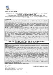
not perform pH monitoring. Barium study showed<br />
two diverticula located in the distal third of the<br />
esophagus. The average sizes of the pouches were<br />
4 cm and 2 cm (figure 1). The diverticula were<br />
located 6-7 cm above the cardia measured by<br />
endoscopy and a hiatal hernia was also diagnosed.<br />
No findings suggestive of primary esophageal<br />
motility disorder like dilatation of the esophagus,<br />
retained food, and resistance at the level of<br />
gastroesophageal junction or tertiary contractions<br />
were recorded during endoscopy so the symptoms<br />
of the patient were thought to be related with the<br />
compression of the larger diverticulum to the<br />
distal esophagus. Following a complete<br />
preoperative evaluation, the patient was scheduled<br />
for laparoscopic operation. The position of the<br />
patient, surgeon and the trocar sites were the same<br />
as for the laparoscopic treatment of functional<br />
diseases of the esophagogastric junction.<br />
The patient was placed on the operating table in<br />
the lithotomy position with a 30º reverse<br />
Trendelenburg. The surgeon was standing<br />
between the legs. Pneumoperitoneum was<br />
established and five operating ports were placed<br />
as usual from the abdomen. A 30º-angled scope<br />
was used. After dividing phreno-esophageal<br />
membrane, the diaphragmatic crura were exposed.<br />
The esophagus was isolated and completely<br />
encircled with a rubber tape for traction. Blunt<br />
dissection was carried out in the mediastinum<br />
until 8-10 cm above the diaphragmatic crura<br />
staying close to the esophageal surface. At the<br />
same time the larger diverticular pouch was<br />
identified by endoscopy and isolated up to the<br />
superior margin of its neck and then resected with<br />
a linear endoscopic stapler with the nasogastric<br />
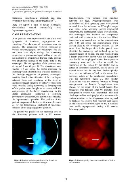
tube inside the esophageal lumen. Intraoperative<br />
endoscopy was used in order to avoid the<br />
narrowing of the lumen by the stapler and to<br />
detect an incomplete resection, also to check the<br />
stapled suture line for any leak. Endoscopically<br />
there was no evidence of leak at the suture line<br />
therefore suture of the esophageal musculature<br />
was not performed (figure 2). The smaller<br />
diverticulum was not resected because of being<br />
asymptomatic. A Toupet fundoplication was<br />
chosen for the repair of the hiatal hernia. The<br />
procedure was finished after 95 minutes. The<br />
postoperative course was uneventful. She had<br />
check-up swallow radiography with water-soluble<br />
contrast medium on the 6th postoperative day and<br />
no leakage was shown. She resumed oral intake<br />
on the same day and discharged on day 8. She has<br />
been totally asymptomatic during a 10 months<br />
follow-up period.<br />
Figure 1: Barium study images showed the diverticula<br />
located in the distal third of the esophagus.<br />
Figure 2: Endoscopically there was no evidence of leak at<br />
the suture line<br />
74