Laboratory testing for LA
Laboratory testing for LA
Laboratory testing for LA

Create successful ePaper yourself
Turn your PDF publications into a flip-book with our unique Google optimized e-Paper software.
Diagnosing lupus<br />
anticoagulants<br />
Emmanuel J Favaloro, Haematology, ICPMR, Westmead Hospital<br />
ISLH, Toronto, May, 2013
Disclosures<br />
No disclosures or conflicts of interest<br />
2
Dedication<br />
Dedicated to memory of Prof Jerry Koutts.<br />
November 27, 1944 - April 2, 2013<br />
3
Talk overview – ‘attack of the acronyms’<br />
‣ APS (antiphospholipid [antibody]<br />
syndrome)<br />
‣ aPL (antiphospholipid [antibodies])<br />
‣ <strong>LA</strong> (Lupus anticoagulants<br />
[antibodies]/inhibitor)<br />
4
Talk overview – ‘attack of the acronyms’<br />
‣ APTT (Activated Partial Thromboplastin Time)<br />
‣ dRVVT (dilute Russell Viper Venom Time)<br />
‣ SCT (Silica Clotting Time)<br />
5
Talk overview – ‘attack of the acronyms’<br />
‣ aCL (anticardiolipin [antibodies])<br />
‣ a 2GPI (anti-beta 2 glycoprotein I [antibodies])<br />
‣ aPT (anti-prothrombin [antibodies])<br />
‣ aPS (anti-phosphatidylserine [antibodies])<br />
‣ aPS/PT (anti-phosphatidylserine-prothrombin<br />
[antibodies] complex)<br />
6
APS – more acronyms<br />
‣ PAPS - Primary Antiphospholipid Syndrome (APS not<br />
associated with another disease process)<br />
‣ SAPS - Secondary Antiphospholipid Syndrome (APS<br />
associated with another disease process; e.g., SLE).<br />
‣ CAPS - Catastrophic Antiphospholipid Syndrome (an<br />
aggressive <strong>for</strong>m of APS; widespread thrombosis, multiple<br />
organ sites; significant morbidity and/or mortality).<br />
‣ SNAPS - Seronegative Antiphospholipid Syndrome<br />
(patients with typical clinical manifestations of APS but<br />
negative <strong>for</strong> a range of aPL tests/assays).<br />
Favaloro & Wong. Autoimmunity Highlights. 2010; 1:5-14<br />
7
APS (antiphospholipid syndrome)<br />
‣ An auto-immune disease associated with the<br />
presence of antiphospholipid antibodies (aPL)<br />
‣ May present with a wide variety of clinical<br />
manifestations:<br />
‣ Haematologists – may see this as a prothrombotic<br />
disorder (& perhaps as thrombocytopaenia)<br />
‣ Obs & Gyn – pregnancy morbidity<br />
‣ Dermatologists – dermatological presentations<br />
‣ Neurologist - migraine, memory loss, chorea<br />
‣ other clinical specialists = other clinical presentations<br />
Favaloro & Wong. Autoimmunity Highlights. 2010; 1:5-14<br />
8
Revised classification criteria <strong>for</strong> APS.*<br />
APS is present if at least one of the following clinical<br />
criteria (and one of the laboratory criteria) are met:<br />
1. Clinical criteria<br />
a. Vascular thrombosis:<br />
One or more clinical episodes of arterial, venous, or small vessel thrombosis, in<br />
any tissue or organ. Thrombosis must be confirmed by objective validated criteria.<br />
b. Pregnancy morbidity:<br />
(i) One or more unexplained deaths of a morphologically normal fetus at or<br />
beyond the 10th week of gestation, with normal fetal morphology, or<br />
(ii) One or more premature births of a morphologically normal neonate be<strong>for</strong>e<br />
the 34th week of gestation because of eclampsia, severe preeclampsia, or<br />
placental insufficiency, or<br />
(iii) Three or more unexplained consecutive spontaneous abortions be<strong>for</strong>e the<br />
10th week of gestation, with exclusion of anatomic, hormonal and chromosomal<br />
causes.<br />
* Summarised from Miyakis et al. JTH 2006;4:295-306.<br />
‘APS classification criteria’ to establish ‘definite APS’ <strong>for</strong> inclusion in<br />
research studies; however, popularly used as ‘APS diagnostic criteria’<br />
9
Revised classification criteria <strong>for</strong> APS.*<br />
APS is present if at least one of the following (clinical<br />
criteria and one of the) laboratory criteria are met:<br />
2. <strong>Laboratory</strong> criteria**<br />
a. <strong>LA</strong> present detected according to ISTH SSC guidelines***<br />
b. aCL antibody of IgG and/or IgM isotype, present in medium or high titer.<br />
c. a 2GPI antibody of IgG and/or IgM isotype.<br />
**aPL must be detected on two or more occasions at least 12<br />
weeks apart. aCL & a 2GPI should be “measured by a<br />
standardized ELISA according to recommended procedures”.<br />
* Miyakis et al. JTH 2006;4:295-306<br />
*** Pengo et al. JTH 2009;7:1737-40<br />
10
Revised classification criteria <strong>for</strong> APS.*<br />
APS is present if at least one of the following (clinical<br />
criteria and one of the) laboratory criteria are met:<br />
2. <strong>Laboratory</strong> criteria<br />
a. <strong>LA</strong> present detected according to ISTH SSC guidelines**<br />
• May soon be challenged by ‘CLSI guidelines’<br />
• What about the British guidelines?***<br />
b. aCL antibody of IgG and/or IgM isotype, present in medium or high titer.<br />
• IgM?<br />
• No standard accepted definition of medium or high titer<br />
c. a 2GPI antibody of IgG and/or IgM isotype.<br />
• IgM?<br />
• No standard accepted definition of medium or high titer<br />
d. aCL & a 2GPI should be “measured by a standardized ELISA according to<br />
recommended procedures”.<br />
• standardized ELISA? No such beast.<br />
* Miyakis et al. JTH 2006;4:295-306<br />
** Pengo et al. JTH 2009;7:1737-40<br />
*** Keeling et al. BJH 2012;157:47-58<br />
11
<strong>Laboratory</strong> <strong>testing</strong> to identify <strong>LA</strong> requires*:<br />
(1) Prolongation of at least one of two phospholipid dependent<br />
clotting assays based on different principles and coagulation<br />
pathways (‘screening’);<br />
(2) Evidence that prolongation of the screening test(s)<br />
demonstrates phospholipid dependence by using a similar second<br />
test(s) using altered concentrations and/or composition of<br />
phospholipids (‘confirmation’);<br />
(3) Mixing assays are recommended; if per<strong>for</strong>med, these must<br />
show evidence of inhibitory activity by the effect of patient plasma<br />
on an equal volume of normal pooled plasma (‘mixing’);<br />
(4) <strong>LA</strong> should be distinguished from other causes of prolonged<br />
clotting times that may mask, mimic, or coexist with <strong>LA</strong>, such as<br />
anticoagulant therapies or other coagulopathies (‘exclusion’).<br />
* annotated from ISTH SSC** & CLSI guidelines<br />
(**Pengo et al. JTH 2009;7:1737-40 & prior)<br />
12
<strong>Laboratory</strong> <strong>testing</strong> <strong>for</strong> <strong>LA</strong>:<br />
‣ Two screening tests ‘negative’ in order to exclude <strong>LA</strong>;<br />
recommended*:<br />
‣ APTT (Activated Partial Thromboplastin Time) &<br />
‣ dRVVT (dilute Russell Viper Venom Time)<br />
‣ Either one ‘positive’ in order to identify <strong>LA</strong><br />
‣ Possible that no two labs use exactly the same <strong>LA</strong> procedure<br />
‣ APTT: reagents are <strong>LA</strong> ‘sensitive’ (variably) vs <strong>LA</strong> insensitive (variably)<br />
‣ dRVVT – some reagents are more sensitive to <strong>LA</strong> than others<br />
‣ APTT (mix vs no mix)<br />
‣ dRVVT (mix or no mix)<br />
‣ Mix (1:1 or other?)<br />
‣ Mix plasma (normal) used (commercial, in house, lyophilised, frozen)<br />
‣ Normal ranges – 20, 40 or more individuals? +/- 2SD, +/- 3 SD, >99 th percentile, etc?<br />
‣ Ratio, normalised ratio, % correction, Rosner index/ICA<br />
‣ Ratio – denominator = normal plasma value or median of reference interval (etc)<br />
‣ etc, etc<br />
* Pengo et al. JTH 2009;7:1737-40<br />
13
<strong>Laboratory</strong> <strong>testing</strong> <strong>for</strong> <strong>LA</strong> - Cut-off values :<br />
‣ Individual screen & confirm assays:<br />
‣ ISTH SSC* & CLSI guidelines – require ‘local validation’ of<br />
cut-off value<br />
‣ ISTH SSC: based on <strong>testing</strong> of ‘at least 40 adult healthy<br />
donors 99 th percentile<br />
‣ CLSI recommendation – will be different<br />
‣ source of normal donors?<br />
‣ are hospital ‘normals’ true normals?<br />
‣ exclude outliers/<strong>LA</strong> positive samples?<br />
‣ statistical anomaly of 40 donors vs 99 th percentile <strong>for</strong> nonnormal<br />
distributions.<br />
* Pengo et al. JTH 2009;7:1737-40<br />
** McGlasson & Fritsma. STH 2013; 39:315-319<br />
*** Kershaw & Orellana. STH 2013;39:283-290<br />
14
<strong>Laboratory</strong> <strong>testing</strong> <strong>for</strong> <strong>LA</strong> - Cut-off values :<br />
‣ Screen & confirm assay cut-offs:<br />
‣ Support <strong>for</strong> ‘generic’ ratio of (around) 1.2*<br />
‣ 200 normal subjects (40 <strong>for</strong> each of 5 centres)<br />
‣ 6 <strong>LA</strong> functional assays (same procedure, reagent<br />
lot and analyser type at each site).<br />
‣ dRVVT <strong>LA</strong> screen and confirm assays, SCT (silica<br />
clotting time) screen and confirm assays, based on<br />
APTT, with low and high phospholipid content<br />
‣ 2 mixing assays (dRVVT & SCT)<br />
‣ IL reagents/ ACL top<br />
* Pradella et al. Clin Chem Lab Med 2013; 51(2): 379–385<br />
15
<strong>Laboratory</strong> <strong>testing</strong> <strong>for</strong> <strong>LA</strong> - Cut-off values <strong>for</strong> screening:<br />
* Pradella et al. Clin Chem Lab Med 2013; 51(2): 379–385<br />
16
<strong>Laboratory</strong> <strong>testing</strong> <strong>for</strong> <strong>LA</strong> - Cut-off values :<br />
Pradella et al. Clin Chem Lab Med 2013; 51(2): 379–85<br />
17
<strong>Laboratory</strong> <strong>testing</strong> <strong>for</strong> <strong>LA</strong> - Cut-off values <strong>for</strong> <strong>LA</strong> pos/neg:<br />
‣ Ratio of screen/confirm:<br />
‣ Most labs probably use 1.2 used as ‘generic’ normalised ratio<br />
‣ Many reagent manufacturers mention a ‘generic’ ratio of 1.2<br />
‣ ‘Generic’ ratio of 1.2 recently ‘validated’*<br />
‣ Values as low as 1.06 are used by some labs<br />
‣ Too low a value = increase false positive; too high a value =<br />
increase false negative.<br />
‣ Alternatives using mixing such as % correction/Rosner<br />
index (ICA – index of circulating anticoagulant)<br />
‣ ICA = [(b-c)/a] x 100 (where a, b, c = clotting times of patient,<br />
mixture, normal, respectively)<br />
‣ For Rosner index/ICA - local determination of value (generally<br />
between 10-20%)**<br />
* McGlasson & Fritsma. STH 2013; 39:315-319<br />
** Kershaw & Orellana. STH 2013;39:283-290<br />
18
Cross laboratory <strong>testing</strong> of <strong>LA</strong> (RCPA QAP)<br />
Favaloro et al. STH 2012;38:404–11<br />
19
<strong>Laboratory</strong> <strong>testing</strong> <strong>for</strong> <strong>LA</strong> - Cut-off values :<br />
‣ Ratio of dRVVT screen/confirm:<br />
‣ ‘Generic’ ratio of 1.2 recently ‘validated’*<br />
‣ Normal subjects (n = 42) vs <strong>LA</strong>-positive plasmas (n<br />
= 43) vs 6 DRVVT <strong>LA</strong> screen and confirm systems on<br />
STAR instrument.<br />
‣ Diagnostica Stago, Precision BioLogic, Siemens<br />
Healthcare, Tcoag, Instrumentation Laboratories<br />
(Werfen), Sekisui Diagnostics (previously American<br />
Diagnostics).<br />
‣ Uncorrected screen/confirm ratios and screen/<br />
confirm ratios that were normalized using MRI and<br />
mean PNP results<br />
* McGlasson & Fritsma. STH 2013; 39:315-319<br />
20
<strong>Laboratory</strong> <strong>testing</strong> <strong>for</strong> <strong>LA</strong> - Cut-off values :<br />
‣ Ratio of dRVVT screen/confirm:<br />
‣ Similar mean screen/confirm ratios <strong>for</strong> normal specimens <strong>for</strong><br />
all systems (all close to 1.0).<br />
* McGlasson & Fritsma. STH 2013; 39:315-319<br />
21
<strong>Laboratory</strong> <strong>testing</strong> <strong>for</strong> <strong>LA</strong> - Cut-off values :<br />
‣ Ratio of dRVVT screen/confirm:<br />
‣ ‘Grand mean action limit, MRI + 3 SD, derived from the local normal<br />
plasmas (1.2) confirmed manufacturers’ recommended ‘cut-off’.<br />
McGlasson & Fritsma. STH<br />
2013; 39:315-319<br />
x6 1.16 1.14 1.20<br />
x5 1.16 1.16 1.21 (Exclude SK)<br />
22
<strong>Laboratory</strong> <strong>testing</strong> <strong>for</strong> <strong>LA</strong> - Cut-off values :<br />
‣ Ratio of dRVVT screen/confirm:<br />
‣ Similar mean dRVVT screen/confirm ratios <strong>for</strong> <strong>LA</strong>-positive specimens<br />
(1.91 to 2.04) <strong>for</strong> 5/6 systems (Sekisui outlier @ 1.2 – ie relatively insensitive<br />
to <strong>LA</strong>).<br />
McGlasson & Fritsma. STH 2013; 39:315-319<br />
23
<strong>Laboratory</strong> <strong>testing</strong> <strong>for</strong> <strong>LA</strong> –<br />
To mix or not to mix – that is the question!:<br />
‣ Latest ISTH SSC guidelines vague on requirement <strong>for</strong> mixing studies<br />
(‘recommended’ but not ‘mandated’)*<br />
‣ Later clarification from SSC guideline authors ‘strong recommendation’ <strong>for</strong><br />
mixing studies**<br />
‣ New CLSI guidelines appear to reduce ‘relative importance’ of mixing<br />
studies.<br />
‣ Integrated test systems (in theory) ‘not requiring mixing’ becoming very<br />
popular.<br />
‣ Mixing reported to ‘dilute’ ‘weak’ <strong>LA</strong> and potentially lead to false negative<br />
<strong>LA</strong>.<br />
‣ Mixing adds complexity and cost to test process (more <strong>testing</strong> &<br />
requirement <strong>for</strong> normal plasma pool).<br />
‣ Not mixing can lead to false negative <strong>LA</strong> in some cases of strong <strong>LA</strong>.<br />
* Pengo et al. JTH 2009;7:1737-40<br />
** Tripodi & Pengo. JTH 2011;9:2126-7<br />
** Tripodi. STH 2012;38:385–9.<br />
24
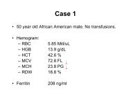
<strong>Laboratory</strong> <strong>testing</strong> <strong>for</strong> <strong>LA</strong> –<br />
To mix or not to mix – that is the question!:<br />
‣ Not mixing can lead to false negative <strong>LA</strong> in some cases of strong <strong>LA</strong>.<br />
‣ Case 1: ‘diagnostic challenge’<br />
‣ Very strong <strong>LA</strong>, dispatched to 93 RCPA QAP participants<br />
‣ Participants were blind to presence of <strong>LA</strong> and asked to per<strong>for</strong>m ‘usual <strong>testing</strong> as<br />
if this sample was received <strong>for</strong> a specific patient investigation’.<br />
‣ Clinical in<strong>for</strong>mation provided:<br />
‣ Sample referred from a satellite facility<br />
‣ 76 year old female patient, B-cell lymphoma diagnosed 4years ago<br />
‣ Put on 3-month follow-up with no specific therapy.<br />
‣ During recent follow-up visit identified to have increasing abdominal distension, night<br />
sweats, weight loss and a significant drop in haemoglobin.<br />
‣ Further clinical investigation might require exploratory surgery.<br />
‣ Routine coagulation tests per<strong>for</strong>med at satellite facility showed prolongation of both<br />
prothrombin time (PT) and activated partial thromboplastin time (APTT). Mixing tests did<br />
not show any substantial correction.<br />
‣ Satellite facility is otherwise unable to provide any additional in<strong>for</strong>mation<br />
Favaloro et al. JTH 2010; 8: 2828–31.<br />
Bonar et al. Pathology 2012;44:240–7<br />
25
<strong>Laboratory</strong> <strong>testing</strong> <strong>for</strong> <strong>LA</strong> –<br />
To mix or not to mix – that is the question!:<br />
Favaloro et al. JTH 2010; 8: 2828–31.<br />
Bonar et al. Pathology 2012;44:240–7<br />
26
<strong>Laboratory</strong> <strong>testing</strong> <strong>for</strong> <strong>LA</strong> –<br />
To mix or not to mix – that is the question!:<br />
Favaloro et al. JTH 2010; 8: 2828–31.<br />
Bonar et al. Pathology 2012;44:240–7<br />
27
<strong>Laboratory</strong> <strong>testing</strong> <strong>for</strong> <strong>LA</strong> –<br />
To mix or not to mix – that is the question!:<br />
Favaloro et al. STH 2012;38:404–11<br />
28
<strong>Laboratory</strong> <strong>testing</strong> <strong>for</strong> <strong>LA</strong> –<br />
Mix then what? That is another question!<br />
APTT as example<br />
‣ 1:1 mixing tests<br />
‣ 55 samples with factor deficiencies<br />
(solid triangles) vs 44 samples with<br />
inhibitors, lupus anticoagulants,<br />
unfractionated heparin, or dabigatran<br />
(open circles).<br />
‣ STA-R & TriniCLOT S<br />
‣ Subtracting the NPP CT from the<br />
1:1mix CT<br />
‣ Corrections within 4 seconds of the NPP<br />
consistent with factor deficiency<br />
‣ More than 8 seconds difference<br />
consistent with an ‘inhibitor’.<br />
‣ Between 4-8 s, inhibitors and factor<br />
deficiencies could not be distinguished.<br />
Kershaw & Orellana. STH 2013;39:283–90.<br />
29
<strong>Laboratory</strong> <strong>testing</strong> <strong>for</strong> <strong>LA</strong> –<br />
Mix then what? That is another question!<br />
APTT as example<br />
‣ Same test, same samples, same<br />
method, different approach.<br />
‣ Ratio of the 1:1 mix APTT to normal<br />
pool APTT<br />
‣ Ratio below 1.1 excludes inhibitors<br />
‣ Ratio above 1.2 suggests the presence<br />
of an ‘inhibitor’.<br />
‣ Between 1.1-1.2, inhibitors and factor<br />
deficiencies could not be distinguished.<br />
Kershaw & Orellana. STH 2013;39:283–90.<br />
30
<strong>Laboratory</strong> <strong>testing</strong> <strong>for</strong> <strong>LA</strong> –<br />
Mix then what? That is another question!<br />
APTT as example<br />
‣ Same test, same samples, same<br />
method, different approach.<br />
‣ Index of circulating anticoagulant<br />
(ICA, or Rosner index).<br />
‣ Level of < 5% consistent with factor<br />
deficiency.<br />
‣ Level of > 11% consistent with the<br />
presence of an inhibitor.<br />
‣ Between 5-11%, inhibitors and factor<br />
deficiencies could not be distinguished.<br />
‣ Cut-off of 15% is shown <strong>for</strong> comparison.<br />
Kershaw & Orellana. STH 2013;39:283–90.<br />
31
<strong>Laboratory</strong> <strong>testing</strong> <strong>for</strong> <strong>LA</strong> –<br />
Mix then what? That is another question!<br />
APTT as example<br />
‣ Same test, same samples, same<br />
method, different approach.<br />
‣ Percent correction <strong>for</strong>mula of<br />
Chang<br />
‣ All factor deficiencies showed a level of<br />
>72% correction.<br />
‣ Level of 72% correction.<br />
Kershaw & Orellana. STH 2013;39:283–90.<br />
32
<strong>Laboratory</strong> diagnosis of <strong>LA</strong> – Summary 1<br />
‣ Follow the guidelines*<br />
‣ But which ones? (ISTH SSC, UK BCSH, CLSI).<br />
‣ Screening by APTT, SCT, dRVVT - nor/abn cut-off :<br />
‣ Ratio will be somewhere between 1.10 and 1.25 (I like 1.15 or 1.20)<br />
‣ ICA will be somewhere between 5-15% (11-12%?)<br />
‣ Difference cut-off will be somewhere between 4-10 sec<br />
‣ No single cut-off value will be 100% sensitive & 100% specific<br />
‣ Ratio of dRVVT screen/confirm – <strong>LA</strong> neg /<strong>LA</strong> pos cut-off:<br />
‣ 1.2 is a good starting point <strong>for</strong> a normalised ratio (Stago, Precision<br />
BioLogic, Siemens, Tcoag, IL)<br />
‣ Value in ‘inter-laboratory’ harmonisation<br />
‣ Sekisui Diagnostics assay not recommended<br />
* Pengo et al. JTH 2009;7:1737-40<br />
Keeling et al. BJH 2012;157:47-58<br />
http://www.clsi.org/standards-development/public-documents/<br />
33
<strong>Laboratory</strong> diagnosis of <strong>LA</strong> – Summary 2<br />
‣ Follow manufacturer instructions<br />
‣ Validate manufacturer claims:<br />
‣ To keep them ‘honest’ (they sometimes make mistakes)<br />
‣ Be warned that validation of cut-off targets with 1.2)<br />
‣ ‘Weak’ <strong>LA</strong> can be missed if mixing per<strong>for</strong>med<br />
‣ ‘Strong’ <strong>LA</strong> can be missed if mixing not per<strong>for</strong>med<br />
‣ Recommendation <strong>for</strong> labs not mixing<br />
‣ Per<strong>for</strong>m mixing study if non-mixed screen and confirm assays<br />
(eg dRVVT) equally prolonged (ie normal ratio) – you may have a<br />
strong <strong>LA</strong>.<br />
* Pengo et al. JTH 2009;7:1737-40<br />
Keeling et al. BJH 2012;157:47-58<br />
http://www.clsi.org/standards-development/public-documents/<br />
34
Clinical utility of aPL <strong>testing</strong>:<br />
‣ <strong>LA</strong> correlates with thrombosis risk more than solid phase assays<br />
‣ Multiple positivity correlates with adverse events risk more than<br />
single positivity<br />
Annual incidence of<br />
thrombosis according to<br />
aPL profile<br />
Galli et al. Blood; 2003; 101:1827-32 & 102:2717-23<br />
Galli. STH;2012;38:348-52<br />
Pengo et al. STH;2012;38:322-7<br />
35
An algorithm <strong>for</strong> identification of APS<br />
Favaloro. IJLH 2013. 35; 269-74.<br />
36
Acknowledgments<br />
‣ Meeting organisers<br />
‣ RCPAQAP Haematology,<br />
‣ Diagnostic Haemostasis laboratory staff<br />
‣ Soma Mohammed<br />
‣ Jane McDonald<br />
‣ Ella Grezchnik<br />
37