Laboratory testing for LA
Laboratory testing for LA
Laboratory testing for LA

You also want an ePaper? Increase the reach of your titles
YUMPU automatically turns print PDFs into web optimized ePapers that Google loves.
Diagnosing lupus<br />
anticoagulants<br />
Emmanuel J Favaloro, Haematology, ICPMR, Westmead Hospital<br />
ISLH, Toronto, May, 2013
Disclosures<br />
No disclosures or conflicts of interest<br />
2
Dedication<br />
Dedicated to memory of Prof Jerry Koutts.<br />
November 27, 1944 - April 2, 2013<br />
3
Talk overview – ‘attack of the acronyms’<br />
‣ APS (antiphospholipid [antibody]<br />
syndrome)<br />
‣ aPL (antiphospholipid [antibodies])<br />
‣ <strong>LA</strong> (Lupus anticoagulants<br />
[antibodies]/inhibitor)<br />
4
Talk overview – ‘attack of the acronyms’<br />
‣ APTT (Activated Partial Thromboplastin Time)<br />
‣ dRVVT (dilute Russell Viper Venom Time)<br />
‣ SCT (Silica Clotting Time)<br />
5
Talk overview – ‘attack of the acronyms’<br />
‣ aCL (anticardiolipin [antibodies])<br />
‣ a 2GPI (anti-beta 2 glycoprotein I [antibodies])<br />
‣ aPT (anti-prothrombin [antibodies])<br />
‣ aPS (anti-phosphatidylserine [antibodies])<br />
‣ aPS/PT (anti-phosphatidylserine-prothrombin<br />
[antibodies] complex)<br />
6
APS – more acronyms<br />
‣ PAPS - Primary Antiphospholipid Syndrome (APS not<br />
associated with another disease process)<br />
‣ SAPS - Secondary Antiphospholipid Syndrome (APS<br />
associated with another disease process; e.g., SLE).<br />
‣ CAPS - Catastrophic Antiphospholipid Syndrome (an<br />
aggressive <strong>for</strong>m of APS; widespread thrombosis, multiple<br />
organ sites; significant morbidity and/or mortality).<br />
‣ SNAPS - Seronegative Antiphospholipid Syndrome<br />
(patients with typical clinical manifestations of APS but<br />
negative <strong>for</strong> a range of aPL tests/assays).<br />
Favaloro & Wong. Autoimmunity Highlights. 2010; 1:5-14<br />
7
APS (antiphospholipid syndrome)<br />
‣ An auto-immune disease associated with the<br />
presence of antiphospholipid antibodies (aPL)<br />
‣ May present with a wide variety of clinical<br />
manifestations:<br />
‣ Haematologists – may see this as a prothrombotic<br />
disorder (& perhaps as thrombocytopaenia)<br />
‣ Obs & Gyn – pregnancy morbidity<br />
‣ Dermatologists – dermatological presentations<br />
‣ Neurologist - migraine, memory loss, chorea<br />
‣ other clinical specialists = other clinical presentations<br />
Favaloro & Wong. Autoimmunity Highlights. 2010; 1:5-14<br />
8
Revised classification criteria <strong>for</strong> APS.*<br />
APS is present if at least one of the following clinical<br />
criteria (and one of the laboratory criteria) are met:<br />
1. Clinical criteria<br />
a. Vascular thrombosis:<br />
One or more clinical episodes of arterial, venous, or small vessel thrombosis, in<br />
any tissue or organ. Thrombosis must be confirmed by objective validated criteria.<br />
b. Pregnancy morbidity:<br />
(i) One or more unexplained deaths of a morphologically normal fetus at or<br />
beyond the 10th week of gestation, with normal fetal morphology, or<br />
(ii) One or more premature births of a morphologically normal neonate be<strong>for</strong>e<br />
the 34th week of gestation because of eclampsia, severe preeclampsia, or<br />
placental insufficiency, or<br />
(iii) Three or more unexplained consecutive spontaneous abortions be<strong>for</strong>e the<br />
10th week of gestation, with exclusion of anatomic, hormonal and chromosomal<br />
causes.<br />
* Summarised from Miyakis et al. JTH 2006;4:295-306.<br />
‘APS classification criteria’ to establish ‘definite APS’ <strong>for</strong> inclusion in<br />
research studies; however, popularly used as ‘APS diagnostic criteria’<br />
9
Revised classification criteria <strong>for</strong> APS.*<br />
APS is present if at least one of the following (clinical<br />
criteria and one of the) laboratory criteria are met:<br />
2. <strong>Laboratory</strong> criteria**<br />
a. <strong>LA</strong> present detected according to ISTH SSC guidelines***<br />
b. aCL antibody of IgG and/or IgM isotype, present in medium or high titer.<br />
c. a 2GPI antibody of IgG and/or IgM isotype.<br />
**aPL must be detected on two or more occasions at least 12<br />
weeks apart. aCL & a 2GPI should be “measured by a<br />
standardized ELISA according to recommended procedures”.<br />
* Miyakis et al. JTH 2006;4:295-306<br />
*** Pengo et al. JTH 2009;7:1737-40<br />
10
Revised classification criteria <strong>for</strong> APS.*<br />
APS is present if at least one of the following (clinical<br />
criteria and one of the) laboratory criteria are met:<br />
2. <strong>Laboratory</strong> criteria<br />
a. <strong>LA</strong> present detected according to ISTH SSC guidelines**<br />
• May soon be challenged by ‘CLSI guidelines’<br />
• What about the British guidelines?***<br />
b. aCL antibody of IgG and/or IgM isotype, present in medium or high titer.<br />
• IgM?<br />
• No standard accepted definition of medium or high titer<br />
c. a 2GPI antibody of IgG and/or IgM isotype.<br />
• IgM?<br />
• No standard accepted definition of medium or high titer<br />
d. aCL & a 2GPI should be “measured by a standardized ELISA according to<br />
recommended procedures”.<br />
• standardized ELISA? No such beast.<br />
* Miyakis et al. JTH 2006;4:295-306<br />
** Pengo et al. JTH 2009;7:1737-40<br />
*** Keeling et al. BJH 2012;157:47-58<br />
11
<strong>Laboratory</strong> <strong>testing</strong> to identify <strong>LA</strong> requires*:<br />
(1) Prolongation of at least one of two phospholipid dependent<br />
clotting assays based on different principles and coagulation<br />
pathways (‘screening’);<br />
(2) Evidence that prolongation of the screening test(s)<br />
demonstrates phospholipid dependence by using a similar second<br />
test(s) using altered concentrations and/or composition of<br />
phospholipids (‘confirmation’);<br />
(3) Mixing assays are recommended; if per<strong>for</strong>med, these must<br />
show evidence of inhibitory activity by the effect of patient plasma<br />
on an equal volume of normal pooled plasma (‘mixing’);<br />
(4) <strong>LA</strong> should be distinguished from other causes of prolonged<br />
clotting times that may mask, mimic, or coexist with <strong>LA</strong>, such as<br />
anticoagulant therapies or other coagulopathies (‘exclusion’).<br />
* annotated from ISTH SSC** & CLSI guidelines<br />
(**Pengo et al. JTH 2009;7:1737-40 & prior)<br />
12
<strong>Laboratory</strong> <strong>testing</strong> <strong>for</strong> <strong>LA</strong>:<br />
‣ Two screening tests ‘negative’ in order to exclude <strong>LA</strong>;<br />
recommended*:<br />
‣ APTT (Activated Partial Thromboplastin Time) &<br />
‣ dRVVT (dilute Russell Viper Venom Time)<br />
‣ Either one ‘positive’ in order to identify <strong>LA</strong><br />
‣ Possible that no two labs use exactly the same <strong>LA</strong> procedure<br />
‣ APTT: reagents are <strong>LA</strong> ‘sensitive’ (variably) vs <strong>LA</strong> insensitive (variably)<br />
‣ dRVVT – some reagents are more sensitive to <strong>LA</strong> than others<br />
‣ APTT (mix vs no mix)<br />
‣ dRVVT (mix or no mix)<br />
‣ Mix (1:1 or other?)<br />
‣ Mix plasma (normal) used (commercial, in house, lyophilised, frozen)<br />
‣ Normal ranges – 20, 40 or more individuals? +/- 2SD, +/- 3 SD, >99 th percentile, etc?<br />
‣ Ratio, normalised ratio, % correction, Rosner index/ICA<br />
‣ Ratio – denominator = normal plasma value or median of reference interval (etc)<br />
‣ etc, etc<br />
* Pengo et al. JTH 2009;7:1737-40<br />
13
<strong>Laboratory</strong> <strong>testing</strong> <strong>for</strong> <strong>LA</strong> - Cut-off values :<br />
‣ Individual screen & confirm assays:<br />
‣ ISTH SSC* & CLSI guidelines – require ‘local validation’ of<br />
cut-off value<br />
‣ ISTH SSC: based on <strong>testing</strong> of ‘at least 40 adult healthy<br />
donors 99 th percentile<br />
‣ CLSI recommendation – will be different<br />
‣ source of normal donors?<br />
‣ are hospital ‘normals’ true normals?<br />
‣ exclude outliers/<strong>LA</strong> positive samples?<br />
‣ statistical anomaly of 40 donors vs 99 th percentile <strong>for</strong> nonnormal<br />
distributions.<br />
* Pengo et al. JTH 2009;7:1737-40<br />
** McGlasson & Fritsma. STH 2013; 39:315-319<br />
*** Kershaw & Orellana. STH 2013;39:283-290<br />
14
<strong>Laboratory</strong> <strong>testing</strong> <strong>for</strong> <strong>LA</strong> - Cut-off values :<br />
‣ Screen & confirm assay cut-offs:<br />
‣ Support <strong>for</strong> ‘generic’ ratio of (around) 1.2*<br />
‣ 200 normal subjects (40 <strong>for</strong> each of 5 centres)<br />
‣ 6 <strong>LA</strong> functional assays (same procedure, reagent<br />
lot and analyser type at each site).<br />
‣ dRVVT <strong>LA</strong> screen and confirm assays, SCT (silica<br />
clotting time) screen and confirm assays, based on<br />
APTT, with low and high phospholipid content<br />
‣ 2 mixing assays (dRVVT & SCT)<br />
‣ IL reagents/ ACL top<br />
* Pradella et al. Clin Chem Lab Med 2013; 51(2): 379–385<br />
15
<strong>Laboratory</strong> <strong>testing</strong> <strong>for</strong> <strong>LA</strong> - Cut-off values <strong>for</strong> screening:<br />
* Pradella et al. Clin Chem Lab Med 2013; 51(2): 379–385<br />
16
<strong>Laboratory</strong> <strong>testing</strong> <strong>for</strong> <strong>LA</strong> - Cut-off values :<br />
Pradella et al. Clin Chem Lab Med 2013; 51(2): 379–85<br />
17
<strong>Laboratory</strong> <strong>testing</strong> <strong>for</strong> <strong>LA</strong> - Cut-off values <strong>for</strong> <strong>LA</strong> pos/neg:<br />
‣ Ratio of screen/confirm:<br />
‣ Most labs probably use 1.2 used as ‘generic’ normalised ratio<br />
‣ Many reagent manufacturers mention a ‘generic’ ratio of 1.2<br />
‣ ‘Generic’ ratio of 1.2 recently ‘validated’*<br />
‣ Values as low as 1.06 are used by some labs<br />
‣ Too low a value = increase false positive; too high a value =<br />
increase false negative.<br />
‣ Alternatives using mixing such as % correction/Rosner<br />
index (ICA – index of circulating anticoagulant)<br />
‣ ICA = [(b-c)/a] x 100 (where a, b, c = clotting times of patient,<br />
mixture, normal, respectively)<br />
‣ For Rosner index/ICA - local determination of value (generally<br />
between 10-20%)**<br />
* McGlasson & Fritsma. STH 2013; 39:315-319<br />
** Kershaw & Orellana. STH 2013;39:283-290<br />
18
Cross laboratory <strong>testing</strong> of <strong>LA</strong> (RCPA QAP)<br />
Favaloro et al. STH 2012;38:404–11<br />
19
<strong>Laboratory</strong> <strong>testing</strong> <strong>for</strong> <strong>LA</strong> - Cut-off values :<br />
‣ Ratio of dRVVT screen/confirm:<br />
‣ ‘Generic’ ratio of 1.2 recently ‘validated’*<br />
‣ Normal subjects (n = 42) vs <strong>LA</strong>-positive plasmas (n<br />
= 43) vs 6 DRVVT <strong>LA</strong> screen and confirm systems on<br />
STAR instrument.<br />
‣ Diagnostica Stago, Precision BioLogic, Siemens<br />
Healthcare, Tcoag, Instrumentation Laboratories<br />
(Werfen), Sekisui Diagnostics (previously American<br />
Diagnostics).<br />
‣ Uncorrected screen/confirm ratios and screen/<br />
confirm ratios that were normalized using MRI and<br />
mean PNP results<br />
* McGlasson & Fritsma. STH 2013; 39:315-319<br />
20
<strong>Laboratory</strong> <strong>testing</strong> <strong>for</strong> <strong>LA</strong> - Cut-off values :<br />
‣ Ratio of dRVVT screen/confirm:<br />
‣ Similar mean screen/confirm ratios <strong>for</strong> normal specimens <strong>for</strong><br />
all systems (all close to 1.0).<br />
* McGlasson & Fritsma. STH 2013; 39:315-319<br />
21
<strong>Laboratory</strong> <strong>testing</strong> <strong>for</strong> <strong>LA</strong> - Cut-off values :<br />
‣ Ratio of dRVVT screen/confirm:<br />
‣ ‘Grand mean action limit, MRI + 3 SD, derived from the local normal<br />
plasmas (1.2) confirmed manufacturers’ recommended ‘cut-off’.<br />
McGlasson & Fritsma. STH<br />
2013; 39:315-319<br />
x6 1.16 1.14 1.20<br />
x5 1.16 1.16 1.21 (Exclude SK)<br />
22
<strong>Laboratory</strong> <strong>testing</strong> <strong>for</strong> <strong>LA</strong> - Cut-off values :<br />
‣ Ratio of dRVVT screen/confirm:<br />
‣ Similar mean dRVVT screen/confirm ratios <strong>for</strong> <strong>LA</strong>-positive specimens<br />
(1.91 to 2.04) <strong>for</strong> 5/6 systems (Sekisui outlier @ 1.2 – ie relatively insensitive<br />
to <strong>LA</strong>).<br />
McGlasson & Fritsma. STH 2013; 39:315-319<br />
23
<strong>Laboratory</strong> <strong>testing</strong> <strong>for</strong> <strong>LA</strong> –<br />
To mix or not to mix – that is the question!:<br />
‣ Latest ISTH SSC guidelines vague on requirement <strong>for</strong> mixing studies<br />
(‘recommended’ but not ‘mandated’)*<br />
‣ Later clarification from SSC guideline authors ‘strong recommendation’ <strong>for</strong><br />
mixing studies**<br />
‣ New CLSI guidelines appear to reduce ‘relative importance’ of mixing<br />
studies.<br />
‣ Integrated test systems (in theory) ‘not requiring mixing’ becoming very<br />
popular.<br />
‣ Mixing reported to ‘dilute’ ‘weak’ <strong>LA</strong> and potentially lead to false negative<br />
<strong>LA</strong>.<br />
‣ Mixing adds complexity and cost to test process (more <strong>testing</strong> &<br />
requirement <strong>for</strong> normal plasma pool).<br />
‣ Not mixing can lead to false negative <strong>LA</strong> in some cases of strong <strong>LA</strong>.<br />
* Pengo et al. JTH 2009;7:1737-40<br />
** Tripodi & Pengo. JTH 2011;9:2126-7<br />
** Tripodi. STH 2012;38:385–9.<br />
24
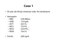
<strong>Laboratory</strong> <strong>testing</strong> <strong>for</strong> <strong>LA</strong> –<br />
To mix or not to mix – that is the question!:<br />
‣ Not mixing can lead to false negative <strong>LA</strong> in some cases of strong <strong>LA</strong>.<br />
‣ Case 1: ‘diagnostic challenge’<br />
‣ Very strong <strong>LA</strong>, dispatched to 93 RCPA QAP participants<br />
‣ Participants were blind to presence of <strong>LA</strong> and asked to per<strong>for</strong>m ‘usual <strong>testing</strong> as<br />
if this sample was received <strong>for</strong> a specific patient investigation’.<br />
‣ Clinical in<strong>for</strong>mation provided:<br />
‣ Sample referred from a satellite facility<br />
‣ 76 year old female patient, B-cell lymphoma diagnosed 4years ago<br />
‣ Put on 3-month follow-up with no specific therapy.<br />
‣ During recent follow-up visit identified to have increasing abdominal distension, night<br />
sweats, weight loss and a significant drop in haemoglobin.<br />
‣ Further clinical investigation might require exploratory surgery.<br />
‣ Routine coagulation tests per<strong>for</strong>med at satellite facility showed prolongation of both<br />
prothrombin time (PT) and activated partial thromboplastin time (APTT). Mixing tests did<br />
not show any substantial correction.<br />
‣ Satellite facility is otherwise unable to provide any additional in<strong>for</strong>mation<br />
Favaloro et al. JTH 2010; 8: 2828–31.<br />
Bonar et al. Pathology 2012;44:240–7<br />
25
<strong>Laboratory</strong> <strong>testing</strong> <strong>for</strong> <strong>LA</strong> –<br />
To mix or not to mix – that is the question!:<br />
Favaloro et al. JTH 2010; 8: 2828–31.<br />
Bonar et al. Pathology 2012;44:240–7<br />
26
<strong>Laboratory</strong> <strong>testing</strong> <strong>for</strong> <strong>LA</strong> –<br />
To mix or not to mix – that is the question!:<br />
Favaloro et al. JTH 2010; 8: 2828–31.<br />
Bonar et al. Pathology 2012;44:240–7<br />
27
<strong>Laboratory</strong> <strong>testing</strong> <strong>for</strong> <strong>LA</strong> –<br />
To mix or not to mix – that is the question!:<br />
Favaloro et al. STH 2012;38:404–11<br />
28
<strong>Laboratory</strong> <strong>testing</strong> <strong>for</strong> <strong>LA</strong> –<br />
Mix then what? That is another question!<br />
APTT as example<br />
‣ 1:1 mixing tests<br />
‣ 55 samples with factor deficiencies<br />
(solid triangles) vs 44 samples with<br />
inhibitors, lupus anticoagulants,<br />
unfractionated heparin, or dabigatran<br />
(open circles).<br />
‣ STA-R & TriniCLOT S<br />
‣ Subtracting the NPP CT from the<br />
1:1mix CT<br />
‣ Corrections within 4 seconds of the NPP<br />
consistent with factor deficiency<br />
‣ More than 8 seconds difference<br />
consistent with an ‘inhibitor’.<br />
‣ Between 4-8 s, inhibitors and factor<br />
deficiencies could not be distinguished.<br />
Kershaw & Orellana. STH 2013;39:283–90.<br />
29
<strong>Laboratory</strong> <strong>testing</strong> <strong>for</strong> <strong>LA</strong> –<br />
Mix then what? That is another question!<br />
APTT as example<br />
‣ Same test, same samples, same<br />
method, different approach.<br />
‣ Ratio of the 1:1 mix APTT to normal<br />
pool APTT<br />
‣ Ratio below 1.1 excludes inhibitors<br />
‣ Ratio above 1.2 suggests the presence<br />
of an ‘inhibitor’.<br />
‣ Between 1.1-1.2, inhibitors and factor<br />
deficiencies could not be distinguished.<br />
Kershaw & Orellana. STH 2013;39:283–90.<br />
30
<strong>Laboratory</strong> <strong>testing</strong> <strong>for</strong> <strong>LA</strong> –<br />
Mix then what? That is another question!<br />
APTT as example<br />
‣ Same test, same samples, same<br />
method, different approach.<br />
‣ Index of circulating anticoagulant<br />
(ICA, or Rosner index).<br />
‣ Level of < 5% consistent with factor<br />
deficiency.<br />
‣ Level of > 11% consistent with the<br />
presence of an inhibitor.<br />
‣ Between 5-11%, inhibitors and factor<br />
deficiencies could not be distinguished.<br />
‣ Cut-off of 15% is shown <strong>for</strong> comparison.<br />
Kershaw & Orellana. STH 2013;39:283–90.<br />
31
<strong>Laboratory</strong> <strong>testing</strong> <strong>for</strong> <strong>LA</strong> –<br />
Mix then what? That is another question!<br />
APTT as example<br />
‣ Same test, same samples, same<br />
method, different approach.<br />
‣ Percent correction <strong>for</strong>mula of<br />
Chang<br />
‣ All factor deficiencies showed a level of<br />
>72% correction.<br />
‣ Level of 72% correction.<br />
Kershaw & Orellana. STH 2013;39:283–90.<br />
32
<strong>Laboratory</strong> diagnosis of <strong>LA</strong> – Summary 1<br />
‣ Follow the guidelines*<br />
‣ But which ones? (ISTH SSC, UK BCSH, CLSI).<br />
‣ Screening by APTT, SCT, dRVVT - nor/abn cut-off :<br />
‣ Ratio will be somewhere between 1.10 and 1.25 (I like 1.15 or 1.20)<br />
‣ ICA will be somewhere between 5-15% (11-12%?)<br />
‣ Difference cut-off will be somewhere between 4-10 sec<br />
‣ No single cut-off value will be 100% sensitive & 100% specific<br />
‣ Ratio of dRVVT screen/confirm – <strong>LA</strong> neg /<strong>LA</strong> pos cut-off:<br />
‣ 1.2 is a good starting point <strong>for</strong> a normalised ratio (Stago, Precision<br />
BioLogic, Siemens, Tcoag, IL)<br />
‣ Value in ‘inter-laboratory’ harmonisation<br />
‣ Sekisui Diagnostics assay not recommended<br />
* Pengo et al. JTH 2009;7:1737-40<br />
Keeling et al. BJH 2012;157:47-58<br />
http://www.clsi.org/standards-development/public-documents/<br />
33
<strong>Laboratory</strong> diagnosis of <strong>LA</strong> – Summary 2<br />
‣ Follow manufacturer instructions<br />
‣ Validate manufacturer claims:<br />
‣ To keep them ‘honest’ (they sometimes make mistakes)<br />
‣ Be warned that validation of cut-off targets with 1.2)<br />
‣ ‘Weak’ <strong>LA</strong> can be missed if mixing per<strong>for</strong>med<br />
‣ ‘Strong’ <strong>LA</strong> can be missed if mixing not per<strong>for</strong>med<br />
‣ Recommendation <strong>for</strong> labs not mixing<br />
‣ Per<strong>for</strong>m mixing study if non-mixed screen and confirm assays<br />
(eg dRVVT) equally prolonged (ie normal ratio) – you may have a<br />
strong <strong>LA</strong>.<br />
* Pengo et al. JTH 2009;7:1737-40<br />
Keeling et al. BJH 2012;157:47-58<br />
http://www.clsi.org/standards-development/public-documents/<br />
34
Clinical utility of aPL <strong>testing</strong>:<br />
‣ <strong>LA</strong> correlates with thrombosis risk more than solid phase assays<br />
‣ Multiple positivity correlates with adverse events risk more than<br />
single positivity<br />
Annual incidence of<br />
thrombosis according to<br />
aPL profile<br />
Galli et al. Blood; 2003; 101:1827-32 & 102:2717-23<br />
Galli. STH;2012;38:348-52<br />
Pengo et al. STH;2012;38:322-7<br />
35
An algorithm <strong>for</strong> identification of APS<br />
Favaloro. IJLH 2013. 35; 269-74.<br />
36
Acknowledgments<br />
‣ Meeting organisers<br />
‣ RCPAQAP Haematology,<br />
‣ Diagnostic Haemostasis laboratory staff<br />
‣ Soma Mohammed<br />
‣ Jane McDonald<br />
‣ Ella Grezchnik<br />
37