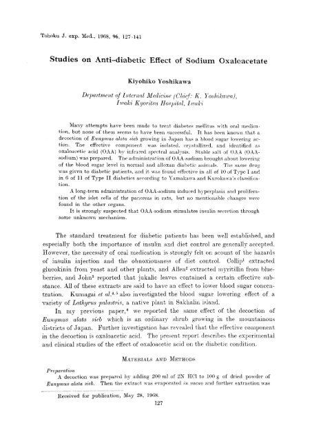
lucki Kyoritsu Hospital, I waki Many attempts have be - Medicina ...
lucki Kyoritsu Hospital, I waki Many attempts have be - Medicina ...
lucki Kyoritsu Hospital, I waki Many attempts have be - Medicina ...

Create successful ePaper yourself
Turn your PDF publications into a flip-book with our unique Google optimized e-Paper software.
Tohoku J. exp. Med .., 1968, 96, 127-141<br />
Studies on Anti-dia<strong>be</strong>tic Effect of Sodium Oxaleacetate<br />
Kiyohiko Yoshikawa<br />
Department of Internal Medicine (Chief: K. Yosh-ikawa),<br />
<strong>lucki</strong> <strong>Kyoritsu</strong> <strong>Hospital</strong>, I <strong>waki</strong><br />
<strong>Many</strong> <strong>attempts</strong> <strong>have</strong> <strong>be</strong>en made to treat dia<strong>be</strong>tes mellitus with oral medic<br />
ation, but none of them seems to <strong>have</strong> <strong>be</strong>en successful. It has <strong>be</strong>en known that a<br />
decoction of Eunymus alata sieb growing in Japan has a blood sugar lowering ac<br />
tion. The effective component was isolated, crystallized, and identified as<br />
oxaloacetic acid (OAA) by infrared spectral analysiṣ Stable salt of O AA (OAA<br />
sodium) was prepared. The administration of OAA-sodium brought about lowering<br />
of the blood sugar level in normal and alloxan dia<strong>be</strong>tic animals. The same drug<br />
was given to dia<strong>be</strong>tic patients, and it was found effective in all of 10 of Type I and<br />
in 6 of 11 of Type II dia<strong>be</strong>tics according to Yamakawand Kurokawa's classifica<br />
tion<br />
Ȧ long-term administration of OAA-sodium induced hyperplasiand prolifera<br />
tion of the islet cells of the pancreas in rats, but no mentionable changes were<br />
found in the other organs.<br />
It is strongly suspected that OAA-sodium stimulates insulin secretion through<br />
some unknown mechanism.<br />
The standard treatment for dia<strong>be</strong>tic patients has <strong>be</strong>en well established, and<br />
especially both the importance of insulin and diet control are generally accepted.<br />
However, the necessity of oral medication is strongly felt on acount of the hazards<br />
of insulin injection and the obnoxiousness of diet control. Collip1 extracted<br />
glucokinin from yeast and other plants, and Allen2 extracted myritillin from blue<br />
<strong>be</strong>rries, and John3 reported that jukalic leaves contained a certain effective sub<br />
stance. All of these extracts are said to <strong>have</strong> an effect to lower blood sugar concen<br />
tration. Kumagai et al.`s,' also investigated the blood sugar lowering effect of a<br />
variety of Lathyrus paalustris, a native plant in Sakhalin island.<br />
In my previous paper,' we reported the same effect of the decoction of<br />
Eunymus alata sieb which is an ordinary shrub growing in the mountainous<br />
districts of Japan. Further investigation has revealed that the effective component<br />
in the decoction is oxaloacetic acid. The present report descri<strong>be</strong>s the experimental<br />
and clinical studies of the effect of oxaloacetic acid on the dia<strong>be</strong>tic condition.<br />
MATERIALS AND METHODS<br />
Preparation<br />
A decoction was prepared by adding 200 ml of 2N HCl to 100 g of dried powder of<br />
Eunymus alata sieb. Then the extract was evaporated in vacuo and further extraction was<br />
Received for publication, May 28, 1968.<br />
127
128 K. Yoshikawa<br />
made with a Soxhlet's extractor using alcohol. The final extract thus obtained was tested<br />
and proved to <strong>be</strong> effective in lowering the blood sugar in rabbits. After this procedure,<br />
further extraction was made with ether and finally a crystalline substance was obtained.<br />
Infrared spectral analysis demonstrated that the crystal was oxaloacetic acid. Since<br />
oxaloacetic acid is unstable, its sodium salt was used in the animal experiment as well as in<br />
the clinical investigation.<br />
Measurement of O AA concentration<br />
The principle of measurement of OAA concentration is based on the fact that OAA is an<br />
acid and dissociates when anilin or Co2+ is added, while ketoglutaric acid, pyruvic acid<br />
and others <strong>have</strong> no such reaction.<br />
Method of analysis<br />
1) Five ml of 10% aqueous solution of trichloroacetic acid was added to 2-3g of the<br />
sample to remove proteinous components. After centrifugation the supernatant was<br />
filtered. After washing with 5 ml of acid solution, the precipitate was filtered again.<br />
Furthermore, the precipitate on filter paper was rinsed with a small quantity of water.<br />
2) The filtrate and the washings were mixed together and the mixture was divided into<br />
halves, each of which was poured onto a 50 ml funnel-shaped separator.<br />
3) A 15% aqueous solution of CoC12.6H2O was added to each of the solutions at a<br />
ratio of 1:5, and the mixture was left to stand for 30 minutes.<br />
4) One ml of 2-4-dinitrophenyl hydrazine solution (0.1 g/2N HCl 100ml) was added<br />
to the solution and the mixture was left to stand for 30 minutes.<br />
5) Then both the solutions were treated according to the following procedures; 10 ml<br />
of ethyl acetate was added to the solution and the mixture was shaken enough to make keto<br />
acid, from which hydrazone and excess hydrazine were extracted. The water layer was<br />
discarded and the ester layer was washed 2-3 times with water. Ten ml of Na2C03-NaHCO3<br />
solution (50 g Na2CO3 anhydride and 5g NaHCO3 per liter) was added and the mixture was<br />
shaken vigorously. Hydrazone alone diffused into the water phase. After the phase separa<br />
tion, most of the ester layer was sucked away with a pipet. The water phase was<br />
washed 3-4 times with 10 ml of carbon tetrachloride until the carbon tetrachloride phase<br />
<strong>be</strong>came colorless. Then the water phase was transferred into a 25 ml measuring flask,<br />
and Na2CO3-NaHCO3 solution was added to make it accurately 25 ml. The mixture was<br />
poured into a cell 1 cm in depth, and the optical density at 384 m,u was measured.<br />
6) The difference in the reading <strong>be</strong>tween both mixtures was proportionate to the<br />
quantity of oxaloacetic acid. (The mixture including Co2+ contained more oxaloacetic acid<br />
than the other, and the optical density lowered, when no difference in the reading was noted<br />
in the absence of oxaloacetic acid). Oxaloaeetic acid was determined on the basis of the<br />
standard curve of oxaloacetic acid-hydrazine derivative.<br />
Two hundred mg of sodium oxaloacetate, measured by the above procedures, were<br />
given orally to two dia<strong>be</strong>tic patients and a healthy subject. The values <strong>be</strong>fore administra<br />
tion, at 30 minutes and at one hour after administration were obtained as follows:<br />
Subject Before At 30 min At I hour<br />
K.S. (dia<strong>be</strong>tic) 0 0 3.1ƒÊg/100 ml<br />
Y.O. (normal) 0 0 2.2ƒÈg/l00ml<br />
M.Y. (dia<strong>be</strong>tic) 0 0 1.5ƒÊg/100 ml<br />
RESULTS<br />
1) Insulin-like activity (ILA) of oxaloacetic acid<br />
Cross-bred male Wistar rats 110-120 g in weight were fed. on high-carbo<br />
hydrate diet consisting mainly of wheat for at least one week under the same condi<br />
tion. After fasting for 24 hours, 3 rats were given a blow against the head, and
Anti-dia<strong>be</strong>tic Effect of Sodium Oxaloacetate 120<br />
killed by cutting both the carotid arteries. The adipose tissue of the epididymis<br />
was quickly resected with minimal damage to the tissue. Here, care must <strong>be</strong> taken<br />
to leave the thick root adjacent to the epididymis and the large blood vessels on the<br />
side of the epididymis. The resected tissue was soaked in Krebs-Ringer bicarbonate<br />
buffer (pH 7.4, 21°C), and after 15-20 minutes the tissue on the side of the peripheral<br />
lobulation was divided into eighteen pieces with even thickness. The water on the<br />
surface of these pieces was sucked up with filter paper and the wet tissue was weigh<br />
ed on a torsion balance. Each piece was 80 to 90 mg in weight.<br />
Two kinds of medium, KRG and KRBG, were prepared. The former con<br />
sisted of Krebs-Ringer bicarbonate buffer (pH 7.4) and glucose at a concentration<br />
of 200 mg/100 ml, and the latter was ten-fold dilution of serum in KRG solution.<br />
Two ml of KRG solution was poured into flasks each and the same amount of KRBG<br />
solution into other 3 flasks. Six pieces of the tissue were put into those 6 flasks.<br />
In order to provide aerobic condition a gas mixture of 95% 02 and 5% CO2 was<br />
filled in the flasks which were connected with the Warburg apparatus. Then,<br />
the contents in. the flasks were incubated for 120 minutes at 37.5°C at a shak<br />
ing frequency of 110/min. Glucose was measured <strong>be</strong>fore and after incubation<br />
by Fujita-Iwatake's method. On the assumption that glucose mg/g wet tissue<br />
weight is the amount of glucose taken up in the tissue, the difference in the<br />
glucose uptake <strong>be</strong>tween KRG and KRBG was considered to <strong>be</strong> due to insulin-like<br />
activity of the sample serum.<br />
To examine the effect of oxaloacetic acid on d-glucose uptake, 0.2 nil of OAA<br />
diluted with KRB buffer at a concentration of 1 mg/100 ml was added to 2 ml of<br />
KRBG solution. After incubation, the glucose uptake was calculated for the<br />
determination of OAA effect.<br />
So far as 10 dia<strong>be</strong>tic cases were concerned, the d-glucose uptake ranged gener<br />
ally from 0.62 to 5.43 mg/100 ml/g in fasting stage and from 3.72 to 9.29 when OAA<br />
was added to the serum (Table 1).<br />
On the other hand, 15 normal subjects showed an increase in the d-glucose<br />
uptake ranging from 0.27 to 12.36 mg/100 ml/g in fasting stage and from 0.67 to<br />
11.57 mg/100 ml/g when OAA was added to the serum, but so far as ILA was con<br />
cerned, 11 among 15 cases showed an increase, while the other 4 cases showed no<br />
change (Table 2).<br />
TABLE 1. Effect of OAA upon glucose uptake in dia<strong>be</strong>tics (mg/100 ml/s)
130 . Yoshikawa K<br />
TABLE 2. d-Glucose uptake in normal subjects (mg/100 ml/g)<br />
Fig. 1. Effect of OAA upon glucose uptake (mg/100 ml/g).<br />
Significant differences were observed <strong>be</strong>tween the two cases, i.e., when OAA<br />
alone was added to the buffer, and when serum and OAA were added. In the<br />
former case, there was no change in ILA. This suggests that OAA has no ILA<br />
increasing effect by itself, but has an effect on the insulin-like activity in the<br />
serum (Fig. 1).<br />
2) Hypoglycemic effect in normal dogs<br />
According to the method reported by Kobayashi,7 healthy adult dogs were<br />
anesthetized by nembutal administration, and 480 ml of 2% OAA-Na solution<br />
was infused into the pancreaticoduodenal artery with constant infusion technic at<br />
a rate of 1 ml per minute. Repeated sampling of the blood was made and blood<br />
sugar was measured. The blood sugar level lowered precipitously from the pre<br />
infusion level of 92 mg/100 ml to 46 mg/100 ml in 6 hours and to 30 mg/100 ml in<br />
8 hours. Control animals showed a pre-infusion value of 98 mg/100 ml on the<br />
average, and 80 and 76 mg/100 ml respectively 6 and 8 hours after infusion of<br />
saline only (Fig. 2).
Anti-dia<strong>be</strong>tic Effect of Sodium Oxaloacetate 131<br />
Fig. 2. Hypoglycemic effect of continued infusion of OAA-Na into the pancreatico<br />
duodenalis in normal dogs.<br />
3) Effect of intravenous administration of OAA-Na to rabbits<br />
In 6 fasting healthy rabbits, OAA-Na was injected in a dose of 20 mg into the<br />
auricular vein, and serial determinations of blood glucose were done<br />
As shown in Table 1, normal rabbits fasting for 24 hours showed a decrease of<br />
blood glucose at 8 hours of fasting ranging from 8 to 21% and averaging 12% of<br />
the control level. In the rabbits treated with OAA, the rate of decrease was 24%<br />
to 45%, averaging 33%. The difference <strong>be</strong>tween these two groups is statistically<br />
significant. It is also noted that there were two types of the blood glucose curve,<br />
one showing initial temporal increase followed by a rapid decrease and the other<br />
showing a gradual decrease (Table 3).<br />
TABLE 3. Hypoglycemic effect of intravenous administration of OAA-Na<br />
4) Effect of OAA-Na administration in alloxan dia<strong>be</strong>tic rabbits<br />
Six rabbits treated with alloxan were fed on standard diet and urinary sugar<br />
excretion was measured. When the daily urinary output of sugar reached a<br />
constant level, OAA-Na was given orally in a dose of 200 mg per kg of body<br />
weight. As shown in Table 2, significant decrease of blood sugar and urine sugar
132 K. Yoshikawa<br />
TABLE 4. Effect of OAA-Na administration in alloxan dia<strong>be</strong>tic rabbits<br />
output as well as the increase of body weight was observed in all but one rabbit<br />
(Table<br />
4)_<br />
5) Clinical trial of OAA-Na (Table 5)<br />
As various factors are known to influence the status of dia<strong>be</strong>tic patients , the<br />
evaluation of any treatment will <strong>be</strong> difficult if these factors are unstable. There<br />
fore, clinical trial of OAA-Na was made in patients who had <strong>be</strong>en treated by diet<br />
and insulin administration and their symptoms were stabilized. A dose from 200<br />
mg to 1,000 mg per day in three divided doses was orally given after meal.<br />
The criteria for the evaluation were as follows:<br />
a) For Type I dia<strong>be</strong>tics according to Yamakawa and Kurokawa's classifica<br />
tion,' fasting blood sugar less than 120 nig/100 ml and 24-hour urinary output of<br />
TABLE 5.<br />
Clinical effect of OAA-Na
Anti-dia<strong>be</strong>tic Effect of Sodium Oxaloacetate 133<br />
sugar less than 10 g were considered to show an excellent effect of OAA-Na. For<br />
Type II dia<strong>be</strong>tics, fasting blood sugar less than 150 mg/ 100 nil and urinary output<br />
of sugar less than 10 g/day were evaluated. Blood sugar was measured by Fujita<br />
Iwatake's method.9<br />
b) A considerable improvement <strong>be</strong>low the criteria stated above was regarded<br />
as <strong>be</strong>ing effective.<br />
The result of the clinical trials is summarized in Table 5. Namely, 15 male<br />
and 6 female dia<strong>be</strong>tic patients, from 15 to 70 years of age, were given OAA-Na<br />
orally, and the administration was remarkably effective in II patients, effective in<br />
5 patients and ineffective in 5 patients, As shown in Table 6, all 10 patients of<br />
Type I dia<strong>be</strong>tes responded favorably to OAA-Na administration. On the other<br />
hand, in cases of Type II it was remarkably effective in 2, effective in 5 and<br />
ineffective in 5 cases. As shown in Table 7, it is noteworthy that the patients who<br />
showed a remarkable effect were all over 40 years of age.<br />
When the constitution of patients was divided into slim, normal and o<strong>be</strong>se,<br />
the administration of OAA-Na was most effective in the o<strong>be</strong>se type as shown in<br />
Table 8. The relationship <strong>be</strong>tween the effectiveness of OAA-Na administration and<br />
the fasting blood sugar level <strong>be</strong>fore the administration is summarized in Table 9.<br />
As naturally expected, the more severe the dia<strong>be</strong>tic condition, the less effective the<br />
administration of OAA-Na.<br />
The patients were divided into three groups depending upon the duration of<br />
the clinical course of the disease. As shown in Table 10, the administration of<br />
on dia<strong>be</strong>tics /oral administration)
134 K. Yoshikawa<br />
TABLE 6. Clinical effect and dia<strong>be</strong>tic types<br />
TABLE 7. Clinical effect and age<br />
TABLE 8.<br />
Clinical effect and the fasting blood sugar<br />
FBS: fasting blood sugar<br />
TABLE 9. Body types and clinical effect
Anti-dia<strong>be</strong>tic Effect of Sodium Oxaloaeetate 135<br />
TABLE 10. Period of the disease and clinical effect<br />
OAA-Na was remarkably effective in 8 of 9 cases under one year, 3 of 9 cases of 1 to<br />
3 years, and none of 3 cases over 3 years. The administration of OAA-Na had no<br />
influence on the liver function tests and the blood acetone or cholesterol level.<br />
6) Results of administration of OAA-Nce in rats<br />
To 40 rats of Wistar strain OAA-Na was given in an oral dose of 5 mg per day<br />
by mouth for 40 days and the effect was examined. The pancreas of these rats<br />
demonstrated proliferation and hyperplasia of the islet cells (Figs. 3 and 4). In<br />
another 40 rats of the same strain given OAA-Na in an oral dose of 10 mg per day<br />
for four months, the same results were obtained (Fig. 5). On the other hand, the<br />
pancreatic islets showed various changes. Some islets were decreased in size and<br />
hyperemic, alpha cells <strong>be</strong>ing atrophic, while <strong>be</strong>ta cells were hypertrophic and<br />
stained densely (Figs. 6 and 7). In the animal group, to which OAA-Na had <strong>be</strong>en<br />
given subcutaneously in a dose of 2 mg for a week, alpha cells were atrophic, but<br />
<strong>be</strong>ta cells showed no change (Fig. 8). In 40 Wistar strain rats, to which OAA-Na<br />
was given for 24 hours, alpha cells showed atrophy and vacuoles and <strong>be</strong>ta cells<br />
hypertrophy (Fig. 9). The liver, hypophysis, adrenals and gonadal glands showed<br />
no particular changes.<br />
DISCUSSION<br />
A num<strong>be</strong>r of investigations <strong>have</strong> <strong>be</strong>en reported of blood sugar lowering<br />
substances which are contained in plants.<br />
In my previous communications we <strong>have</strong> reported the blood sugar lowering<br />
effect of the decoction of Euaymus alata sieb. A further analysis of the decoction<br />
has led to the conclusion that OAA contained in the decoction was the effective<br />
substance. OAA was stabilized when its sodium salt was made.<br />
The critical evaluation of effectiveness of a new anti-dia<strong>be</strong>tic agent is in general<br />
based on a decrease in blood sugar, urinary sugar output and insulin requirement.<br />
OAA-Na was found to <strong>be</strong> effective in treating dia<strong>be</strong>tics, especially those <strong>be</strong>long<br />
ing to the so-called. Type I. In our clinical investigation, OAA-Na was given only<br />
to those patients with a long-standing dia<strong>be</strong>tic condition, and therefore a higher
136 K. Yoshikawa<br />
rate of effectiveness will <strong>be</strong> expected when milder cases are subjected to the treat<br />
ment.<br />
The mechanism of the effectiveness of OAA-Na is still obscure. Rice and<br />
Evance10 reported that the addition of oxaloacetic acid to the homogenized major<br />
pectoral muscle of pigeon enhanced the metabolism of pyruvic acid.<br />
Acetyl-CoA produced from pyruvic acid condenses with oxaloacetate to form<br />
citrate, which is then oxidized in the TCA cycle to water and C02. It is also well<br />
established that in dia<strong>be</strong>tic patients the intermediates of the TCA cycle, especially<br />
oxaloacetate, are decreased. Frohman et al.11 reported that intermediates of the TCA<br />
cycle are decreased in the muscle of alloxan dia<strong>be</strong>tic rats. In our experiment, the<br />
administration of OAA-Na to alloxan dia<strong>be</strong>tic rabbits caused a decrease of blood<br />
sugar and urine glucose and an increase of body weight. Acetone body disappeared<br />
in these dia<strong>be</strong>tic animals when administered with OAA-Na. In normal dogs, the<br />
administration of OAA-Na induced significant hypoglycemia. Long-term admin<br />
istration of OAA-Na in rats caused hypoglycemia and proliferation of the islet cells<br />
of the pancreas. These findings suggest that OAA-Na stimulates the islet cells,<br />
regulates the abnormal metabolic process, and enhances the secretion of insulin,<br />
and thereby controls the dia<strong>be</strong>tic condition. Further investigation is needed<br />
in determining whether OAA-Na per se stimulates the <strong>be</strong>ta cells or other inter<br />
mediates of the TCA cycle are responsible.<br />
According to Maske,12 insulin possesses a specific and strong ability to form<br />
chelate complexes with zinc and with some other metals, and insulin is stored by the<br />
interaction of zinc within the granules. Various metabolites, such as citrate,<br />
oxaloacetate, glutathione, cysteine, histidine and organic phosphorus compounds,<br />
form a stronger complex with zinc than they do with insulin. This reaction was<br />
considered, according to Maske, to consist in bringing insoluble insulin into soluble<br />
one by releasing the hormone from insoluble zinc-insulin complexes. The anti<br />
dia<strong>be</strong>tic effect of the decoction of Eunymus alata sieb and of OAA-Na isolated there<br />
from can <strong>be</strong> understood from this point of view, although the exact mechanism<br />
needs further investigation.<br />
Acknowledgment<br />
My gratitudes are due to Emeritus Prof. Toshio Kurokawa, Tohoku University,<br />
Director of the Cancer Institute <strong>Hospital</strong>, Tokyo for his direction throughout this study.<br />
References<br />
1) Collip, J.B. Glucokinin. A new hormone present in plant tissue. J. biol. Chem.,<br />
1923, 56, 513-543.<br />
2) Allen, F.M. Blue<strong>be</strong>rry leaf extract. Physiologic and clinical properties in relation to<br />
carbohydrate metabolism. J. Amer. Med. Ass., 1927, 89, 1577-1581.<br />
3) John, H.J. A trial of eucalyptus infusion in dia<strong>be</strong>tes. J. Metab. Res., 1922, 1, 489-495.<br />
4) Kumagai, T., Fujise, S., Yagi, S., Moriya, M. & Endo, H. On the effect of Lathyrus<br />
in the treatment of dia<strong>be</strong>tes mellitus and on the effective fraction of its extract. Sogo<br />
Igaku (Jap.), 1946, 3, 385-389.<br />
5) Kumagai, T., Kumagai, K., Oikawa, Y. & Endo, H. On the blood sugar lowering
Anti-dia<strong>be</strong>tic Effect of Sodium Oxaloacetate 137<br />
effect of oxalic acid. Sogo Igaku (Jap.), 1949, 6, 768-770.<br />
6) Yoshikawa, K. Studies on the treatment of dia<strong>be</strong>tes mellitus by the decoction of<br />
eunymus alata sieb. Jap. J. Gastroent. (Jap.), 1958, 55. 23.<br />
7) Kobayashi, Y. Chemical treatment of dia<strong>be</strong>tes mellitus. Collection of submitted<br />
reports of government sponsored scientific research by Japanese Ministry of Education.<br />
Supplement (Jap.), 1953.<br />
8) Kurokawa, T. On the dia<strong>be</strong>tes mellitus. Folia endocr. jap. (Jap.), 1951, 27, 141-166.<br />
9) Fujita, S. & Iwatake, D. Quantitative determination of true blood sugar by enzymatic<br />
method. Biochem.. Z., 1931, 242, 43-60.<br />
10) Rice, L. & Evans, E.A.Jr. The in vitro effect of insulin in pigeon breast muscle.<br />
Science, 1943, 97, 470-471.<br />
11) Frohman, C.E., Orten, J. M. & Smith, A.H. Levels of acids of the citric acid cycle<br />
in tissues of normal and dia<strong>be</strong>tic rats. J. biol. Chena., 1951, 193, 803-807.<br />
12) Maske, H. Role of zink in the insulin secretion. Dia<strong>be</strong>tes, edited by R.H. Williams,<br />
Paul B. Hoe<strong>be</strong>r, New York, 1962, pp. 46-63.
138 K. Yoshikawa<br />
Fig. 3. The pancreatic islet of a normal rat shows significant hypertrophy after a long term<br />
administration of OAA-Na. Gomori stain, 10•~60.<br />
Fig. 4. The pancreatic islets of normal rats after long term administration of OAA-Na.<br />
The islet's cells increased in num<strong>be</strong>r and size. Gomori stain, 10•~10.<br />
Fig. 5. The pancreatic islets of normal rats after long term administration of OAA-Na<br />
are shown. Gomori, 10•~10. Two islets were conjugated to form a larger islet.
Anti-dia<strong>be</strong>tic Effect of Sodium Oxaloacetate 130
140 K. Yoshikawa<br />
Fig. 6. The pancreatic islets of normal rats after oral administration of OAA-Na, with<br />
daily doses of 2 mg for 4 months. # cells are dense and hypertrophied.<br />
Fig. 7. The pancreatic islet of normal rats after oral administration of OAA-Na with<br />
daily doses of 10 mg for 4 months. The islet are small. ƒÀ cells are hypertrophied and<br />
j granules are stained densely. a cells are decreased in num<strong>be</strong>r and atrophic.<br />
Fig. 8. The pancreatic islet of normal rats after subcutaneous administration of OAA-Na<br />
for 4 months. a, and fi cells are atiopbic and scme nuelei are destroyed.<br />
Fig. 9. The pancreatic islet of normal rats after subcutaneous administration of OAA-Na<br />
for 4 months. The islet is hyperemic. ƒÀ cells decrease in size and encircle the islet.<br />
ƒÀ cells are densely stained.
Anti-dia<strong>be</strong>tic Effect of Sodium Oxaloacetate 141