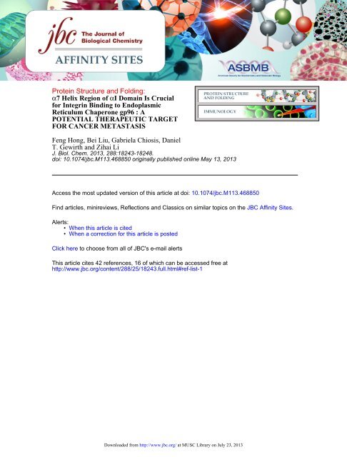
T. Gewirth and Zihai Li Feng Hong, Bei Liu, Gabriela Chiosis, Daniel ...
T. Gewirth and Zihai Li Feng Hong, Bei Liu, Gabriela Chiosis, Daniel ...
T. Gewirth and Zihai Li Feng Hong, Bei Liu, Gabriela Chiosis, Daniel ...

You also want an ePaper? Increase the reach of your titles
YUMPU automatically turns print PDFs into web optimized ePapers that Google loves.
Protein Structure <strong>and</strong> Folding:<br />
α7 Helix Region of αI Domain Is Crucial<br />
for Integrin Binding to Endoplasmic<br />
Reticulum Chaperone gp96 : A<br />
POTENTIAL THERAPEUTIC TARGET<br />
FOR CANCER METASTASIS<br />
<strong>Feng</strong> <strong>Hong</strong>, <strong>Bei</strong> <strong>Li</strong>u, <strong>Gabriela</strong> <strong>Chiosis</strong>, <strong>Daniel</strong><br />
T. <strong>Gewirth</strong> <strong>and</strong> <strong>Zihai</strong> <strong>Li</strong><br />
J. Biol. Chem. 2013, 288:18243-18248.<br />
doi: 10.1074/jbc.M113.468850 originally published online May 13, 2013<br />
Access the most updated version of this article at doi: 10.1074/jbc.M113.468850<br />
Find articles, minireviews, Reflections <strong>and</strong> Classics on similar topics on the JBC Affinity Sites.<br />
Alerts:<br />
• When this article is cited<br />
• When a correction for this article is posted<br />
Click here to choose from all of JBC's e-mail alerts<br />
This article cites 42 references, 16 of which can be accessed free at<br />
http://www.jbc.org/content/288/25/18243.full.html#ref-list-1<br />
Downloaded from http://www.jbc.org/ at MUSC <strong>Li</strong>brary on July 23, 2013
THE JOURNAL OF BIOLOGICAL CHEMISTRY VOL. 288, NO. 25, pp. 18243–18248, June 21, 2013<br />
© 2013 by The American Society for Biochemistry <strong>and</strong> Molecular Biology, Inc. Published in the U.S.A.<br />
7 Helix Region of I Domain Is Crucial for Integrin Binding<br />
to Endoplasmic Reticulum Chaperone gp96<br />
A POTENTIAL THERAPEUTIC TARGET FOR CANCER METASTASIS *<br />
Received for publication, March 12, 2013, <strong>and</strong> in revised form, April 30, 2013 Published, JBC Papers in Press, May 13, 2013, DOI 10.1074/jbc.M113.468850<br />
<strong>Feng</strong> <strong>Hong</strong> ‡ , <strong>Bei</strong> <strong>Li</strong>u ‡ , <strong>Gabriela</strong> <strong>Chiosis</strong> § , <strong>Daniel</strong> T. <strong>Gewirth</strong> , <strong>and</strong> <strong>Zihai</strong> <strong>Li</strong> ‡1<br />
From the ‡ Department of Microbiology & Immunology, Medical University of South Carolina, Charleston, South Carolina 29425,<br />
the § Hauptman-Woodward Medical Research Institute, Buffalo, New York 14203, <strong>and</strong> the Department of Molecular<br />
Pharmacology & Chemistry, Memorial Sloan Kettering Cancer Center, New York, New York 10065<br />
Background: Integrins are chaperoned by gp96, but the binding region to gp96 remains unknown.<br />
Results: Deletion of 7 helix from I domain of integrin abolished the interaction between integrin <strong>and</strong> gp96. Targeting this<br />
region by corresponding peptide blocked cell invasion.<br />
Conclusion: We successfully mapped the binding region of integrin to gp96.<br />
Significance: This study will facilitate the development of new cancer therapeutics.<br />
Integrins play important roles in regulating a diverse array of<br />
cellular functions crucial to the initiation, progression, <strong>and</strong><br />
metastasis of tumors. Previous studies have shown that a majority<br />
of integrins are folded by the endoplasmic reticulum chaperone<br />
gp96. Here, we demonstrate that the dimerization of integrin<br />
L <strong>and</strong> 2 is highly dependent on gp96. The I domain (AID),<br />
a lig<strong>and</strong> binding domain shared by seven integrin -subunits, is a<br />
critical region for integrin binding to gp96. Deletion of AID significantly<br />
reduced the interaction between integrin L <strong>and</strong> gp96.<br />
Overexpression of AID intracellularly decreased surface expression<br />
of gp96 clients (integrins <strong>and</strong> Toll-like receptors) <strong>and</strong> cancer<br />
cell invasion. The 7 helix region is crucial for AID binding to gp96.<br />
A cell-permeable 7 helix peptide competitively inhibited the<br />
interaction between gp96 <strong>and</strong> integrins <strong>and</strong> blocked cell invasion.<br />
Thus, targeting the binding site of 7 helix of AID on gp96 is potentially<br />
a new strategy for treatment of cancer metastasis.<br />
Integrins are a large family of cell surface type I transmembrane<br />
receptors that mediate adhesion to the extracellular<br />
matrix <strong>and</strong> immunoglobulin superfamily molecules. At least 24<br />
integrin heterodimers are formed by the combination of 18<br />
-subunits <strong>and</strong> 8 -subunits (1). A wide variety of integrins<br />
have been shown to promote cancer cell proliferation, invasion,<br />
<strong>and</strong> survival. For example, in melanoma, the V subunit has<br />
been found to be strongly expressed in both benign <strong>and</strong> malignant<br />
lesions, whereas the 3 subunit is exclusively expressed in<br />
vertical growth stage <strong>and</strong> metastatic disease (2, 3). In addition,<br />
increased expression of the integrin 64 stimulates the survival<br />
of breast cancer cells (4, 5), <strong>and</strong> elevated expression of<br />
* This work was supported by National Institutes of Health Grants AI070603<br />
<strong>and</strong> AI077283 (to Z. L.) <strong>and</strong> by the Flow Cytometry Shared Resource, Hollings<br />
Cancer Center, Medical University of South Carolina Grant P30<br />
CA138313.<br />
1 Abney Chair Remembering Sally Abney Rose in Stem Cell Biology & Therapy<br />
<strong>and</strong> supported by the SmartState Endowed Chair Program of South Carolina.<br />
To whom correspondence should be addressed: Dept. of Microbiology &<br />
Immunology, Medical University of South Carolina, 86 Jonathan Lucas St.,<br />
Charleston, SC 29425. Tel.: 843-792-1034; E-mail: zihai@musc.edu.<br />
integrin 51 correlates with decreased survival in patients<br />
with lymph node-negative non-small-cell lung carcinoma (6).<br />
Moreover, integrin L is up-regulated in CD44 stimulationinduced<br />
adhesion of colon cancer cells (7), <strong>and</strong> integrin L, X,<br />
1, 2, <strong>and</strong> ICAM are highly expressed in marginal zone B-cell<br />
lymphoma (8, 9). Furthermore, integrins on cancer stem cells<br />
have also been reported to play essential roles for cancer initiation<br />
<strong>and</strong> progression (10). In recent years, novel insights into<br />
the mechanisms that regulate tumor progression have led to the<br />
development of integrin-based therapeutics for cancer treatment.<br />
Integrin inhibitors, including antibodies, peptides, <strong>and</strong><br />
nonpeptidic molecules, are considered to have direct <strong>and</strong> indirect<br />
antitumor effects by restricting tumor growth <strong>and</strong> blocking<br />
angiogenesis. Several inhibitors have shown promise in preclinical<br />
studies <strong>and</strong> phase I <strong>and</strong> phase II trials, but phase III trials<br />
have reached no clinically significant results (11–13). Vitaxin, a<br />
specific monoclonal antibody that targets the v3 integrin,<br />
has shown significant antiangiogenetic effects in preclinical<br />
studies <strong>and</strong> phase I/II trials (14–16). However, phase III trials<br />
have thus far shown no significant clinical benefits. Cilengitide<br />
is an L-arginine-glycine-L-aspartic acid-based peptide which<br />
antagonizes V3 integrins <strong>and</strong> has been administered to<br />
patients with cancers of the breast, lung, <strong>and</strong> head <strong>and</strong> neck, but<br />
the results of those trials were not sufficiently encouraging to<br />
indicate further use in clinical practice (17, 18). Thus, novel<br />
integrin inhibitors for cancer therapy need to be discovered.<br />
gp96 (also known as grp94, endoplasmin, <strong>and</strong> HSP90b1) is<br />
the ER-resident member of the Hsp90 family. Its expression is<br />
up-regulated by metabolic stress or the unfolded protein<br />
response, which results from the accumulation of misfolded<br />
proteins in the ER 2 (19–21). gp96 has been implicated in cancer<br />
biology. Clinically, gp96 expression correlates with advanced<br />
stage <strong>and</strong> poor survival in a variety of cancers <strong>and</strong> is closely<br />
linked to cancer growth <strong>and</strong> metastasis in melanoma, breast,<br />
prostate, multiple myeloma, lung cancer, <strong>and</strong> colon cancer (22–<br />
2 The abbreviations used are: ER, endoplasmic reticulum; AID, I domain; TLR,<br />
Toll-like receptor; KD, knockdown; EV, empty vector; TAT, trans-activating<br />
transduction protein.<br />
JUNE 21, 2013•VOLUME 288•NUMBERDownloaded 25 from http://www.jbc.org/ at MUSC <strong>Li</strong>brary on July 23, JOURNAL 2013 OF BIOLOGICAL CHEMISTRY 18243
Definition of Binding Region of Integrin to gp96<br />
29). gp96 has also been found to confer decreased sensitivity to<br />
x-ray irradiation (30), <strong>and</strong> it is required for the canonical Wnt<br />
pathway (31).<br />
Recently, our group showed that the maturation of a majority<br />
of integrins is dependent on gp96, which folds integrins in the<br />
ER <strong>and</strong> controls their surface expression (32–34). In addition,<br />
we have identified a C-terminal loop structure formed by residues<br />
652–678 of gp96 that constitutes the critical client-binding<br />
domain for chaperoning both integrins <strong>and</strong> the Toll-like<br />
receptor (35). Interestingly, we previously showed that all of the<br />
integrin subunits that contain the I domain (AID) are gp96-<br />
dependent, suggesting that this domain may play an important<br />
role in the gp96-mediated cell surface expression of integrins<br />
(33). In this study, we have confirmed this hypothesis <strong>and</strong>, furthermore,<br />
demonstrated that AID is a critical region for integrin<br />
binding to gp96. Finally, we show that the AID-based TATtagged<br />
peptide inhibitor disrupts the interaction between<br />
integrins <strong>and</strong> gp96 <strong>and</strong> blocks cancer cell invasion.<br />
EXPERIMENTAL PROCEDURES<br />
Cell <strong>Li</strong>nes—All gp96 mutant-transduced PreB leukemia cell<br />
lines were generated from parental gp96-null E4.126 PreB cell<br />
line, which was a kind gift from Brian Seed (Harvard Univeristy).<br />
RAW 264.7 leukemia cell <strong>and</strong> HCT116 colon cancer cell<br />
lines were purchased from ATCC. Phoenix Eco packaging cell<br />
line from ATCC was used for retrovirus production. All culture<br />
conditions have been previously described in Ref. 36.<br />
Antibodies, Reagents, <strong>and</strong> Peptides—gp96 N terminus antibody<br />
9G10 <strong>and</strong> gp96 C terminus antibody SPA851 were purchased<br />
from Enzo <strong>Li</strong>fe Sciences <strong>and</strong> detected both endogenous<br />
<strong>and</strong> overexpressed proteins. -Actin antibody, Myc (9E10), <strong>and</strong><br />
FLAG antibody were from Sigma Aldrich. HA antibody (clone<br />
16B12) was purchased from Covance, Inc. Biotin-conjugated<br />
anti-mouse CD11a (clone M174), CD49d (clone R1–2), CD18<br />
(clone M18/2), TLR2 (clone 6C2), <strong>and</strong> TLR4 (clone MTS510)<br />
antibodies used for flow cytometry were purchased from eBioscience<br />
<strong>and</strong> detected endogenous proteins. TAT-7 peptide,<br />
containing TAT sequence (YGRKKRRQRRR) <strong>and</strong> amino acids<br />
316–327 of integrin L, was synthesized by NEO Biolab to<br />
98% purity as verified by HPLC <strong>and</strong> mass spectrometry.<br />
Other reagents were obtained from Sigma-Aldrich unless otherwise<br />
specified. H39, a gp96-specific Hsp90 inhibitor of the<br />
purine scaffold class, was synthesized using the protocol<br />
described in Ref. 37.<br />
Constructs <strong>and</strong> Site-directed Mutagenesis—Wild type murine<br />
integrin L <strong>and</strong> 2 cDNA were used as templates for all<br />
PCR. Primers for integrin L are 5- ATTAGCGGCCGCGC-<br />
CACCATGAGTTTCCGGATTGCGGG-3 <strong>and</strong> 5-TAATGC-<br />
GGCCGCTTAAGCATAATCTGGAACATCATATGGATA-<br />
GTCCTTGTCACTCTCCCGGAGG-3. Primers for integrin<br />
2 are 5-ATTAGCGGCCGCGCCACCATGCTGGGCCCA-<br />
CACTCACTG-3 <strong>and</strong> 5-TAATGCGGCCGCCTACAGATC-<br />
CTCTTCTGAGATGAGTTTTTGTTCGCTTTCAGCAAA-<br />
CTTGGGGTTCATG-3. Integrin L AID were constructed<br />
by fusion PCR utilizing respective primers with Pfu (Invitrogen).<br />
All constructs were subcloned into MigR1 retroviral vector<br />
for retrovirus production as described previously (38).<br />
Retrovirus Production <strong>and</strong> Transduction—MigR1-integrin<br />
L, 2, or AID plasmids were transfected into Phoenix Eco cell<br />
line using <strong>Li</strong>pofectamine 2000 (Invitrogen). Six hours after<br />
transfection, medium was replaced by prewarmed fresh culture<br />
medium. Virus-containing medium was collected at 48 h after<br />
transfection. To facilitate the virus adhesion, spin transduction<br />
was performed at 1800 g for 1.5 h at 32 °C in the presence of<br />
8 g/ml hexadimethrine bromide (Sigma).<br />
Blasticidin Selection—A blasticidin-resistant gene was bicistronically<br />
expressed downstream of the target gene in the<br />
MigR1 vector. All transduced PreB or RAW 264.7 cells were<br />
selected for a week in RPMI or DMEM culture medium containing<br />
10 g/ml blasticidin to ensure a relatively homogenous population<br />
<strong>and</strong> comparable expression levels between all mutants.<br />
Pulse-Chase Experiment—HA-tagged integrin L-overexpressing<br />
RAW 264.7 (WT <strong>and</strong> gp96 KD) cells were incubated<br />
with methionine- <strong>and</strong> cysteine-free medium for 2 h, followed by<br />
pulsing with 110 Ci [ 35 S]methionine at 37 °C for 1 h, <strong>and</strong><br />
chased at 0, 1, 2, <strong>and</strong> 4 h. Cells were washed with PBS <strong>and</strong> lysed<br />
in PBS containing 5% SDS. Cells were freeze thawed three times<br />
to enhance lysis. 200 g of lysate were immunoprecipitated<br />
by using anti-HA antibody, followed by SDS-PAGE <strong>and</strong><br />
autoradiography.<br />
Flow Cytometry—All staining protocol, flow cytometry instrumentation,<br />
as well as data analysis were performed as<br />
described previously without significant modifications (34, 36,<br />
39). For cell surface staining, single cell suspension of living<br />
cells was obtained <strong>and</strong> washed with FACS buffer twice. Fc<br />
receptor blocking with or without serum blocking was performed<br />
depending on individual primary antibody used for<br />
staining. Primary <strong>and</strong> secondary antibodies staining were performed<br />
stepwise, with FACS buffer washing in between steps.<br />
Propidium iodide was used to gate out dead cells. Stained cells<br />
were acquired on a FACS Calibur or FACS verse (BD Biosciences)<br />
<strong>and</strong> analyzed using the FlowJo software (Tree Star).<br />
GST Pulldown Assay—AID of mouse integrin <strong>and</strong> deletion<br />
mutants of 7 helix region of AID were subcloned into pGEXpMagEmcs<br />
vector. GST fusion proteins were isolated on glutathione-Sepharose<br />
4B beads (Amersham Biosciences). Cell<br />
lysate was incubated with GST alone or with GST-AID in the<br />
presence of 20 mM HEPES, pH 7.2, 50 mM KCl, 5 mM MgCl 2 ,20<br />
mM Na 2 MO 4 , 0.5% Nonidet P-40, <strong>and</strong> 1 mM ATP, followed by<br />
incubation with glutathione-Sepharose 4B beads at 4 °C overnight,<br />
<strong>and</strong> then washed three times, boiled in Laemmli buffer,<br />
<strong>and</strong> resolved by SDS-PAGE.<br />
Invasion Assay—Cells (1 10 5 ) were seeded in the upper<br />
chamber of a 1% gelatin-coated Transwell membrane (Corning).<br />
At 15 h, cells were fixed in 90% ethanol for 10 min <strong>and</strong><br />
stained with 1% crystal violet for 10 min. Cells in the lower<br />
chamber were eluted with 10% acetic acid for 10 min, <strong>and</strong> the<br />
cell number was determined by OD at 595 nm.<br />
Statistical Analysis—The Student’s t test was used for statistical<br />
analysis. p 0.05 was considered significant.<br />
RESULTS<br />
Formation of the Integrin Heterodimer Is gp96-dependent—<br />
To test whether gp96 is required for formation of the integrin<br />
heterodimer, we used shRNA to knock down gp96 in RAW<br />
18244 JOURNAL OF BIOLOGICAL CHEMISTRY Downloaded from http://www.jbc.org/ at MUSC <strong>Li</strong>brary on July 23, 2013 VOLUME 288•NUMBER 25•JUNE 21, 2013
Definition of Binding Region of Integrin to gp96<br />
FIGURE 2. I domain is critical for L integrin to interact with gp96. A, AID<br />
binds to gp96 in vitro. Murine B cell lysates were incubated with GST or GST-<br />
AID, recovered by glutathione-Sepharose 4B, <strong>and</strong> then resolved by SDS-<br />
PAGE. The associated gp96 <strong>and</strong> GST-AID were detected by immunoblot.<br />
Equal amount of lysate were used as indicated by -actin immunoblot. B,WT<br />
L-HA or AID deletion mutant (AID) were transiently transfected into<br />
HEK293T cells. L precipitates (IP:HA) were resolved by SDS-PAGE <strong>and</strong> immunoblotted<br />
for indicated proteins. The expression level of L-HA <strong>and</strong> AID<br />
mutant in the whole cell lysates (WCL) were shown. C, 7 helix is the critical<br />
region of AID to bind to gp96. Sequential deletion mutants of AID were fused<br />
with GST. GST pulldown assay was carried out. GST-AID deletion mutants <strong>and</strong><br />
gp96 were detected by immunoblot. FL, full-length integrin L.<br />
FIGURE 1. Integrin L-2 interaction is gp96-dependent. A, RAW264.7<br />
cells were transduced with either EV or gp96 shRNA (KD), <strong>and</strong> then levels of<br />
endogenous L <strong>and</strong> 2 were immunoblotted. Surface expression of L <strong>and</strong><br />
2 was analyzed by flow cytometry. B, HA-tagged integrin L <strong>and</strong> Myctagged<br />
2 were overexpressed in EV-transduced wild type (EV) <strong>and</strong> gp96<br />
knockdown (KD-1, KD-2) RAW264.7 cells. Immunoprecipitation of HA-tagged<br />
integrin L from EV <strong>and</strong> gp96 KD cells was done, followed by immunoblot (IB)<br />
for indicated proteins. Whole cell lysates (WCL) were used as control. Is0 indicated<br />
immunoprecipitation with isotype control antibody. Exp., exposure. C,<br />
immunoprecipitation of Myc-tagged integrin 2 from gp96 EV <strong>and</strong> KD (KD-1)<br />
cells, followed by immunoblot for indicated proteins. Whole cell lysates (WCL)<br />
were used as control. D, total lysates of HA-tagged L-overexpressed EVtransduced<br />
<strong>and</strong> KD-1 RAW 264.7 cells were untreated or treated with<br />
endoglycosidase H (Endo H) or peptide-N-glycosidase F (PNGase F), followed<br />
by immunoblot for integrin L using anti-HA antibody. E, EV <strong>and</strong> KD-1 cells<br />
were untreated or treated with 5 g/ml tunicamycin (Tuni) for 12 h, followed<br />
by immunoprecipitation for indicated proteins. WCLs were used as control.<br />
F, HA-tagged L-overexpressed EV-transduced WT <strong>and</strong> KD-1 RAW 264.7 cells<br />
were pulse labeled with [ 35 S]Met, followed by chasing with cold Met for the<br />
indicated time point <strong>and</strong> immunoprecipitation (IP) for L-HA. The precipitated<br />
proteins were analyzed by SDS-PAGE <strong>and</strong> autoradiography.<br />
264.7 macrophages. We found that both total <strong>and</strong> surface<br />
expression of L <strong>and</strong> 2 were reduced in gp96 knockdown<br />
RAW 264.7 cells (KD), comparing with that in wild type cells<br />
transduced with empty vector (EV) (Fig. 1A). We further overexpressed<br />
HA-tagged integrin L <strong>and</strong> Myc-tagged integrin 2<br />
in EV-transduced WT or two KD RAW 264.7 leukemia cell<br />
lines (KD1 <strong>and</strong> KD2). We found that the level of L-HA in KD<br />
cells was much less than that in EV-transduced WT cells (Fig.<br />
1B). The dimerization of L-HA <strong>and</strong> 2-Myc was also reduced<br />
dramatically in gp96 KD RAW 264.7 cells, compared with that<br />
in EV-transduced WT cells (Fig. 1B). Immunoprecipitation of<br />
2-Myc failed to pull down L-HA in gp96 KD cells, indicating<br />
inefficient dimerization between integrin L <strong>and</strong> 2 in gp96 KD<br />
cells (Fig. 1C). This suggests that gp96 is required for integrin<br />
L binding to 2. However, L-HA presented as a doublet in<br />
both EV-transduced WT <strong>and</strong> KD RAW 264.7 cells (Fig. 1, B <strong>and</strong><br />
D). The top b<strong>and</strong> was the major form in EV-transduced WT<br />
cells, whereas the lower b<strong>and</strong> was dominant in KD RAW 264.7<br />
cells. The top b<strong>and</strong> was shown to be resistant to endoglycosidase<br />
H treatment, suggesting that this is the matured cell surface<br />
form of L-HA, whereas the lower b<strong>and</strong> was sensitive to<br />
endoglycosidase H, indicating it as the immature ER form of<br />
L-HA (Fig. 1D). Additionally, both b<strong>and</strong>s were sensitive to<br />
peptide-N-glycosidase F, which cleaves the entire N-linked glycan.<br />
The immature ER L-HA was also sensitive to tunicamycin,<br />
an N-linked glycosylation inhibitor, causing reduction in<br />
binding to gp96 even though tunicamycin induced gp96 upregulation<br />
via unfolded protein response. However, the<br />
matured cell surface L-HA was resistant to this blockade <strong>and</strong><br />
had no change in forming the dimerization with 2-Myc (Fig.<br />
1E). Our previous study showed that 5% of gp96 was superglycosylated<br />
<strong>and</strong> preferentially binds to its clientele such as<br />
TLR9. Massively increased gp96 upon tunicamycin treatment<br />
was deglycosylated <strong>and</strong> failed to interact with TLR9 (34). All of<br />
these observations suggest that N-linked glycosylation on both<br />
gp96 <strong>and</strong> its clients are required for their optimal interaction.<br />
We also performed the pulse-chase experiment to follow the<br />
newly synthesized L-HA in gp96 KD cells. In EV-transduced<br />
WT cells, the mature L-HA started to show up 1 h after chasing<br />
<strong>and</strong> had completely changed to the mature form 4 h later.<br />
However, in gp96 KD cells (KD), the level of L-HA was dramatically<br />
reduced after 4-hour chasing, <strong>and</strong> a majority of<br />
L-HA remained immature (Fig. 1F).<br />
AID Is Crucial for the Interaction between Integrins <strong>and</strong><br />
gp96—To determine whether AID is required for AID-containing<br />
integrin binding to gp96, we generated GST-tagged AID<br />
JUNE 21, 2013•VOLUME 288•NUMBERDownloaded 25 from http://www.jbc.org/ at MUSC <strong>Li</strong>brary on July 23, JOURNAL 2013 OF BIOLOGICAL CHEMISTRY 18245
Definition of Binding Region of Integrin to gp96<br />
FIGURE 3. Overexpression of AID results in reduced surface expression of<br />
multiple integrins <strong>and</strong> cell invasion. A, confirmation of expression of FLAG-<br />
AID in RAW 264.7 macrophages by immunoblot. B, reduced surface expression<br />
of multiple gp96 clients (blue histogram) by flow cytometry. Red histograms<br />
represent isotype controls. Numbers represent mean fluorescence<br />
intensity of integrin or TLR stain as indicated. C, invasion potential of EVtransduced<br />
or AID-overexpressing RAW 264.7 leukemia cells through an<br />
8-m diameter Transwell membrane after 15 h of incubation. *, p 0.03.<br />
proteins from six AID-contained integrins including 1, 2,<br />
D, E, L, <strong>and</strong> M subunits. We found that all six GST-tagged<br />
AID proteins bound to gp96 (Fig. 2A). Moreover, when AID<br />
was deleted from integrin L, the deletion resulted in significantly<br />
reduced interaction between integrin L <strong>and</strong> gp96 (Fig.<br />
2B). These results suggested that AID is a major binding region<br />
for integrin association with gp96. To further define which<br />
region of AID is critical for binding gp96, sequential deletion<br />
mutants of AID were generated. 7 helix is composed of 12<br />
amino acids. Deletion of this region (7) resulted in failure of<br />
AID to bind to gp96, indicating that 7 is integral to the binding<br />
of AID to gp96 (Fig. 2C).<br />
AID Overexpression Decreased Cell Invasion in Vitro—If AID<br />
is needed for integrin binding to gp96, then intracellular<br />
expression of isolated AID mini-protein in the ER should competitively<br />
bind to gp96, thereby reducing gp96 binding <strong>and</strong> surface<br />
expression of multiple endogenous clienteles. To test this<br />
hypothesis, we overexpressed FLAG-tagged AID in RAW 264.7<br />
cells by retroviral-mediated transduction (Fig. 3A) <strong>and</strong> found<br />
that surface expression of integrin L, along with M, 2,<br />
TLR2, <strong>and</strong> TLR4, was indeed decreased (Fig. 3B). In addition,<br />
AID-overexpressing cells also showed decreased cell invasion<br />
in a Transwell system (Fig. 3C).<br />
Cell-permeable TAT-7 Peptide Blocked Interaction between<br />
gp96 <strong>and</strong> Integrin L—Because the 7 helix region is critical for<br />
AID binding to gp96, we synthesized a cell-permeable TATtagged<br />
7 helix peptide to test whether or not it competes with<br />
the endogenous integrin L. TAT is an HIV protein that plays a<br />
pivotal role in both the HIV-1 replication cycle <strong>and</strong> in the<br />
pathogenesis of HIV-1 infection. An HIV TAT-derived peptide<br />
enables the intracellular delivery of cargos of various sizes <strong>and</strong><br />
physicochemical properties, including small particles, proteins,<br />
peptides, <strong>and</strong> nucleic acids (40). We performed a competition<br />
experiment by incubating cells with this TAT-7 peptide for<br />
24 h prior to cell lysis. We then performed IP analysis to examine<br />
the interaction between gp96 <strong>and</strong> HA-tagged L integrin.<br />
We found that TAT-7 peptide inhibited the ability of gp96 to<br />
FIGURE 4. 7 helix peptide blocked interaction between gp96 <strong>and</strong> L <strong>and</strong><br />
surface expression of multiple integrins. A, immunoprecipitation (IP) of<br />
gp96 was carried out after 10 M TAT-7 helix peptide treatment for 12 h,<br />
followed by immunoblot for gp96 <strong>and</strong> L-HA. Expression level of indicated<br />
proteins in whole cell lysates (WCL) were verified. -Actin is shown as a loading<br />
control. B, PreB cells were treated with PBS or 10 M TAT-7 helix peptide<br />
for 12 h, <strong>and</strong> then surface expression of integrin L, M, 4, <strong>and</strong> 1 was<br />
measured by flow cytometry. Numbers represent mean fluorescence intensity<br />
of integrin stain. C, CD44-stimulated L expression was inhibited by cell permeable<br />
7 helix peptide. HCT116 cells were pretreated with 10 M TAT-7<br />
peptide for 12 h <strong>and</strong> then incubated with control 2nd antibody or CD44 crosslink<br />
antibody for additional 12 h. Cells were harvested, <strong>and</strong> flow cytometry<br />
was carried out for cell surface integrins. Colors are as follows: blue, IgG control;<br />
red, non-cross-linked; green, CD44 cross-link; orange, CD44 cross-link <br />
TAT-7 peptide.<br />
interact with L-HA (Fig. 4A). This further supports the suggestion<br />
that there is a direct interaction between gp96 <strong>and</strong> the<br />
AID of L integrin through the 7 helix region. In further support<br />
of this hypothesis, we also found that TAT-7 peptide<br />
partially blocked surface expression of integrin L, M, <strong>and</strong> 4,<br />
but not 1 (Fig. 4B).<br />
CD44 cross-linking on cancer cells has been shown to<br />
increase the cell surface expression of integrin L, resulting in<br />
increased cancer invasion (7). To determine whether the 7<br />
helix peptide reduces CD44 cross-linking induced surface<br />
expression of integrin L, we treated the human colon cancer<br />
cell line, HCT116, with 10 M TAT-tagged 7 helix peptide.<br />
Such treatment resulted in complete abrogation of CD44-stimulated<br />
surface up-regulation of L (Fig. 4C).<br />
TAT-7 Helix Peptide Prevented Cell Invasion in Vitro—<br />
Next, we tested whether TAT-7 helix peptide can inhibit cell<br />
survival <strong>and</strong> invasion. As shown in Fig. 5A, a PreB leukemia cell<br />
line was treated with the indicated doses of TAT-7 helix peptide,<br />
which had little effect on cell survival. However, when<br />
PreB <strong>and</strong> RAW 264.7 cells were pretreated with 10 M of<br />
TAT-7 helix peptide <strong>and</strong> then incubated in a Transwell system,<br />
cell invasion showed significant compromise, compared<br />
with PBS-treated cells (Fig. 5B). This reduced invasion was also<br />
18246 JOURNAL OF BIOLOGICAL CHEMISTRY Downloaded from http://www.jbc.org/ at MUSC <strong>Li</strong>brary on July 23, 2013 VOLUME 288•NUMBER 25•JUNE 21, 2013
Definition of Binding Region of Integrin to gp96<br />
FIGURE 5. 7 helix peptide blocked cell invasion. A, PreB leukemia cells<br />
were treated with the indicated concentrations of TAT-7 helix peptide.<br />
3-(4,5-Dimethylthiazol-2-yl)-2,5-diphenyltetrazolium bromide assay was carried<br />
out. B, PreB <strong>and</strong> RAW264.7 cells were pretreated with PBS or 10 M<br />
TAT-7 helix peptide for 12 h <strong>and</strong> then were incubated in a Transwell chamber<br />
for an additional 15 h to measure cell invasion. *, p 0.05. C, RPMI 8226<br />
myeloma cells were treated with PBS, 10 M TAT-7 helix peptide, 5 M H39<br />
or TAT-7 plus H39 for 12 h, <strong>and</strong> then the Transwell assay was performed. *,<br />
p 0.05. D, HCT116 cells were pretreated with 10 M TAT-7 peptide for 12 h<br />
<strong>and</strong> then seeded into a Transwell chamber <strong>and</strong> incubated with control 2nd<br />
antibody (Ab) or CD44 antibody with/without a 12-h pretreatment of TAT-7<br />
peptide for 12 h. The numbers of invaded cells were counted. *, p 0.05.<br />
observed in CD44 antibody-treated HCT116 cells with a pretreatment<br />
of the TAT-7 helix peptide (Fig. 5D). We also tested<br />
whether this novel peptide inhibitor can potentiate the antitumor<br />
effect of H39, a gp96-specific Hsp90 inhibitor of the<br />
purine scaffold class (41). H39 inhibits gp96 by directly binding<br />
to the ATP-binding pocket but not the client-binding domain<br />
of gp96. We found that the TAT-7 helix peptide <strong>and</strong> gp96-<br />
specific inhibitor, H39, had at least an additive effect on preventing<br />
invasion of RPMI 8226 human myeloma cells (Fig. 5C).<br />
DISCUSSION<br />
Many integrin-based inhibitors have thus far been introduced<br />
to the field for cancer therapy. However, these inhibitors<br />
only showed promising results in some preclinical studies,<br />
phase I/II clinical trials but largely failed during clinical phase<br />
III trials (11–17). The failure of these phase III trials can be<br />
ascribed to three causes. 1) It is difficult to deliver the antibodies<br />
or peptides to tumors in humans even though preclinical<br />
studies show that the drugs have benefits in animal models. 2)<br />
Integrin blockade is incomplete due to dose, affinity, or accessibility<br />
problems. 3) Most of the inhibitors block the function of<br />
a single integrin, <strong>and</strong> it is possible that blocking multiple integrins<br />
could have better therapeutic effects. However, this approach<br />
has proven to be difficult because most of the current<br />
integrin inhibitors are designed to compete with the lig<strong>and</strong>s<br />
that bind to specific integrins. Such a strategy still allows for<br />
some lig<strong>and</strong> binding to other integrins that could trigger the<br />
outside-in signaling cascade in tumor cells. Our study is the first<br />
to show that AID is required for the interaction between integrin<br />
<strong>and</strong> gp96 (Fig. 2, A <strong>and</strong> B <strong>and</strong> that the 7 helix of AID is<br />
critical for binding to gp96 (Fig. 2C). Of particular interest, gp96<br />
plays a key role in the folding <strong>and</strong> cell surface expression of<br />
multiple integrin subunits, including 1, 2, 4, D, L, M,<br />
X, V, E, 2, 5, 6, 7, <strong>and</strong> 8 (32, 34, 35, 42), many of<br />
which are critically required for tumor growth <strong>and</strong> metastasis<br />
(2–9). In this study, we found that competitive blocking of the<br />
gp96-integrin interaction by TAT-7 helix peptide decreased<br />
surface expression <strong>and</strong> maturation of not only integrin L but<br />
also of other integrins (i.e. M <strong>and</strong> 4) (Fig. 4, B <strong>and</strong> C). Our<br />
unique strategy thus allows us to target multiple integrins<br />
simultaneously, which is based on integrin substrate-derived<br />
peptide to occupy the client-binding site of gp96 to impair maturation<br />
of other gp96 clients. We have previously demonstrated<br />
that the residues 652–678 of client-binding domain of gp96 are<br />
critical for its binding to both integrins <strong>and</strong> TLRs (35). Thus, it<br />
is tempting to speculate that TAT-7 helix peptide binds <strong>and</strong><br />
blocks the 652–678 region of the client-binding domain. Further<br />
structural studies should not only define the structural<br />
basis of gp96-integrin interaction but also facilitate the rational<br />
design of inhibitors against this pathway for cancer therapy.<br />
As a proof-of-principle experiment, we found that TAT-7<br />
helix peptide caused reduction of cell surface expression of<br />
multiple integrins (Fig. 4, B <strong>and</strong> C), as well as blocked cancer cell<br />
invasion in vitro (Fig. 5). Further studies are necessary to<br />
improve the druggability of this compound, including enhancing<br />
its intracellular delivery, its binding affinity to gp96, <strong>and</strong> its<br />
in vivo bioavailability <strong>and</strong> anti-cancer activity. Notwithst<strong>and</strong>ing,<br />
chaperone-based <strong>and</strong> client-specific inhibitors could<br />
potentially hold a promise as a new class of therapeutics against<br />
cancer in the future.<br />
Acknowledgments—We thank the past <strong>and</strong> present members in the <strong>Li</strong><br />
laboratory for insightful discussions during the course of this work.<br />
REFERENCES<br />
1. Barczyk, M., Carracedo, S., <strong>and</strong> Gullberg, D. (2010) Integrins. Cell Tissue<br />
Res. 339, 269–280<br />
2. Albelda, S. M., Mette, S. A., Elder, D. E., Stewart, R., Damjanovich, L.,<br />
Herlyn, M., <strong>and</strong> Buck, C. A. (1990) Integrin distribution in malignant<br />
melanoma: association of the 3 subunit with tumor progression. Cancer<br />
Res. 50, 6757–6764<br />
3. Natali, P. G., Hamby, C. V., Felding-Habermann, B., <strong>Li</strong>ang, B., Nicotra,<br />
M. R., Di Filippo, F., Giannarelli, D., Temponi, M., <strong>and</strong> Ferrone, S. (1997)<br />
Clinical significance of v 3 integrin <strong>and</strong> intercellular adhesion molecule-1<br />
expression in cutaneous malignant melanoma lesions. Cancer Res.<br />
57, 1554–1560<br />
4. Weaver, V. M., Lelièvre, S., Lakins, J. N., Chrenek, M. A., Jones, J. C.,<br />
Giancotti, F., Werb, Z., <strong>and</strong> Bissell, M. J. (2002) 4 integrin-dependent<br />
formation of polarized three-dimensional architecture confers resistance<br />
to apoptosis in normal <strong>and</strong> malignant mammary epithelium. Cancer Cell<br />
2, 205–216<br />
5. Guo, W., Pylayeva, Y., Pepe, A., Yoshioka, T., Muller, W. J., Inghirami, G.,<br />
<strong>and</strong> Giancotti, F. G. (2006) 4 integrin amplifies ErbB2 signaling to promote<br />
mammary tumorigenesis. Cell 126, 489–502<br />
6. Dingemans, A. M., van den Boogaart, V., Vosse, B. A., van Suylen, R. J.,<br />
Griffioen, A. W., <strong>and</strong> Thijssen, V. L. (2010) Integrin expression profiling<br />
identifies integrin 5 <strong>and</strong> 1 as prognostic factors in early stage non-small<br />
cell lung cancer. Mol. Cancer 9, 152<br />
7. Fujisaki, T., Tanaka, Y., Fujii, K., Mine, S., Saito, K., Yamada, S., Yamashita,<br />
U., Irimura, T., <strong>and</strong> Eto, S. (1999) CD44 stimulation induces integrinmediated<br />
adhesion of colon cancer cell lines to endothelial cells by upregulation<br />
of integrins <strong>and</strong> c-Met <strong>and</strong> activation of integrins. Cancer Res.<br />
59, 4427–4434<br />
8. Vincent, A. M., Cawley, J. C., <strong>and</strong> Burthem, J. (1996) Integrin function in<br />
chronic lymphocytic leukemia. Blood 87, 4780–4788<br />
JUNE 21, 2013•VOLUME 288•NUMBERDownloaded 25 from http://www.jbc.org/ at MUSC <strong>Li</strong>brary on July 23, JOURNAL 2013 OF BIOLOGICAL CHEMISTRY 18247
Definition of Binding Region of Integrin to gp96<br />
9. Matos, D. M., Rizzatti, E. G., Garcia, A. B., Gallo, D. A., <strong>and</strong> Falcão,R.P.<br />
(2006) Adhesion molecule profiles of B-cell non-Hodgkin’s lymphomas in<br />
the leukemic phase. Braz. J. Med. Biol. Res. 39, 1349–1355<br />
10. Pontier, S. M., <strong>and</strong> Muller, W. J. (2009) Integrins in mammary-stem-cell<br />
biology <strong>and</strong> breast-cancer progression–a role in cancer stem cells? J. Cell<br />
Sci. 122, 207–214<br />
11. Bolli, N., De Marco, M. F., Martelli, M. P., Bigerna, B., Pucciarini, A., Rossi,<br />
R., Mannucci, R., Manes, N., Pettirossi, V., Pileri, S. A., Nicoletti, I., <strong>and</strong><br />
Falini, B. (2009) A dose-dependent tug of war involving the NPM1 leukaemic<br />
mutant, nucleophosmin, <strong>and</strong> ARF. Leukemia 23, 501–509<br />
12. Makrilia, N., Kollias, A., Manolopoulos, L., <strong>and</strong> Syrigos, K. (2009) Cell<br />
adhesion molecules: role <strong>and</strong> clinical significance in cancer. Cancer Invest.<br />
27, 1023–1037<br />
13. Desgrosellier, J. S., <strong>and</strong> Cheresh, D. A. (2010) Integrins in cancer: biological<br />
implications <strong>and</strong> therapeutic opportunities. Nat. Rev. Cancer 10, 9–22<br />
14. Brooks, P. C., Montgomery, A. M., Rosenfeld, M., Reisfeld, R. A., Hu, T.,<br />
Klier, G., <strong>and</strong> Cheresh, D. A. (1994) Integrin v3 antagonists promote<br />
tumor regression by inducing apoptosis of angiogenic blood vessels. Cell<br />
79, 1157–1164<br />
15. Gutheil, J. C., Campbell, T. N., Pierce, P. R., Watkins, J. D., Huse, W. D.,<br />
Bodkin, D. J., <strong>and</strong> Cheresh, D. A. (2000) Targeted antiangiogenic therapy<br />
for cancer using Vitaxin: a humanized monoclonal antibody to the integrin<br />
v3. Clin. Cancer Res. 6, 3056–3061<br />
16. McNeel, D. G., Eickhoff, J., Lee, F. T., King, D. M., Alberti, D., Thomas,<br />
J. P., Friedl, A., Kolesar, J., Marnocha, R., Volkman, J., Zhang, J., Hammershaimb,<br />
L., Zwiebel, J. A., <strong>and</strong> Wilding, G. (2005) Phase I trial of a monoclonal<br />
antibody specific for v3 integrin (MEDI-522) in patients with<br />
advanced malignancies, including an assessment of effect on tumor perfusion.<br />
Clin. Cancer Res. 11, 7851–7860<br />
17. Burkhart, D. J., Kalet, B. T., Coleman, M. P., Post, G. C., <strong>and</strong> Koch, T. H.<br />
(2004) Doxorubicin-formaldehyde conjugates targeting v3 integrin.<br />
Mol. Cancer Ther. 3, 1593–1604<br />
18. Raguse, J. D., Gath, H. J., Bier, J., Riess, H., <strong>and</strong> Oettle, H. (2004) Cilengitide<br />
(EMD 121974) arrests the growth of a heavily pretreated highly vascularised<br />
head <strong>and</strong> neck tumour. Oral Oncol. 40, 228–230<br />
19. Yang, Y., <strong>and</strong> <strong>Li</strong>, Z. (2005) Roles of heat shock protein gp96 in the ER<br />
quality control: redundant or unique function? Mol. Cells 20, 173–182<br />
20. Eletto, D., Dersh, D., <strong>and</strong> Argon, Y. (2010) GRP94 in ER quality control <strong>and</strong><br />
stress responses. Semin. Cell Dev. Biol. 21, 479–485<br />
21. <strong>Li</strong>, X., Zhang, K., <strong>and</strong> <strong>Li</strong>, Z. (2011) Unfolded protein response in cancer:<br />
the physician’s perspective. J. Hematol. Oncol. 4, 8<br />
22. Zheng, H. C., Takahashi, H., <strong>Li</strong>, X. H., Hara, T., Masuda, S., Guan, Y. F.,<br />
<strong>and</strong> Takano, Y. (2008) Overexpression of GRP78 <strong>and</strong> GRP94 are markers<br />
for aggressive behavior <strong>and</strong> poor prognosis in gastric carcinomas. Hum.<br />
Pathol. 39, 1042–1049<br />
23. Missotten, G. S., Journée-de Korver, J. G., de Wolff-Rouendaal, D.,<br />
Keunen, J. E., Schlingemann, R. O., <strong>and</strong> Jager, M. J. (2003) Heat shock<br />
protein expression in the eye <strong>and</strong> in uveal melanoma. Invest. Ophthalmol.<br />
Vis. Sci. 44, 3059–3065<br />
24. Hodorova, I., Rybarova, S., Solar, P., Vecanova, J., Prokopcakova, L., Bohus,<br />
P., Solarova, Z., Mellova, Y., <strong>and</strong> Schmidtova, K. (2008) Gp96 <strong>and</strong> its<br />
different expression in breast carcinomas. Neoplasma 55, 31–35<br />
25. Wu, M., Bai, X., Xu, G., Wei, J., Zhu, T., Zhang, Y., <strong>Li</strong>, Q., <strong>Li</strong>u, P., Song, A.,<br />
Zhao, L., Gang, C., Han, Z., Wang, S., Zhou, J., Lu, Y., <strong>and</strong> Ma, D. (2007)<br />
Proteome analysis of human <strong>and</strong>rogen-independent prostate cancer cell<br />
lines: variable metastatic potentials correlated with vimentin expression.<br />
Proteomics 7, 1973–1983<br />
26. Shen, C., Hui-Zhao, Wang, D., Jiang, G., Wang, J., <strong>and</strong> Zhang, G. (2002)<br />
Molecular cloning, identification <strong>and</strong> analysis of lung squamous cell carcinoma-related<br />
genes. Lung Cancer 38, 235–241<br />
27. Heike, M., Frenzel, C., Meier, D., <strong>and</strong> Galle, P. R. (2000) Expression of<br />
stress protein gp96, a tumor rejection antigen, in human colorectal cancer.<br />
Int. J. Cancer 86, 489–493<br />
28. Obeng, E. A., Carlson, L. M., Gutman, D. M., Harrington, W. J., Jr., Lee,<br />
K. P., <strong>and</strong> Boise, L. H. (2006) Proteasome inhibitors induce a terminal<br />
unfolded protein response in multiple myeloma cells. Blood 107,<br />
4907–4916<br />
29. Usmani, S. Z., Bona, R. D., <strong>Chiosis</strong>, G., <strong>and</strong> <strong>Li</strong>, Z. (2010) The anti-myeloma<br />
activity of a novel purine scaffold HSP90 inhibitor PU-H71 is via inhibition<br />
of both HSP90A <strong>and</strong> HSP90B1. J. Hematol. Oncol. 3, 40<br />
30. <strong>Li</strong>n, C. Y., <strong>Li</strong>n, T. Y., Wang, H. M., Huang, S. F., Fan, K. H., <strong>Li</strong>ao, C. T.,<br />
Chen, I. H., Lee, L. Y., <strong>Li</strong>, Y. L., Chen, Y. J., Cheng, A. J., <strong>and</strong> Chang, J. T.<br />
(2011) GP96 is over-expressed in oral cavity cancer <strong>and</strong> is a poor prognostic<br />
indicator for patients receiving radiotherapy. Radiat. Oncol. 6, 136<br />
31. <strong>Li</strong>u, B., Staron, M., <strong>Hong</strong>, F., Wu, B. X., Sun, S., Morales, C., Crosson, C. E.,<br />
Tomlinson, S., Kim, I., Wu, D., <strong>and</strong> <strong>Li</strong>, Z. (2013) Essential roles of grp94 in<br />
gut homeostasis via chaperoning canonical Wnt pathway. Proc. Natl.<br />
Acad. Sci. U.S.A. 110, 6877–6882<br />
32. <strong>Li</strong>u, B., <strong>and</strong> <strong>Li</strong>, Z. (2008) Endoplasmic reticulum HSP90b1 (gp96, grp94)<br />
optimizes B-cell function via chaperoning integrin <strong>and</strong> TLR but not immunoglobulin.<br />
Blood 112, 1223–1230<br />
33. Staron, M., Yang, Y., <strong>Li</strong>u, B., <strong>Li</strong>, J., Shen, Y., Zúñiga-Pflücker, J. C., Aguila,<br />
H. L., Goldschneider, I., <strong>and</strong> <strong>Li</strong>, Z. (2010) gp96, an endoplasmic reticulum<br />
master chaperone for integrins <strong>and</strong> Toll-like receptors, selectively regulates<br />
early T <strong>and</strong> B lymphopoiesis. Blood 115, 2380–2390<br />
34. Yang, Y., <strong>Li</strong>u, B., Dai, J., Srivastava, P. K., Zammit, D. J., Lefrançois, L., <strong>and</strong><br />
<strong>Li</strong>, Z. (2007) Heat shock protein gp96 is a master chaperone for toll-like<br />
receptors <strong>and</strong> is important in the innate function of macrophages. Immunity<br />
26, 215–226<br />
35. Wu, S., <strong>Hong</strong>, F., <strong>Gewirth</strong>, D., Guo, B., <strong>Li</strong>u, B., <strong>and</strong> <strong>Li</strong>, Z. (2012) The<br />
molecular chaperone gp96/GRP94 interacts with Toll-like receptors <strong>and</strong><br />
integrins via its C-terminal hydrophobic domain. J. Biol. Chem. 287,<br />
6735–6742<br />
36. <strong>Li</strong>u, B., Yang, Y., Qiu, Z., Staron, M., <strong>Hong</strong>, F., <strong>Li</strong>, Y., Wu, S., <strong>Li</strong>, Y., Hao, B.,<br />
Bona, R., Han, D., <strong>and</strong> <strong>Li</strong>, Z. (2010) Folding of Toll-like receptors by the<br />
HSP90 paralogue gp96 requires a substrate-specific cochaperone. Nat.<br />
Commun. 1, 79<br />
37. He, H., Zatorska, D., Kim, J., Aguirre, J., Llauger, L., She, Y., Wu, N.,<br />
Immormino, R. M., <strong>Gewirth</strong>, D. T., <strong>and</strong> <strong>Chiosis</strong>, G. (2006) Identification of<br />
potent water soluble purine-scaffold inhibitors of the heat shock protein<br />
90. J. Med. Chem. 49, 381–390<br />
38. <strong>Li</strong>u, B., Staron, M., <strong>and</strong> <strong>Li</strong>, Z. (2012) Murine but not human basophil<br />
undergoes cell-specific proteolysis of a major endoplasmic reticulum<br />
chaperone. PLoS One 7, e39442<br />
39. Staron, M., Wu, S., <strong>Hong</strong>, F., Stojanovic, A., Du, X., Bona, R., <strong>Li</strong>u, B., <strong>and</strong> <strong>Li</strong>,<br />
Z. (2011) Heat-shock protein gp96/grp94 is an essential chaperone for the<br />
platelet glycoprotein Ib-IX-V complex. Blood 117, 7136–7144<br />
40. Zhao, M., <strong>and</strong> Weissleder, R. (2004) Intracellular cargo delivery using tat<br />
peptide <strong>and</strong> derivatives. Med. Res. Rev. 24, 1–12<br />
41. Taldone, T., <strong>and</strong> <strong>Chiosis</strong>, G. (2009) Purine-scaffold Hsp90 inhibitors.<br />
Curr. Top. Med. Chem. 9, 1436–1446<br />
42. Morales, C., Wu, S., Yang, Y., Hao, B., <strong>and</strong> <strong>Li</strong>, Z. (2009) Drosophila glycoprotein<br />
93 Is an ortholog of mammalian heat shock protein gp96 (grp94,<br />
HSP90b1, HSPC4) <strong>and</strong> retains disulfide bond-independent chaperone<br />
function for TLRs <strong>and</strong> integrins. J. Immunol. 183, 5121–5128<br />
18248 JOURNAL OF BIOLOGICAL CHEMISTRY Downloaded from http://www.jbc.org/ at MUSC <strong>Li</strong>brary on July 23, 2013 VOLUME 288•NUMBER 25•JUNE 21, 2013